OPA1 Antibody (1E8-1D9) - BSA Free
Novus Biologicals | Catalog # NBP1-71656
![Western Blot: OPA1 Antibody (1E8-1D9)BSA Free [NBP1-71656]](https://resources.rndsystems.com/images/products/OPA1-Antibody-1E8-1D9-Western-Blot-NBP1-71656-img0009.jpg "Western Blot: OPA1 Antibody (1E8-1D9)BSA Free [NBP1-71656]")
Key Product Details
Species Reactivity
Validated:
Human, Mouse, Rat, Bovine, Chinese Hamster
Cited:
Human, Mouse
Applications
Validated:
Immunohistochemistry, Immunohistochemistry-Paraffin, Western Blot, Immunocytochemistry/ Immunofluorescence, Simple Western
Cited:
Western Blot, Simple Western, IF/IHC
Label
Unconjugated
Antibody Source
Monoclonal Mouse IgG1 kappa Clone # 1E8-1D9
Format
BSA Free
Loading...
Product Specifications
Immunogen
Human OPA1 [Swiss-Prot# O60313].
Localization
Mitochondrion inner membrane; Single-pass membrane protein. Mitochondrion intermembrane space
Clonality
Monoclonal
Host
Mouse
Isotype
IgG1 kappa
Scientific Data Images for OPA1 Antibody (1E8-1D9) - BSA Free
Western Blot: OPA1 Antibody (1E8-1D9)BSA Free [NBP1-71656]
Western Blot: OPA1 Antibody (1E8-1D9) [NBP1-71656] - Analysis of OPA1 expression in 1) HeLa 2) MEF 3) HepG2 4) A431 5) CHO 6)PC12 and 7) Ntera2 whole cell lysates using NBP1-71656.![Immunocytochemistry/ Immunofluorescence: OPA1 Antibody (1E8-1D9) - BSA Free [NBP1-71656]](https://resources.rndsystems.com/images/products/OPA1-Antibody-1E8-1D9-Immunocytochemistry-Immunofluorescence-NBP1-71656-img0007.jpg "Immunocytochemistry/ Immunofluorescence: OPA1 Antibody (1E8-1D9) - BSA Free [NBP1-71656]")
Immunocytochemistry/ Immunofluorescence: OPA1 Antibody (1E8-1D9) - BSA Free [NBP1-71656]
Immunocytochemistry/Immunofluorescence: OPA1 Antibody (1E8-1D9) [NBP1-71656] - OPA1 antibody was tested in ARPE-19 cells with FITC (green). Nuclei and actin were counterstained with DAPI (blue) and Phalloidin (red).![Immunohistochemistry: OPA1 Antibody (1E8-1D9) - BSA Free [NBP1-71656]](https://resources.rndsystems.com/images/products/OPA1-Antibody-1E8-1D9-Immunohistochemistry-NBP1-71656-img0008.jpg "Immunohistochemistry: OPA1 Antibody (1E8-1D9) - BSA Free [NBP1-71656]")
Immunohistochemistry: OPA1 Antibody (1E8-1D9) - BSA Free [NBP1-71656]
Immunohistochemistry: OPA1 Antibody (1E8-1D9) [NBP1-71656] - Analysis of OPA1 on mouse skin using NBP1-71656.![Simple Western: OPA1 Antibody (1E8-1D9)BSA Free [NBP1-71656]](https://resources.rndsystems.com/images/products/OPA1-Antibody-1E8-1D9-Simple-Western-NBP1-71656-img0010.jpg "Simple Western: OPA1 Antibody (1E8-1D9)BSA Free [NBP1-71656]")
Simple Western: OPA1 Antibody (1E8-1D9)BSA Free [NBP1-71656]
Simple Western: OPA1 Antibody (1E8-1D9) [NBP1-71656] - Image shows a specific band for OPA1 in 1.0 mg/mL of HeLa lysate. This experiment was performed under reducing conditions using the 12-230 kDa separation system. -")
Simple Western: OPA1 Antibody (1E8-1D9) -
Simple Western: OPA1 Antibody (1E8-1D9) - BSA Free [NBP1-71656] - Image shows a specific band for OPA1 in 0.1 ug/uL mouse hippocampus tissue lysate. Primary antibody dilution: 1:200. Image from verified customer review. - BSA Free [NBP1-71656] -")
Western Blot: OPA1 Antibody (1E8-1D9) - BSA Free [NBP1-71656] -
Melatonin-induced miR-4516 improves mitochondrial dynamics and enhances PINK1/Parkin-mediated mitophagy in the CKD mouse model. (A) Representative TEM images of mitochondria in renal cortex of CKD mice either treated with melatonin (0.2 mg/kg), or both melatonin and miR-4516 inhibitor (300 nM). Each group received two intraperitoneal injections per week (every 3–4 days)—a total of 4 injections for 2 weeks. All comparisons were made against healthy kidney control (scare bar = 1 μm) (B,C) Measurement of mitochondrial area and number of abnormal mitochondria in renal cortex of each groups (n = 3). (D) The expression of p-DRP1, DRP1, MFN1, and OPA1 in renal cortex of each group. Protein expression level were quantified by densitometry and normalized to DRP1 or alpha -tubulin levels (n = 3). (E) Immunofluorescence staining for LAMP-1 (green) and COX4 (red) in renal cortex of each group. Scare bar = 20 μm. (F) Expression of LC3B-II/LC3B-I ratio and P62 in renal cortex of each groups (n = 3). The values represent mean +/- SEM, * p < 0.05, ** p < 0.01 versus healthy kidney cortex; #p < 0.05, ##p < 0.01 versus PBS; $p < 0.05, $$p < 0.01 versus melatonin. The alpha -tubulin was used as Western blot loading control for whole tissue lysates. Image collected and cropped by CiteAb from the following open publication (https://pubmed.ncbi.nlm.nih.gov/34359852), licensed under a CC-BY license. Not internally tested by Novus Biologicals. - BSA Free [NBP1-71656] -")
Western Blot: OPA1 Antibody (1E8-1D9) - BSA Free [NBP1-71656] -
Melatonin-induced miR-4516 rescues abnormal mitochondrial functions. (A) Representative TEM images for TH1 cells either treated with p-Cresol alone, melatonin under p-Cresol exposure, or miR-4516 inhibitor (50 nM for 48 h) before melatonin treatment, compared with TH1 control (scare bar = 1 μm). (B,C) Measurement of mitochondrial area and number of abnormal mitochondria in each experimental group (n = 3). (D) The effects of melatonin on p-DRP1, DRP1, MFN1, and OPA1 were reversed with miR-4516 inhibitor. Protein expression level was detected using western blot, quantified by densitometry, and normalized to DRP1 or VDAC1 levels (n = 3) respectively. (E,F) Measurement of TMRE (E) and MitoSOX (F) positive cells for each group (n = 3). The values represent mean +/- SEM, * p < 0.05, ** p < 0.01 versus control; #p < 0.05, ##p < 0.01 versus p-Cresol exposure; $$p < 0.01 versus melatonin-treated cells in p-Cresol exposure. The beta -actin or VDAC1 was used as Western blot loading control for whole cell lysates or mitochondrial fraction, respectively. Image collected and cropped by CiteAb from the following open publication (https://pubmed.ncbi.nlm.nih.gov/34359852), licensed under a CC-BY license. Not internally tested by Novus Biologicals.Applications for OPA1 Antibody (1E8-1D9) - BSA Free
Application
Recommended Usage
Immunocytochemistry/ Immunofluorescence
1:50
Immunohistochemistry
1:100
Immunohistochemistry-Paraffin
1:100
Simple Western
1:25
Western Blot
1:1000
Application Notes
In Western blot, multiple protein isoforms can be seen at ~90, 80 and 65 kDa.
In Simple Western only 10 - 15 uL of the recommended dilution is used per data point.
See Simple Western Antibody Database for Simple Western validation: Tested in HeLa lysate 1.0 mg/mL, separated by Size, antibody dilution of 1:25, apparent MW was 93 kDa. Separated by Size-Wes, Sally Sue/Peggy Sue.
Reviewed Applications
Read 2 reviews rated 5 using NBP1-71656 in the following applications:
Formulation, Preparation, and Storage
Purification
Protein G purified
Formulation
PBS
Format
BSA Free
Preservative
0.02% Sodium Azide
Concentration
1.0 mg/ml
Shipping
The product is shipped with polar packs. Upon receipt, store it immediately at the temperature recommended below.
Stability & Storage
Store at 4C short term. Aliquot and store at -20C long term. Avoid freeze-thaw cycles.
Background: OPA1
Long Name
Optic Atrophy Protein 1
Alternate Names
BERHS, EC 3.6.5.5, LargeG, lilr3, MGM1, MTDPS14, NPG, NTG
Gene Symbol
OPA1
UniProt
Additional OPA1 Products
Product Documents for OPA1 Antibody (1E8-1D9) - BSA Free
Certificate of Analysis
To download a Certificate of Analysis, please enter a lot or batch number in the search box below.
Product Specific Notices for OPA1 Antibody (1E8-1D9) - BSA Free
This product is for research use only and is not approved for use in humans or in clinical diagnosis. Primary Antibodies are guaranteed for 1 year from date of receipt.
Citations for OPA1 Antibody (1E8-1D9) - BSA Free
 Powered by Bioz
Customer Reviews for OPA1 Antibody (1E8-1D9) - BSA Free (2)
5 out of 5
2 Customer Ratings
Have you used OPA1 Antibody (1E8-1D9) - BSA Free?
Submit a review and receive an Amazon gift card!
$25/€18/£15/$25CAN/¥2500 Yen for a review with an image
$10/€7/£6/$10CAN/¥1110 Yen for a review without an image
Submit a review
Customer Images

Showing
1
-
2 of
2 reviews
Showing All
Filter By:
-
Application: Simple WesternSample Tested: Brain (hippocampus) tissueSpecies: MouseVerified Customer | Posted 02/06/20230.1 ug/ul hippocampus tissue lysate with OPA1 antibody (1:200)

-
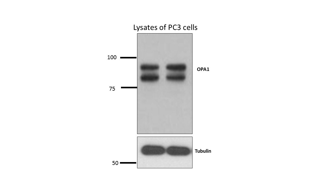
Application: Western BlotSample Tested: Human prostate cancer cell line (PC3)Species: HumanVerified Customer | Posted 07/25/2015OPA1 antibody testing
There are no reviews that match your criteria.
Protocols
View specific protocols for OPA1 Antibody (1E8-1D9) - BSA Free (NBP1-71656):
Immunocytochemistry Protocol
Culture cells to appropriate density in 35 mm culture dishes or 6-well plates.
1. Remove culture medium and wash the cells briefly in PBS. Add 10% formalin to the dish and fix at room temperature for 10 minutes.
2. Remove the formalin and wash the cells in PBS.
3. Permeablize the cells with 0.1% Triton X100 or other suitable detergent for 10 min.
4. Remove the permeablization buffer and wash three times for 10 minutes each in PBS. Be sure to not let the specimen dry out.
5. To block nonspecific antibody binding, incubate in 10% normal goat serum from 1 hour to overnight at room temperature.
6. Add primary antibody at appropriate dilution and incubate overnight at 4C.
7. Remove primary antibody and replace with PBS. Wash three times for 10 minutes each.
8. Add secondary antibody at appropriate dilution. Incubate for 1 hour at room temperature.
9. Remove secondary antibody and replace with PBS. Wash three times for 10 minutes each.
10. Counter stain DNA with DAPi if required.
Immunohistochemistry-Paraffin Embedded Sections
Antigen Unmasking:
Bring slides to a boil in 10 mM sodium citrate buffer (pH 6.0) then maintain at a sub-boiling temperature for 10 minutes. Cool slides on bench-top for 30 minutes (keep slides in the sodium citrate buffer at all times).
Staining:
1. Wash sections in deionized water three times for 5 minutes each.
2. Wash sections in PBS for 5 minutes.
3. Block each section with 100-400 ul blocking solution (1% BSA in PBS) for 1 hour at room temperature.
4. Remove blocking solution and add 100-400 ul diluted primary antibody. Incubate overnight at 4 C.
5. Remove antibody solution and wash sections in wash buffer three times for 5 minutes each.
6. Add 100-400 ul HRP polymer conjugated secondary antibody. Incubate 30 minutes at room temperature.
7. Wash sections three times in wash buffer for 5 minutes each.
8. Add 100-400 ul DAB substrate to each section and monitor staining closely.
9. As soon as the sections develop, immerse slides in deionized water.
10. Counterstain sections in hematoxylin.
11. Wash sections in deionized water two times for 5 minutes each.
12. Dehydrate sections.
13. Mount coverslips.
Western Blot Protocol
1. Perform SDS-PAGE on samples to be analyzed, loading 10-25 ug of total protein per lane.
2. Transfer proteins to PVDF membrane according to the instructions provided by the manufacturer of the membrane and transfer apparatus.
3. Stain the membrane with Ponceau S (or similar product) to assess transfer success, and mark molecular weight standards where appropriate.
4. Rinse the blot TBS -0.05% Tween 20 (TBST).
5. Block the membrane in 5% Non-fat milk in TBST (blocking buffer) for at least 1 hour.
6. Wash the membrane in TBST three times for 10 minutes each.
7. Dilute primary antibody in blocking buffer and incubate overnight at 4C with gentle rocking.
8. Wash the membrane in TBST three times for 10 minutes each.
9. Incubate the membrane in diluted HRP conjugated secondary antibody in blocking buffer (as per manufacturer's instructions) for 1 hour at room temperature.
10. Wash the blot in TBST three times for 10 minutes each (this step can be repeated as required to reduce background).
11. Apply the detection reagent of choice in accordance with the manufacturer's instructions.
Find general support by application which include: protocols, troubleshooting, illustrated assays, videos and webinars.
- Antigen Retrieval Protocol (PIER)
- Antigen Retrieval for Frozen Sections Protocol
- Appropriate Fixation of IHC/ICC Samples
- Cellular Response to Hypoxia Protocols
- Chromogenic IHC Staining of Formalin-Fixed Paraffin-Embedded (FFPE) Tissue Protocol
- Chromogenic Immunohistochemistry Staining of Frozen Tissue
- ClariTSA™ Fluorophore Kits
- Detection & Visualization of Antibody Binding
- Fluorescent IHC Staining of Frozen Tissue Protocol
- Graphic Protocol for Heat-induced Epitope Retrieval
- Graphic Protocol for the Preparation and Fluorescent IHC Staining of Frozen Tissue Sections
- Graphic Protocol for the Preparation and Fluorescent IHC Staining of Paraffin-embedded Tissue Sections
- Graphic Protocol for the Preparation of Gelatin-coated Slides for Histological Tissue Sections
- ICC Cell Smear Protocol for Suspension Cells
- ICC Immunocytochemistry Protocol Videos
- ICC for Adherent Cells
- IHC Sample Preparation (Frozen sections vs Paraffin)
- Immunocytochemistry (ICC) Protocol
- Immunocytochemistry Troubleshooting
- Immunofluorescence of Organoids Embedded in Cultrex Basement Membrane Extract
- Immunofluorescent IHC Staining of Formalin-Fixed Paraffin-Embedded (FFPE) Tissue Protocol
- Immunohistochemistry (IHC) and Immunocytochemistry (ICC) Protocols
- Immunohistochemistry Frozen Troubleshooting
- Immunohistochemistry Paraffin Troubleshooting
- Preparing Samples for IHC/ICC Experiments
- Preventing Non-Specific Staining (Non-Specific Binding)
- Primary Antibody Selection & Optimization
- Protocol for Heat-Induced Epitope Retrieval (HIER)
- Protocol for Making a 4% Formaldehyde Solution in PBS
- Protocol for VisUCyte™ HRP Polymer Detection Reagent
- Protocol for the Fluorescent ICC Staining of Cell Smears - Graphic
- Protocol for the Fluorescent ICC Staining of Cultured Cells on Coverslips - Graphic
- Protocol for the Preparation & Fixation of Cells on Coverslips
- Protocol for the Preparation and Chromogenic IHC Staining of Frozen Tissue Sections
- Protocol for the Preparation and Chromogenic IHC Staining of Frozen Tissue Sections - Graphic
- Protocol for the Preparation and Chromogenic IHC Staining of Paraffin-embedded Tissue Sections
- Protocol for the Preparation and Chromogenic IHC Staining of Paraffin-embedded Tissue Sections - Graphic
- Protocol for the Preparation and Fluorescent ICC Staining of Cells on Coverslips
- Protocol for the Preparation and Fluorescent ICC Staining of Non-adherent Cells
- Protocol for the Preparation and Fluorescent ICC Staining of Stem Cells on Coverslips
- Protocol for the Preparation and Fluorescent IHC Staining of Frozen Tissue Sections
- Protocol for the Preparation and Fluorescent IHC Staining of Paraffin-embedded Tissue Sections
- Protocol for the Preparation of Gelatin-coated Slides for Histological Tissue Sections
- Protocol for the Preparation of a Cell Smear for Non-adherent Cell ICC - Graphic
- R&D Systems Quality Control Western Blot Protocol
- TUNEL and Active Caspase-3 Detection by IHC/ICC Protocol
- The Importance of IHC/ICC Controls
- Troubleshooting Guide: Immunohistochemistry
- Troubleshooting Guide: Western Blot Figures
- Western Blot Conditions
- Western Blot Protocol
- Western Blot Protocol for Cell Lysates
- Western Blot Troubleshooting
- Western Blot Troubleshooting Guide
- View all Protocols, Troubleshooting, Illustrated assays and Webinars
Loading...