Opsin 1 (Medium Wave) Antibody - BSA Free
Novus Biologicals | Catalog # NBP2-29858

Key Product Details
Species Reactivity
Mouse, Rat
Applications
Western Blot, ELISA
Label
Unconjugated
Antibody Source
Polyclonal Chicken
Format
BSA Free
Loading...
Product Specifications
Immunogen
Synthetic peptides from mouse green opsin and zebra finch red opsin.
Specificity
Opsin, red and green. The antibody reacts with a protein of ~40 kDa.
Clonality
Polyclonal
Host
Chicken
Applications for Opsin 1 (Medium Wave) Antibody - BSA Free
Application
Recommended Usage
ELISA
1:100-1:2000
Western Blot
1:500:1:2000
Application Notes
ImmunoBlotting: on mouse brain extract. To minimize background staining it is suggested that you include a higher content of detergent (Tween-20) in your dilution and washing solutions. Immunohistochemistry: not tested. It is recommended that you try the antibody at 1:50-1:1,000.
Reviewed Applications
Read 2 reviews rated 1 using NBP2-29858 in the following applications:
Formulation, Preparation, and Storage
Purification
Immunogen affinity purified
Formulation
PBS
Format
BSA Free
Preservative
0.02% Sodium Azide
Concentration
1 mg/ml
Shipping
The product is shipped with polar packs. Upon receipt, store it immediately at the temperature recommended below.
Stability & Storage
Aliquot and store at -20C or -80C. Avoid freeze-thaw cycles.
Background: Opsin 1 (Medium Wave)
Alternate Names
GCP, GOP, Green cone photoreceptor pigment, Green-sensitive opsin, medium-wave-sensitive opsin 1, OPN1MW, opsin 1 (cone pigments), medium-wave-sensitive 2
Gene Symbol
OPN1MW
UniProt
Additional Opsin 1 (Medium Wave) Products
Product Documents for Opsin 1 (Medium Wave) Antibody - BSA Free
Certificate of Analysis
To download a Certificate of Analysis, please enter a lot or batch number in the search box below.
Product Specific Notices for Opsin 1 (Medium Wave) Antibody - BSA Free
Chicken products cannot be exported to Canada.
Relevant Entrez Gene Numbers: NM_020061.3
This product is for research use only and is not approved for use in humans or in clinical diagnosis. Primary Antibodies are guaranteed for 1 year from date of receipt.
Customer Reviews for Opsin 1 (Medium Wave) Antibody - BSA Free (2)
1 out of 5
2 Customer Ratings
Have you used Opsin 1 (Medium Wave) Antibody - BSA Free?
Submit a review and receive an Amazon gift card!
$25/€18/£15/$25CAN/¥2500 Yen for a review with an image
$10/€7/£6/$10CAN/¥1110 Yen for a review without an image
Submit a review
Customer Images
Showing
1
-
2 of
2 reviews
Showing All
Filter By:
-
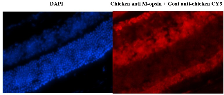
Application: Immunohistochemistry-FrozenSample Tested: mouse retinal tissuesSpecies: MouseVerified Customer | Posted 12/27/2016An image of retinal cryosection that I tried to label with the same antibody by Novus, but I did not get any signal from photoreceptor outer segments where the positive staining should be localized.Cryosection Primary: Chicken anti M-opsin 1:50 Secondary: Goat anti-chicken CY3 secondary 1:100 Procedure 1. Take the sections out from freezer and thaw the slides at room temperature for 10 minutes 2. Surround the tissues with a hydrophobic barrier (proper big lines!) using a barrier marker pen a. Let to dry properly 3. Rinse the sections in 2X in PBS (5 min each) 4. Drain the excess wash buffer 5. Incubate the sections in blocking buffer (10 % NGS) for 1 h at RT a. 50 µl of solution 6. Drain the excess blocking buffer 7. Incubate in primary antibody overnight at 4º C in a wet chamber. a. 0.01 M PBS + 1 % NGS as buffer b. 50 µl of solution 8. Next day rinse the sections in PBS+triton (0.25 %) for three times (5 min each) 9. Incubate with Secondary antibody prepared in PBS for 2 hrs. a. 50 µl of solution 10. Rinse the sections in PBST twice (5 min each) 11. Rinse the sections in PBS twice (5 min each) 12. Drain the excess wash buffer a. Air-dry for few mins 13. Mount with an anti-fade mounting media 14. Visualize using a fluorescence microscope

-
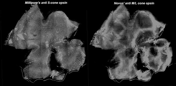
Application: Immunohistochemistry-Whole MountSample Tested: mouse retinal tissuesSpecies: MouseVerified Customer | Posted 12/27/2016Below is a figure of retinal whole mount stained by anti S-cone antibody from Merck Millipore and anti M-cone antibody by Novus. Millipore’s antibody stains cone photoreceptors distinguishly, but Novus’ does not.Tested in an untested application (IHC-whole mount) and earned the Innovator's Reward for this product. Immunostainning of the retinal flat mount Prim. antibodies: Anti M/L-cone opsin, chicken polyclonal, 1/50 dil., 1/100 dil. & 1/200 dil. Sec. antibodies: Goat anti-chicken CY3, 1/100 dil. 1. Fix the eye for 3 hours 2. Dissect retina from the eye 3. Wash overnight in PBS 4. Wash the tissue in PBST 2 times; 10 min each a. Triton-X 0.5 % 5. Block the whole mount in 10% normal goat serum (NGS) solution in PBST (1 hr) 6. Incubate o/n in primary antibody solution at 4 º C, primary ABs made in 1X PBST (0.05M) + 1% NGS 7. Next day wash in PBST + 1 % NGS; 3 X 10 min each 8. Incubate in secondary antibodies for 2-3 h a. Protect from light with tin 9. Wash in PBST + 1 % NGS; 2 X 10 min each 10. Wash once in PBS for at least 5 min 11. Transfer the whole mount on the microscope slide onto a slide retina side up. 12. Remove PBS, add mounting medium. 13. Place coverslip. Observe. If correct… 14. Secure with nail polish Samples are C57BL retinas
There are no reviews that match your criteria.
Protocols
Find general support by application which include: protocols, troubleshooting, illustrated assays, videos and webinars.
- Cellular Response to Hypoxia Protocols
- ELISA Sample Preparation & Collection Guide
- ELISA Troubleshooting Guide
- How to Run an R&D Systems DuoSet ELISA
- How to Run an R&D Systems Quantikine ELISA
- How to Run an R&D Systems Quantikine™ QuicKit™ ELISA
- Quantikine HS ELISA Kit Assay Principle, Alkaline Phosphatase
- Quantikine HS ELISA Kit Principle, Streptavidin-HRP Polymer
- R&D Systems Quality Control Western Blot Protocol
- Sandwich ELISA (Colorimetric) – Biotin/Streptavidin Detection Protocol
- Sandwich ELISA (Colorimetric) – Direct Detection Protocol
- Troubleshooting Guide: ELISA
- Troubleshooting Guide: Western Blot Figures
- Western Blot Conditions
- Western Blot Protocol
- Western Blot Protocol for Cell Lysates
- Western Blot Troubleshooting
- Western Blot Troubleshooting Guide
- View all Protocols, Troubleshooting, Illustrated assays and Webinars
Loading...