PER2 Antibody - BSA Free
Novus Biologicals | Catalog # NBP2-93587
![Western Blot: PER2 AntibodyAzide and BSA Free [NBP2-93587]](https://resources.rndsystems.com/images/products/PER2-Antibody-Western-Blot-NBP2-93587-img0002.jpg "Western Blot: PER2 AntibodyAzide and BSA Free [NBP2-93587]")
Loading...
Key Product Details
Species Reactivity
Human, Mouse, Rat, Amphibian
Applications
Immunohistochemistry, Immunohistochemistry-Paraffin, Immunohistochemistry-Frozen, Western Blot, ELISA, Chromatin Immunoprecipitation (ChIP)
Label
Unconjugated
Antibody Source
Polyclonal Rabbit IgG
Format
BSA Free
Loading...
Product Specifications
Immunogen
Recombinant fusion protein containing a sequence corresponding to amino acids 390-565 of human PER2 (NP_073728.1). GQPFDYSPIRFRARNGEYITLDTSWSSFINPWSRKISFIIGRHKVRVGPLNEDVFAAHPCTEEKALHPSIQELTEQIHRLLLQPVPHSGSSGYGSLGSNGSHEHLMSQTSSSDSNGHEDSRRRRAEICKNGNKTKNRSHYSHESGEQKKKSVTEMQTNPPAEKKAVPAMEKDSLGV
Reactivity Notes
Amphibian reactivity reported from a verified customer review.
Clonality
Polyclonal
Host
Rabbit
Isotype
IgG
Theoretical MW
137 kDa.
Disclaimer note: The observed molecular weight of the protein may vary from the listed predicted molecular weight due to post translational modifications, post translation cleavages, relative charges, and other experimental factors.
Disclaimer note: The observed molecular weight of the protein may vary from the listed predicted molecular weight due to post translational modifications, post translation cleavages, relative charges, and other experimental factors.
Scientific Data Images for PER2 Antibody - BSA Free
Western Blot: PER2 AntibodyAzide and BSA Free [NBP2-93587]
Western Blot: PER2 Antibody [NBP2-93587] - Analysis of extracts of 293T cells, using PER2 antibody at 1:1000 dilution.Secondary antibody: HRP Goat Anti-Rabbit IgG (H+L) at 1:10000 dilution.Lysates/proteins: 25ug per lane. Blocking buffer: 3% nonfat dry milk in TBST.Detection: ECL Basic Kit. Exposure time: 10s.![Immunohistochemistry-Paraffin: PER2 Antibody - Azide and BSA Free [NBP2-93587]](https://resources.rndsystems.com/images/products/PER2-Antibody-Immunohistochemistry-Paraffin-NBP2-93587-img0005.jpg "Immunohistochemistry-Paraffin: PER2 Antibody - Azide and BSA Free [NBP2-93587]")
Immunohistochemistry-Paraffin: PER2 Antibody - Azide and BSA Free [NBP2-93587]
Immunohistochemistry-Paraffin: PER2 Antibody [NBP2-93587] - Mouse testis using PER2 Rabbit pAb at dilution of 1:100 (40x lens).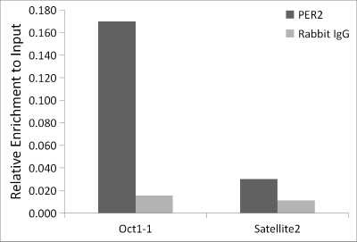
Chromatin Immunoprecipitation: PER2 Antibody [NBP2-93587] - Chromatin immunoprecipitation analysis of extracts of MCF7 cells, using PER2 antibody (NBP2-93587) and rabbit IgG.The amount of immunoprecipitated DNA was checked by quantitative PCR. Histogram was constructed by the ratios of the immunoprecipitated DNA to the input.
![Western Blot: PER2 AntibodyAzide and BSA Free [NBP2-93587]](https://resources.rndsystems.com/images/products/PER2-Antibody-Western-Blot-NBP2-93587-img0001.jpg "Western Blot: PER2 AntibodyAzide and BSA Free [NBP2-93587]")
Western Blot: PER2 AntibodyAzide and BSA Free [NBP2-93587]
Western Blot: PER2 Antibody [NBP2-93587] - Analysis of extracts of Mouse liver, using PER2 antibody at 1:1000 dilution.Secondary antibody: HRP Goat Anti-Rabbit IgG (H+L) at 1:10000 dilution.Lysates/proteins: 25ug per lane. Blocking buffer: 3% nonfat dry milk in TBST.Detection: ECL Basic Kit. Exposure time: 90s.![Immunohistochemistry-Paraffin: PER2 Antibody - Azide and BSA Free [NBP2-93587]](https://resources.rndsystems.com/images/products/PER2-Antibody-Immunohistochemistry-Paraffin-NBP2-93587-img0003.jpg "Immunohistochemistry-Paraffin: PER2 Antibody - Azide and BSA Free [NBP2-93587]")
Immunohistochemistry-Paraffin: PER2 Antibody - Azide and BSA Free [NBP2-93587]
Immunohistochemistry-Paraffin: PER2 Antibody [NBP2-93587] - Rat spleen using PER2 Rabbit pAb at dilution of 1:100 (40x lens).![Immunohistochemistry-Paraffin: PER2 Antibody - Azide and BSA Free [NBP2-93587]](https://resources.rndsystems.com/images/products/PER2-Antibody-Immunohistochemistry-Paraffin-NBP2-93587-img0004.jpg "Immunohistochemistry-Paraffin: PER2 Antibody - Azide and BSA Free [NBP2-93587]")
Immunohistochemistry-Paraffin: PER2 Antibody - Azide and BSA Free [NBP2-93587]
Immunohistochemistry-Paraffin: PER2 Antibody [NBP2-93587] - Human thyroid cancer using PER2 Rabbit pAb at dilution of 1:100 (40x lens).
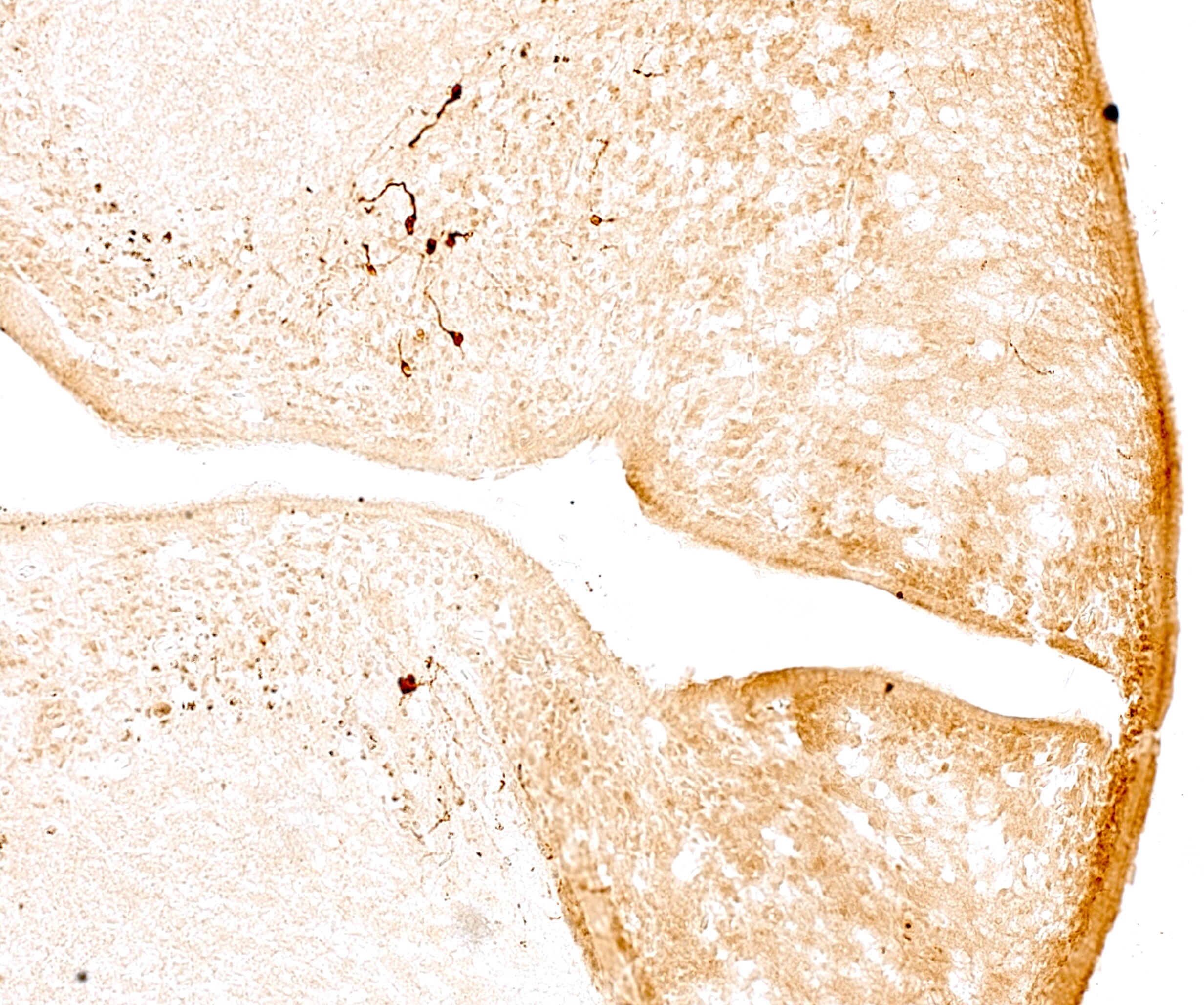
Immunohistochemistry-Frozen: Rabbit Polyclonal PER2 Antibody [NBP2-93587] -
Immunohistochemistry-Frozen: Rabbit Polyclonal PER2 Antibody [NBP2-93587] - 10X magnification. Immunoreactive cell bodies (DAB staining) along the 4th ventricle in 20um from Hyla cinerea. Image from a verified customer review.
Western Blot: PER2 Antibody - BSA Free [NBP2-93587] -
Circadian gene expression varies between pHGGs and pLGGs. (A) Interleaved scatter plots represent bulk RNA sequencing (rsem FPKM) analysis comparing circadian gene expression across pediatric low-grade glioma (pLGG, n = 426, blue) and pediatric high-grade glioma (pHGG, n = 211, red) samples from the Pediatric Brain Tumor Atlas (PBTA, Provisional). Data are expressed as mean +/- SEM, and expression values are log₂-transformed FPKM counts. Statistical analysis was performed using a two-way ANOVA with Benjamini and Hochberg false discovery rate (FDR) to determine differences in gene expression between pHGGs and pLGGs. Each dot represents an individual tumor sample. Asterisks indicate statistically significant differences (*P < 0.05), while “NS" denotes non-significant comparisons. (B) Western blots of four representative pediatric high-grade glioma tissue samples (left) and four pediatric low-grade glioma tissue samples (right) comparing BMAL1, CLOCK, PER2, and REV-ERB alpha protein expression (sections of separate blots displayed; full blots are shown in Supplementary Figure S3). Of note, tissue sample HGG 3 is derived from the same patient as the STN-49 cell line. (C) Normalization to beta -ACTIN control reveals BMAL1 (p < 0.05, 1.57 vs. 0.98) and CLOCK (p < 0.05, 0.98 vs. 0.39) protein expression is higher in high-grade gliomas compared to low-grade gliomas, REV-ERB alpha is lower (p < 0.05, 0.12 vs. 0.55), and there are no significant differences in PER2 protein expression. Data are expressed as mean +/- SEM. Image collected and cropped by CiteAb from the following open publication (https://www.nature.com/articles/s41598-025-17461-9), licensed under a CC-BY license. Not internally tested by Novus Biologicals.Applications for PER2 Antibody - BSA Free
Application
Recommended Usage
Chromatin Immunoprecipitation (ChIP)
5ug antibody for 10ug-15ug of Chromatin
ELISA
Recommended starting concentration is 1 μg/mL.
Immunohistochemistry
1:50 - 1:200
Immunohistochemistry-Frozen
Validated from a verified customer review
Immunohistochemistry-Paraffin
1:50 - 1:200
Western Blot
1:500 - 1:1000
Reviewed Applications
Read 1 review rated 5 using NBP2-93587 in the following applications:
Formulation, Preparation, and Storage
Purification
Affinity purified
Formulation
PBS (pH 7.3), 50% glycerol
Format
BSA Free
Preservative
0.02% Sodium Azide
Concentration
Please see the vial label for concentration. If unlisted please contact technical services.
Shipping
The product is shipped with polar packs. Upon receipt, store it immediately at the temperature recommended below.
Stability & Storage
Store at -20C. Avoid freeze-thaw cycles.
Background: PER2
Alternate Names
Circadian clock protein PERIOD 2, FASPS, hPER2, KIAA0347period 2, period (Drosophila) homolog 2, period circadian protein 2, period circadian protein homolog 2, period homolog 2 (Drosophila)
Gene Symbol
PER2
Additional PER2 Products
Product Documents for PER2 Antibody - BSA Free
Certificate of Analysis
To download a Certificate of Analysis, please enter a lot or batch number in the search box below.
Product Specific Notices for PER2 Antibody - BSA Free
This product is for research use only and is not approved for use in humans or in clinical diagnosis. Primary Antibodies are guaranteed for 1 year from date of receipt.
⚠ WARNING: This product can expose you to chemicals including Methotrexate, which is known to the State of California to cause reproductive toxicity with developmental effects. For more information, go to www.P65Warnings.ca.govCitations for PER2 Antibody - BSA Free
 Powered by Bioz
Customer Reviews for PER2 Antibody - BSA Free (1)
5 out of 5
1 Customer Rating
Have you used PER2 Antibody - BSA Free?
Submit a review and receive an Amazon gift card!
$25/€18/£15/$25CAN/¥2500 Yen for a review with an image
$10/€7/£6/$10CAN/¥1110 Yen for a review without an image
Submit a review
Customer Images
Showing
1
-
1 of
1 review
Showing All
Filter By:
-
Application: Immunohistochemistry-FrozenSample Tested: brain sectionSpecies: OtherVerified Customer | Posted 09/11/202310X magnification. Immunoreactive cell bodies (DAB staining) along the 4th ventricle in 20um from Hyla cinerea.
 Bio-Techne ResponseThis review was submitted through the legacy Novus Innovators Program, reflecting a new species or application tested on a primary antibody.
There are no reviews that match your criteria.
Protocols
Find general support by application which include: protocols, troubleshooting, illustrated assays, videos and webinars.
- Antigen Retrieval Protocol (PIER)
- Antigen Retrieval for Frozen Sections Protocol
- Appropriate Fixation of IHC/ICC Samples
- Cellular Response to Hypoxia Protocols
- ChIP Protocol Video
- Chromatin Immunoprecipitation (ChIP) Protocol
- Chromatin Immunoprecipitation Protocol
- Chromogenic IHC Staining of Formalin-Fixed Paraffin-Embedded (FFPE) Tissue Protocol
- Chromogenic Immunohistochemistry Staining of Frozen Tissue
- ClariTSA™ Fluorophore Kits
- Detection & Visualization of Antibody Binding
- ELISA Sample Preparation & Collection Guide
- ELISA Troubleshooting Guide
- Fluorescent IHC Staining of Frozen Tissue Protocol
- Graphic Protocol for Heat-induced Epitope Retrieval
- Graphic Protocol for the Preparation and Fluorescent IHC Staining of Frozen Tissue Sections
- Graphic Protocol for the Preparation and Fluorescent IHC Staining of Paraffin-embedded Tissue Sections
- Graphic Protocol for the Preparation of Gelatin-coated Slides for Histological Tissue Sections
- How to Run an R&D Systems DuoSet ELISA
- How to Run an R&D Systems Quantikine ELISA
- How to Run an R&D Systems Quantikine™ QuicKit™ ELISA
- IHC Sample Preparation (Frozen sections vs Paraffin)
- Immunofluorescent IHC Staining of Formalin-Fixed Paraffin-Embedded (FFPE) Tissue Protocol
- Immunohistochemistry (IHC) and Immunocytochemistry (ICC) Protocols
- Immunohistochemistry Frozen Troubleshooting
- Immunohistochemistry Paraffin Troubleshooting
- Preparing Samples for IHC/ICC Experiments
- Preventing Non-Specific Staining (Non-Specific Binding)
- Primary Antibody Selection & Optimization
- Protocol for Heat-Induced Epitope Retrieval (HIER)
- Protocol for Making a 4% Formaldehyde Solution in PBS
- Protocol for VisUCyte™ HRP Polymer Detection Reagent
- Protocol for the Preparation & Fixation of Cells on Coverslips
- Protocol for the Preparation and Chromogenic IHC Staining of Frozen Tissue Sections
- Protocol for the Preparation and Chromogenic IHC Staining of Frozen Tissue Sections - Graphic
- Protocol for the Preparation and Chromogenic IHC Staining of Paraffin-embedded Tissue Sections
- Protocol for the Preparation and Chromogenic IHC Staining of Paraffin-embedded Tissue Sections - Graphic
- Protocol for the Preparation and Fluorescent IHC Staining of Frozen Tissue Sections
- Protocol for the Preparation and Fluorescent IHC Staining of Paraffin-embedded Tissue Sections
- Protocol for the Preparation of Gelatin-coated Slides for Histological Tissue Sections
- Quantikine HS ELISA Kit Assay Principle, Alkaline Phosphatase
- Quantikine HS ELISA Kit Principle, Streptavidin-HRP Polymer
- R&D Systems Quality Control Western Blot Protocol
- Sandwich ELISA (Colorimetric) – Biotin/Streptavidin Detection Protocol
- Sandwich ELISA (Colorimetric) – Direct Detection Protocol
- TUNEL and Active Caspase-3 Detection by IHC/ICC Protocol
- The Importance of IHC/ICC Controls
- Troubleshooting Guide: ELISA
- Troubleshooting Guide: Immunohistochemistry
- Troubleshooting Guide: Western Blot Figures
- Western Blot Conditions
- Western Blot Protocol
- Western Blot Protocol for Cell Lysates
- Western Blot Troubleshooting
- Western Blot Troubleshooting Guide
- View all Protocols, Troubleshooting, Illustrated assays and Webinars
Loading...