![Western Blot: PGAM5 AntibodyAzide and BSA Free [NBP2-93600]](https://resources.rndsystems.com/images/products/PGAM5-Antibody-Western-Blot-NBP2-93600-img0007.jpg "Western Blot: PGAM5 AntibodyAzide and BSA Free [NBP2-93600]")
Loading...
Key Product Details
Species Reactivity
Human, Mouse, Rat
Applications
Immunohistochemistry, Immunohistochemistry-Paraffin, Western Blot, Immunocytochemistry/ Immunofluorescence
Label
Unconjugated
Antibody Source
Polyclonal Rabbit IgG
Format
Azide and BSA Free
Loading...
Product Specifications
Immunogen
Recombinant fusion protein containing a sequence corresponding to amino acids 50-146 of human PGAM5 (NP_001164014.1). GARPGPGVWDPNWDRREPLSLINVRKRNVESGEEELASKLDHYKAKATRHIFLIRHSQYHVDGSLEKDRTLTPLGREQAELTGLRLASLGLKFNKIV
Clonality
Polyclonal
Host
Rabbit
Isotype
IgG
Scientific Data Images for PGAM5 Antibody - Azide and BSA Free
Western Blot: PGAM5 AntibodyAzide and BSA Free [NBP2-93600]
Western Blot: PGAM5 Antibody [NBP2-93600] - Analysis of various lysates, using PGAM5 antibody at 1:500 dilution.Secondary antibody: HRP Goat Anti-Rabbit IgG (H+L) at 1:10000 dilution.Lysates/proteins: 25ug per lane. Blocking buffer: 3% nonfat dry milk in TBST.Detection: ECL Basic Kit. Exposure time: 1s.
Immunocytochemistry/ Immunofluorescence: PGAM5 Antibody - Azide and BSA Free [PGAM5] -
Immunocytochemistry/ Immunofluorescence: PGAM5 Antibody - Azide and BSA Free [PGAM5] - Immunofluorescence analysis of MCF7 cells using PGAM5 Rabbit pAb at dilution of 1:20 (40x lens). Secondary antibody: Cy3-conjugated Goat anti-Rabbit IgG (H+L) at 1:500 dilution. Blue: DAPI for nuclear staining.![Immunohistochemistry-Paraffin: PGAM5 Antibody - Azide and BSA Free [NBP2-93600]](https://resources.rndsystems.com/images/products/PGAM5-Antibody-Immunohistochemistry-Paraffin-NBP2-93600-img0003.jpg "Immunohistochemistry-Paraffin: PGAM5 Antibody - Azide and BSA Free [NBP2-93600]")
Immunohistochemistry-Paraffin: PGAM5 Antibody - Azide and BSA Free [NBP2-93600]
Immunohistochemistry-Paraffin: PGAM5 Antibody [NBP2-93600] - Human tonsil using PGAM5 Rabbit pAb (NBP2-93600) at dilution of 1:25 (40x lens). Perform high pressure antigen retrieval with 10 mM citrate buffer pH 6.0 before commencing with IHC staining protocol.![Immunohistochemistry-Paraffin: PGAM5 Antibody - Azide and BSA Free [NBP2-93600]](https://resources.rndsystems.com/images/products/PGAM5-Antibody-Immunohistochemistry-Paraffin-NBP2-93600-img0001.jpg "Immunohistochemistry-Paraffin: PGAM5 Antibody - Azide and BSA Free [NBP2-93600]")
Immunohistochemistry-Paraffin: PGAM5 Antibody - Azide and BSA Free [NBP2-93600]
Immunohistochemistry-Paraffin: PGAM5 Antibody [NBP2-93600] - Human colon carcinoma using PGAM5 Rabbit pAb (NBP2-93600) at dilution of 1:25 (40x lens). Perform high pressure antigen retrieval with 10 mM citrate buffer pH 6.0 before commencing with IHC staining protocol.![Immunohistochemistry-Paraffin: PGAM5 Antibody - Azide and BSA Free [NBP2-93600]](https://resources.rndsystems.com/images/products/PGAM5-Antibody-Immunohistochemistry-Paraffin-NBP2-93600-img0002.jpg "Immunohistochemistry-Paraffin: PGAM5 Antibody - Azide and BSA Free [NBP2-93600]")
Immunohistochemistry-Paraffin: PGAM5 Antibody - Azide and BSA Free [NBP2-93600]
Immunohistochemistry-Paraffin: PGAM5 Antibody [NBP2-93600] - Human liver cancer using PGAM5 Rabbit pAb (NBP2-93600) at dilution of 1:25 (40x lens). Perform high pressure antigen retrieval with 10 mM citrate buffer pH 6.0 before commencing with IHC staining protocol.
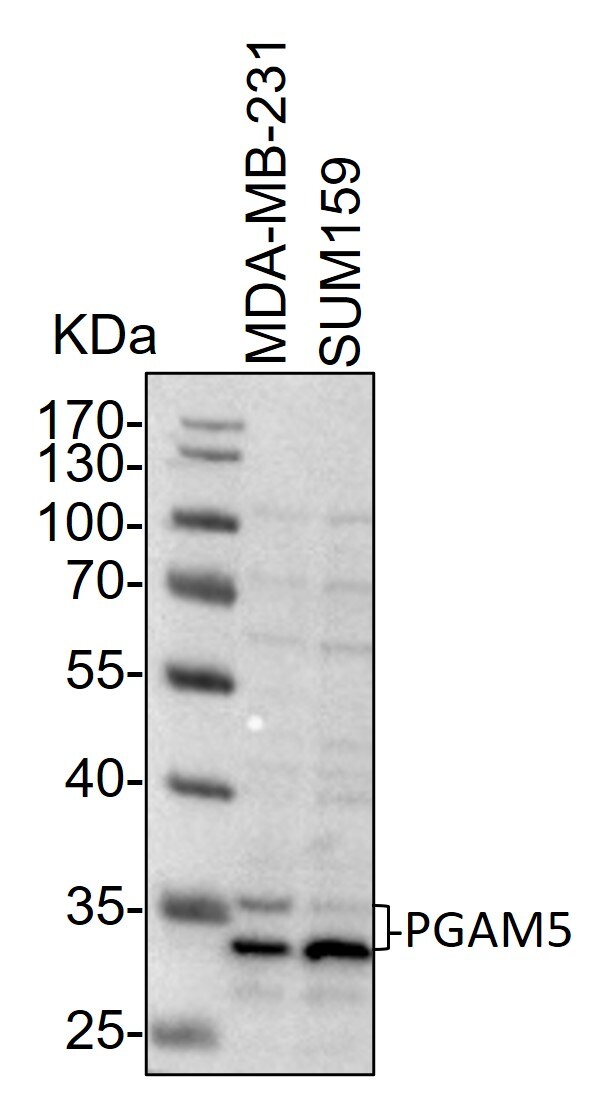
Western Blot: Rabbit Polyclonal PGAM5 Antibody [NBP2-93600] -
Western Blot: Rabbit Polyclonal PGAM5 Antibody [NBP2-93600] - Whole cell lysates from MDA-MB-231 and SUM159 cells were loaded with 50 ug/lane. 10% SDS-PAGE. PGAM5 Antibody (NBP2-93600) was used for primary antibody: 1:1000, 4℃, overnight. Image from a verified customer review.Applications for PGAM5 Antibody - Azide and BSA Free
Application
Recommended Usage
Immunocytochemistry/ Immunofluorescence
1:50 - 1:200
Immunohistochemistry
1:50 - 1:200
Western Blot
1:100 - 1:500
Reviewed Applications
Read 1 review rated 5 using NBP2-93600 in the following applications:
Formulation, Preparation, and Storage
Purification
Affinity purified
Formulation
PBS (pH 7.3), 50% glycerol
Format
Azide and BSA Free
Preservative
0.02% Sodium Azide
Concentration
Please see the vial label for concentration. If unlisted please contact technical services.
Shipping
The product is shipped with polar packs. Upon receipt, store it immediately at the temperature recommended below.
Stability & Storage
Store at -20C. Avoid freeze-thaw cycles.
Background: PGAM5
Alternate Names
Bcl-XL-binding protein v68, BXLBv68, EC 3.1.3.16, MGC5352, phosphoglycerate mutase family member 5BXLBV68, serine/threonine-protein phosphatase PGAM5, mitochondrial
Gene Symbol
PGAM5
Additional PGAM5 Products
Product Documents for PGAM5 Antibody - Azide and BSA Free
Certificate of Analysis
To download a Certificate of Analysis, please enter a lot or batch number in the search box below.
Product Specific Notices for PGAM5 Antibody - Azide and BSA Free
This product is for research use only and is not approved for use in humans or in clinical diagnosis. Primary Antibodies are guaranteed for 1 year from date of receipt.
Customer Reviews for PGAM5 Antibody - Azide and BSA Free (1)
5 out of 5
1 Customer Rating
Have you used PGAM5 Antibody - Azide and BSA Free?
Submit a review and receive an Amazon gift card!
$25/€18/£15/$25CAN/¥2500 Yen for a review with an image
$10/€7/£6/$10CAN/¥1110 Yen for a review without an image
Submit a review
Customer Images
Showing
1
-
1 of
1 review
Showing All
Filter By:
-
Application: Western BlotVerified Customer | Posted 12/20/2023Western Blot: whole cell lysates from MDA-MB-231 and SUM159 cells were loaded with 50 ug/lane. 10% SDS-PAGE. PGAM5 Antibody (NBP2-93600) was used for primary antibody: 1:1000, 4℃, overnight.

There are no reviews that match your criteria.
Protocols
Find general support by application which include: protocols, troubleshooting, illustrated assays, videos and webinars.
- Antigen Retrieval Protocol (PIER)
- Antigen Retrieval for Frozen Sections Protocol
- Appropriate Fixation of IHC/ICC Samples
- Cellular Response to Hypoxia Protocols
- Chromogenic IHC Staining of Formalin-Fixed Paraffin-Embedded (FFPE) Tissue Protocol
- Chromogenic Immunohistochemistry Staining of Frozen Tissue
- ClariTSA™ Fluorophore Kits
- Detection & Visualization of Antibody Binding
- Fluorescent IHC Staining of Frozen Tissue Protocol
- Graphic Protocol for Heat-induced Epitope Retrieval
- Graphic Protocol for the Preparation and Fluorescent IHC Staining of Frozen Tissue Sections
- Graphic Protocol for the Preparation and Fluorescent IHC Staining of Paraffin-embedded Tissue Sections
- Graphic Protocol for the Preparation of Gelatin-coated Slides for Histological Tissue Sections
- ICC Cell Smear Protocol for Suspension Cells
- ICC Immunocytochemistry Protocol Videos
- ICC for Adherent Cells
- IHC Sample Preparation (Frozen sections vs Paraffin)
- Immunocytochemistry (ICC) Protocol
- Immunocytochemistry Troubleshooting
- Immunofluorescence of Organoids Embedded in Cultrex Basement Membrane Extract
- Immunofluorescent IHC Staining of Formalin-Fixed Paraffin-Embedded (FFPE) Tissue Protocol
- Immunohistochemistry (IHC) and Immunocytochemistry (ICC) Protocols
- Immunohistochemistry Frozen Troubleshooting
- Immunohistochemistry Paraffin Troubleshooting
- Preparing Samples for IHC/ICC Experiments
- Preventing Non-Specific Staining (Non-Specific Binding)
- Primary Antibody Selection & Optimization
- Protocol for Heat-Induced Epitope Retrieval (HIER)
- Protocol for Making a 4% Formaldehyde Solution in PBS
- Protocol for VisUCyte™ HRP Polymer Detection Reagent
- Protocol for the Fluorescent ICC Staining of Cell Smears - Graphic
- Protocol for the Fluorescent ICC Staining of Cultured Cells on Coverslips - Graphic
- Protocol for the Preparation & Fixation of Cells on Coverslips
- Protocol for the Preparation and Chromogenic IHC Staining of Frozen Tissue Sections
- Protocol for the Preparation and Chromogenic IHC Staining of Frozen Tissue Sections - Graphic
- Protocol for the Preparation and Chromogenic IHC Staining of Paraffin-embedded Tissue Sections
- Protocol for the Preparation and Chromogenic IHC Staining of Paraffin-embedded Tissue Sections - Graphic
- Protocol for the Preparation and Fluorescent ICC Staining of Cells on Coverslips
- Protocol for the Preparation and Fluorescent ICC Staining of Non-adherent Cells
- Protocol for the Preparation and Fluorescent ICC Staining of Stem Cells on Coverslips
- Protocol for the Preparation and Fluorescent IHC Staining of Frozen Tissue Sections
- Protocol for the Preparation and Fluorescent IHC Staining of Paraffin-embedded Tissue Sections
- Protocol for the Preparation of Gelatin-coated Slides for Histological Tissue Sections
- Protocol for the Preparation of a Cell Smear for Non-adherent Cell ICC - Graphic
- R&D Systems Quality Control Western Blot Protocol
- TUNEL and Active Caspase-3 Detection by IHC/ICC Protocol
- The Importance of IHC/ICC Controls
- Troubleshooting Guide: Immunohistochemistry
- Troubleshooting Guide: Western Blot Figures
- Western Blot Conditions
- Western Blot Protocol
- Western Blot Protocol for Cell Lysates
- Western Blot Troubleshooting
- Western Blot Troubleshooting Guide
- View all Protocols, Troubleshooting, Illustrated assays and Webinars
Loading...