PINK1 Antibody - BSA Free
Novus Biologicals | Catalog # NBP1-49678
![Immunocytochemistry/ Immunofluorescence: PINK1 Antibody - BSA Free [NBP1-49678]](https://resources.rndsystems.com/images/products/PINK1-Antibody-Immunocytochemistry-Immunofluorescence-NBP1-49678-img0002.jpg "Immunocytochemistry/ Immunofluorescence: PINK1 Antibody - BSA Free [NBP1-49678]")
Key Product Details
Species Reactivity
Validated:
Human, Mouse
Cited:
Mouse
Applications
Validated:
Immunohistochemistry, Immunohistochemistry-Paraffin, Western Blot, Immunocytochemistry/ Immunofluorescence, Simple Western
Cited:
Western Blot
Label
Unconjugated
Antibody Source
Polyclonal Rabbit IgG
Format
BSA Free
Loading...
Product Specifications
Immunogen
PINK1 antibody was developed using a synthetic protein made to an internal region of the human PINK1 protein (within residues 350-500). [Swiss-Prot Q9BXM7]
Localization
Mitochondrion outer membrane; Single-pass membrane protein. Cytoplasm - cytosol
Specificity
Reactivity expected for both isotype 1 and 2.
Clonality
Polyclonal
Host
Rabbit
Isotype
IgG
Theoretical MW
62.7 kDa.
Disclaimer note: The observed molecular weight of the protein may vary from the listed predicted molecular weight due to post translational modifications, post translation cleavages, relative charges, and other experimental factors.
Disclaimer note: The observed molecular weight of the protein may vary from the listed predicted molecular weight due to post translational modifications, post translation cleavages, relative charges, and other experimental factors.
Scientific Data Images for PINK1 Antibody - BSA Free
Immunocytochemistry/ Immunofluorescence: PINK1 Antibody - BSA Free [NBP1-49678]
Immunocytochemistry/Immunofluorescence: PINK1 Antibody [NBP1-49678] - PINK1 antibody was tested in HepG2 cells with DyLight 488 (green). Nuclei and alpha-tubulin were counterstained with DAPI (blue) and DyLight 550 (red).![Simple Western: PINK1 AntibodyBSA Free [NBP1-49678]](https://resources.rndsystems.com/images/products/PINK1-Antibody-Simple-Western-NBP1-49678-img0006.jpg "Simple Western: PINK1 AntibodyBSA Free [NBP1-49678]")
Simple Western: PINK1 AntibodyBSA Free [NBP1-49678]
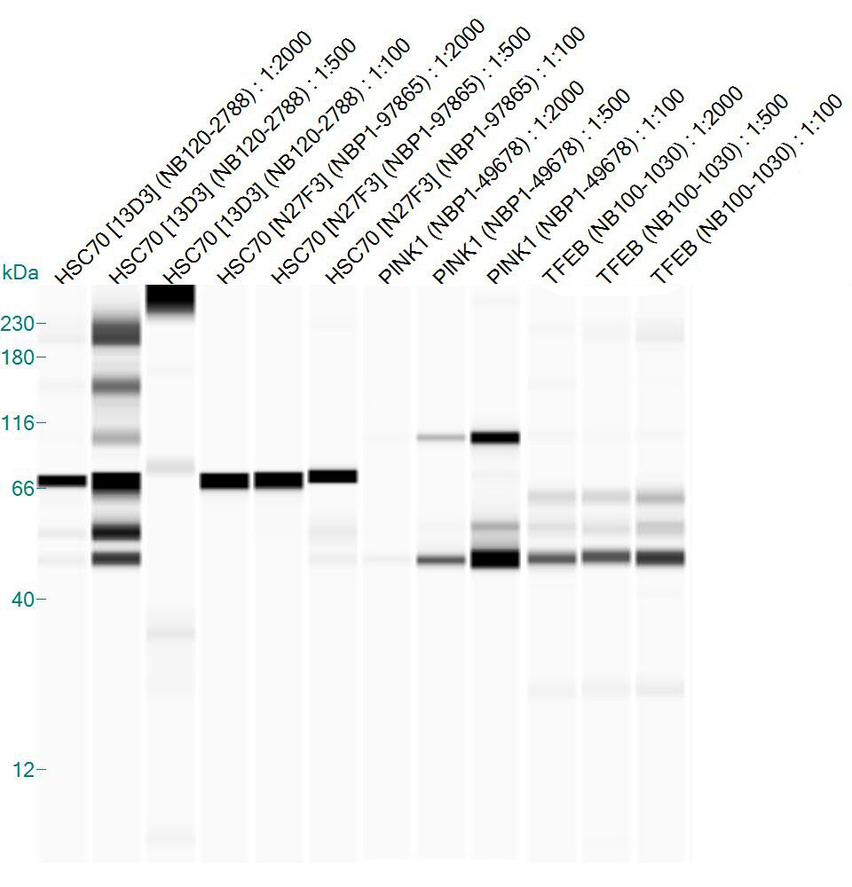
Simple Western: PINK1 Antibody [NBP1-49678] - Lane view shows a specific band for PINK1 at a dilution of 1:50 in 1.0 mg/ml of HeLa lysate. Molecular weight ~61kDa. This experiment was performed under reducing conditions using the 12-230kDa separation system. * Non-specific interaction with the 230 kDa Simple Western standard may be seen with this antibody.![Immunohistochemistry-Paraffin: PINK1 Antibody - BSA Free [NBP1-49678]](https://resources.rndsystems.com/images/products/PINK1-Antibody-Immunohistochemistry-Paraffin-NBP1-49678-img0004.jpg "Immunohistochemistry-Paraffin: PINK1 Antibody - BSA Free [NBP1-49678]")
Immunohistochemistry-Paraffin: PINK1 Antibody - BSA Free [NBP1-49678]
Immunohistochemistry-Paraffin: PINK1 Antibody [NBP1-49678] - Stain in paraffin embedded mouse brain.![Immunocytochemistry/ Immunofluorescence: PINK1 Antibody - BSA Free [NBP1-49678]](https://resources.rndsystems.com/images/products/PINK1-Antibody-Immunocytochemistry-Immunofluorescence-NBP1-49678-img0005.jpg "Immunocytochemistry/ Immunofluorescence: PINK1 Antibody - BSA Free [NBP1-49678]")
Immunocytochemistry/ Immunofluorescence: PINK1 Antibody - BSA Free [NBP1-49678]
Immunocytochemistry/Immunofluorescence: PINK1 Antibody [NBP1-49678] - PINK1 antibody was tested at 1:50 in HeLa cells with DyLight 488 (green). Nuclei and alpha-tubulin were counterstained with DAPI (blue) and DyLight 550 (red). Image objective 40x.Applications for PINK1 Antibody - BSA Free
Application
Recommended Usage
Immunocytochemistry/ Immunofluorescence
1:50-1:1000
Immunohistochemistry
1:100
Immunohistochemistry-Paraffin
1:100
Simple Western
1:50
Western Blot
1-2 ug/ml
Application Notes
This PINK1 antibody is useful for IHC and ICC/IF. Prior to immunostaining paraffin tissues, antigen retrieval with sodium citrate buffer (pH 6.0) is recommended.
In Simple Western only 10 - 15 uL of the recommended dilution is used per data point.
See Simple Western Antibody Database for Simple Western validation: Tested in HeLa lysate 1.0 mg/mL, separated by Size, antibody dilution of 1:50, apparent MW was 61 kDa. Separated by Size-Wes, Sally Sue/Peggy Sue. Unprocessed PINK1 is 63 kDa which undergoes proteolytic processing to generate 55 kDa and 42 kDa cleaved forms, and bands at the mentioned positions may be expected in Western blot application.
Reviewed Applications
Read 1 review rated 4 using NBP1-49678 in the following applications:
Formulation, Preparation, and Storage
Purification
Immunogen affinity purified
Formulation
PBS
Format
BSA Free
Preservative
0.02% Sodium Azide
Concentration
1.0 mg/ml
Shipping
The product is shipped with polar packs. Upon receipt, store it immediately at the temperature recommended below.
Stability & Storage
Store at 4C short term. Aliquot and store at -20C long term. Avoid freeze-thaw cycles.
Background: PINK1
PINK1 (PTEN induced putative kinase 1) protein contains a N-terminal mitochondrial targeting sequence, putative transmembrane helix, linker region, serine (Ser65)/threonine (Thr257) kinase domain and C-terminal segment. PINK1 is translated in the cytosol, then translocated to the outer mitochondrial membrane where it is rapidly cleaved and degraded as a part of normal mitochondrial function. In damaged (depolarized) mitochondria, PINK1 becomes stabilized and accumulates, resulting in the subsequent phosphorylation of numerous proteins on the mitochondrial surface.
When PINK1 is imported into the cell, mitochondrial processing peptidase, presenilin-associated rhomboid-like protease and AFG3L2 cleave PINK1 and tag it for the ubiquitin-proteasome pathway, keeping low PINK1 protein expression at basal conditions (1,2). Accumulation of PINK1 in mitochondria indicate damage. PINK1 maintains mitochondrial function/integrity, provides protection against mitochondrial dysfunction during cellular stress, and is involved in the clearance of damaged mitochondria via selective autophagy (mitophagy) (3). PINK1 has a theoretical molecular weight of 63 kDa and undergoes proteolytic processing to generate at least two cleaved forms (55 kDa and 42 kDa).
Ultimately PARK2 (E3 Ubiquitin Ligase Parkin) is recruited to the damaged mitochondria where it is activated by 1) PINK-mediated phosphorylation of PARK2 at serine 65, and 2) PARK2 interaction with phosphorylated ubiquitin (also phosphorylated by PINK1 on serine 65) (4,5). There is a strong interplay between Parkin and PINK1, where loss-of-function of human PINK1 results in mitochondrial pathology and can be rescued by Parkin (2,4,5). Mutations in either Parkin or PINK1 alter mitochondrial turnover, resulting in the accumulation of defective mitochondria and, ultimately, neurodegeneration in Parkinson's disease. Mutations in the PINK1 gene located within the PARK6 locus on chromosome 1p35-p36 have been identified in patients with early-onset Parkinson's disease (6).
References
1.Rasool, S., Soya, N., Truong, L., Croteau, N., Lukacs, G. L., & Trempe, J. F. (2018). PINK1 autophosphorylation is required for ubiquitin recognition. EMBO Rep, 19(4). doi:10.15252/embr.201744981
2.Shiba-Fukushima, K., Arano, T., Matsumoto, G., Inoshita, T., Yoshida, S., Ishihama, Y.,... Imai, Y. (2014). Phosphorylation of mitochondrial polyubiquitin by PINK1 promotes Parkin mitochondrial tethering. PLoS Genet, 10(12), e1004861. doi:10.1371/journal.pgen.1004861
3.Vives-Bauza, C., Zhou, C., Huang, Y., Cui, M., de Vries, R. L., Kim, J.,... Przedborski, S. (2010). PINK1-dependent recruitment of Parkin to mitochondria in mitophagy. Proc Natl Acad Sci U S A, 107(1), 378-383. doi:10.1073/pnas.0911187107
4.McWilliams, T. G., Barini, E., Pohjolan-Pirhonen, R., Brooks, S. P., Singh, F., Burel, S.,... Muqit, M. M. K. (2018). Phosphorylation of Parkin at serine 65 is essential for its activation in vivo. Open Biol, 8(11). doi:10.1098/rsob.180108
5.Exner, N., Treske, B., Paquet, D., Holmstrom, K., Schiesling, C., Gispert, S.,... Haass, C. (2007). Loss-of-function of human PINK1 results in mitochondrial pathology and can be rescued by parkin. J Neurosci, 27(45), 12413-12418. doi:10.1523/jneurosci.0719-07.2007
6.Valente, E. M., Bentivoglio, A. R., Dixon, P. H., Ferraris, A., Ialongo, T., Frontali, M.,... Wood, N. W. (2001). Localization of a novel locus for autosomal recessive early-onset parkinsonism, PARK6, on human chromosome 1p35-p36. Am J Hum Genet, 68(4), 895-900. doi:10.1086/319522
Long Name
PTEN-induced Putative Kinase 1
Alternate Names
BRPK, PARK6
Gene Symbol
PINK1
UniProt
Additional PINK1 Products
Product Documents for PINK1 Antibody - BSA Free
Certificate of Analysis
To download a Certificate of Analysis, please enter a lot or batch number in the search box below.
Product Specific Notices for PINK1 Antibody - BSA Free
Manufactured by Genomic Antibody Technology™. GAT FAQs
This product is for research use only and is not approved for use in humans or in clinical diagnosis. Primary Antibodies are guaranteed for 1 year from date of receipt.
Related Research Areas
Citations for PINK1 Antibody - BSA Free
 Powered by Bioz
Customer Reviews for PINK1 Antibody - BSA Free (1)
4 out of 5
1 Customer Rating
Have you used PINK1 Antibody - BSA Free?
Submit a review and receive an Amazon gift card!
$25/€18/£15/$25CAN/¥2500 Yen for a review with an image
$10/€7/£6/$10CAN/¥1110 Yen for a review without an image
Submit a review
Customer Images
Showing
1
-
1 of
1 review
Showing All
Filter By:
-
Application: Simple WesternSample Tested: whole brain homogenates from miceSpecies: MouseVerified Customer | Posted 01/21/2016Simple Western: PINK1 expression in mouse brain lysate

There are no reviews that match your criteria.
Protocols
View specific protocols for PINK1 Antibody - BSA Free (NBP1-49678):
Immunocytochemistry Protocol
Culture cells to appropriate density in 35 mm culture dishes or 6-well plates.
1. Remove culture medium and add 10% formalin to the dish. Fix at room temperature for 30 minutes.
2. Remove the formalin and add ice cold methanol. Incubate for 5-10 minutes.
3. Remove methanol and add washing solution (i.e. PBS). Be sure to not let the specimen dry out. Wash three times for 10 minutes.
4. To block nonspecific antibody binding incubate in 10% normal goat serum from 1 hour to overnight at room temperature.
5. Add primary antibody at appropriate dilution and incubate at room temperature from 2 hours to overnight at room temperature.
6. Remove primary antibody and replace with washing solution. Wash three times for 10 minutes.
7. Add secondary antibody at appropriate dilution. Incubate for 1 hour at room temperature.
8. Remove antibody and replace with wash solution, then wash for 10 minutes. Add Hoechst 33258 to wash solution at 1:25,0000 and incubate for 10 minutes. Wash a third time for 10 minutes.
9. Cells can be viewed directly after washing. The plates can also be stored in PBS containing Azide covered in Parafilm (TM). Cells can also be cover-slipped using Fluoromount, with appropriate sealing.
*The above information is only intended as a guide. The researcher should determine what protocol best meets their needs. Please follow safe laboratory procedures.
Immunohistochemistry-Paraffin Embedded Sections
Antigen Unmasking:
Bring slides to a boil in 10 mM sodium citrate buffer (pH 6.0) then maintain at a sub-boiling temperature for 10 minutes. Cool slides on bench-top for 30 minutes.
Staining:
1. Wash sections in deionized water three times for 5 minutes each.
2. Wash sections in wash buffer for 5 minutes.
3. Block each section with 100-400 ul blocking solution for 1 hour at room temperature.
4. Remove blocking solution and add 100-400 ul diluted primary antibody. Incubate overnight at 4C.
5. Remove antibody solution and wash sections in wash buffer three times for 5 minutes each.
6. Add 100-400 ul biotinylated diluted secondary antibody. Incubate 30 minutes at room temperature.
7. Remove secondary antibody solution and wash sections three times with wash buffer for 5 minutes each.
8. Add 100-400 ul Streptavidin-HRP reagent to each section and incubate for 30 minutes at room temperature.
9. Wash sections three times in wash buffer for 5 minutes each.
10. Add 100-400 ul DAB substrate to each section and monitor staining closely.
11. As soon as the sections develop, immerse slides in deionized water.
12. Counterstain sections in hematoxylin.
13. Wash sections in deionized water two times for 5 minutes each.
14. Dehydrate sections.
15. Mount coverslips.
Find general support by application which include: protocols, troubleshooting, illustrated assays, videos and webinars.
- Antigen Retrieval Protocol (PIER)
- Antigen Retrieval for Frozen Sections Protocol
- Appropriate Fixation of IHC/ICC Samples
- Cellular Response to Hypoxia Protocols
- Chromogenic IHC Staining of Formalin-Fixed Paraffin-Embedded (FFPE) Tissue Protocol
- Chromogenic Immunohistochemistry Staining of Frozen Tissue
- ClariTSA™ Fluorophore Kits
- Detection & Visualization of Antibody Binding
- Fluorescent IHC Staining of Frozen Tissue Protocol
- Graphic Protocol for Heat-induced Epitope Retrieval
- Graphic Protocol for the Preparation and Fluorescent IHC Staining of Frozen Tissue Sections
- Graphic Protocol for the Preparation and Fluorescent IHC Staining of Paraffin-embedded Tissue Sections
- Graphic Protocol for the Preparation of Gelatin-coated Slides for Histological Tissue Sections
- ICC Cell Smear Protocol for Suspension Cells
- ICC Immunocytochemistry Protocol Videos
- ICC for Adherent Cells
- IHC Sample Preparation (Frozen sections vs Paraffin)
- Immunocytochemistry (ICC) Protocol
- Immunocytochemistry Troubleshooting
- Immunofluorescence of Organoids Embedded in Cultrex Basement Membrane Extract
- Immunofluorescent IHC Staining of Formalin-Fixed Paraffin-Embedded (FFPE) Tissue Protocol
- Immunohistochemistry (IHC) and Immunocytochemistry (ICC) Protocols
- Immunohistochemistry Frozen Troubleshooting
- Immunohistochemistry Paraffin Troubleshooting
- Preparing Samples for IHC/ICC Experiments
- Preventing Non-Specific Staining (Non-Specific Binding)
- Primary Antibody Selection & Optimization
- Protocol for Heat-Induced Epitope Retrieval (HIER)
- Protocol for Making a 4% Formaldehyde Solution in PBS
- Protocol for VisUCyte™ HRP Polymer Detection Reagent
- Protocol for the Fluorescent ICC Staining of Cell Smears - Graphic
- Protocol for the Fluorescent ICC Staining of Cultured Cells on Coverslips - Graphic
- Protocol for the Preparation & Fixation of Cells on Coverslips
- Protocol for the Preparation and Chromogenic IHC Staining of Frozen Tissue Sections
- Protocol for the Preparation and Chromogenic IHC Staining of Frozen Tissue Sections - Graphic
- Protocol for the Preparation and Chromogenic IHC Staining of Paraffin-embedded Tissue Sections
- Protocol for the Preparation and Chromogenic IHC Staining of Paraffin-embedded Tissue Sections - Graphic
- Protocol for the Preparation and Fluorescent ICC Staining of Cells on Coverslips
- Protocol for the Preparation and Fluorescent ICC Staining of Non-adherent Cells
- Protocol for the Preparation and Fluorescent ICC Staining of Stem Cells on Coverslips
- Protocol for the Preparation and Fluorescent IHC Staining of Frozen Tissue Sections
- Protocol for the Preparation and Fluorescent IHC Staining of Paraffin-embedded Tissue Sections
- Protocol for the Preparation of Gelatin-coated Slides for Histological Tissue Sections
- Protocol for the Preparation of a Cell Smear for Non-adherent Cell ICC - Graphic
- R&D Systems Quality Control Western Blot Protocol
- TUNEL and Active Caspase-3 Detection by IHC/ICC Protocol
- The Importance of IHC/ICC Controls
- Troubleshooting Guide: Immunohistochemistry
- Troubleshooting Guide: Western Blot Figures
- Western Blot Conditions
- Western Blot Protocol
- Western Blot Protocol for Cell Lysates
- Western Blot Troubleshooting
- Western Blot Troubleshooting Guide
- View all Protocols, Troubleshooting, Illustrated assays and Webinars
FAQs for PINK1 Antibody - BSA Free
Showing
1
-
2 of
2 FAQs
Showing All
-
Q: I purchased the PINK1 polyclonal antibody NBP1-49678 for use in Western blot and I detected 3 bands from human and mouse cell lines from 64 kDa to 45 kDa. What should I do to get a cleaner blot?
Many thanksA: Thank you for contacting Novus Biologicals technical services. Don’t despair, PINK1 can display multiple bands on a western blot. Unprocessed PINK1 runs at 63 kDa and there are 2 proteolytic processed fragments of 55 kDa and 48 kDa localized in the cytosol. Also, PINK1 isoform 2 has a molecular weight of 30kDa which correlates with the topological cytoplasmic domain of isoform 1. With this information please re-evaluate your results, and if you still feel unhappy with the results please contact us with any issues or concerns you may have.
-
Q: I’m performing western blots on a neuronal cell line using RIPA buffer, sadly, we are only able to detect the 30 kDa isoform. Do you have any suggestions for detecting the 66 kDa isoform?
A: Hello and thank you for contacting Novus Biological’s tech line. I would recommend that you use an antibody that targets the N-terminal position of the protein between 78 to 110. Antibody NB100-493 targets the topological mitochondrial intermembrane domain, so there may be a better opportunity of detecting the pre-processed protein. Also, I would suggest that you use an overexpression PINK1 lysate as a positive control.
-
Q: I purchased the PINK1 polyclonal antibody NBP1-49678 for use in Western blot and I detected 3 bands from human and mouse cell lines from 64 kDa to 45 kDa. What should I do to get a cleaner blot?
Many thanksA: Thank you for contacting Novus Biologicals technical services. Don’t despair, PINK1 can display multiple bands on a western blot. Unprocessed PINK1 runs at 63 kDa and there are 2 proteolytic processed fragments of 55 kDa and 48 kDa localized in the cytosol. Also, PINK1 isoform 2 has a molecular weight of 30kDa which correlates with the topological cytoplasmic domain of isoform 1. With this information please re-evaluate your results, and if you still feel unhappy with the results please contact us with any issues or concerns you may have.
-
Q: I’m performing western blots on a neuronal cell line using RIPA buffer, sadly, we are only able to detect the 30 kDa isoform. Do you have any suggestions for detecting the 66 kDa isoform?
A: Hello and thank you for contacting Novus Biological’s tech line. I would recommend that you use an antibody that targets the N-terminal position of the protein between 78 to 110. Antibody NB100-493 targets the topological mitochondrial intermembrane domain, so there may be a better opportunity of detecting the pre-processed protein. Also, I would suggest that you use an overexpression PINK1 lysate as a positive control.
Loading...