
PKN3 Antibody - BSA Free
Novus Biologicals | Catalog # NBP1-30102

![Knockout Validated: PKN3 Antibody [NBP1-30102]](https://resources.rndsystems.com/images/products/PKN3-Antibody-Knockout-Validated-NBP1-30102-img0005.jpg "Western Blot: PKN3 Antibody [NBP1-30102]")
![Western Blot: PKN3 Antibody [NBP1-30102]](https://resources.rndsystems.com/images/products/PKN3-Antibody-Western-Blot-NBP1-30102-img0004.jpg "Western Blot: PKN3 Antibody [NBP1-30102]")
Key Product Details
Validated by
Species Reactivity
Validated:
Cited:
Applications
Validated:
Cited:
Label
Antibody Source
Format
Product Specifications
Immunogen
Reactivity Notes
Clonality
Host
Isotype
Theoretical MW
Disclaimer note: The observed molecular weight of the protein may vary from the listed predicted molecular weight due to post translational modifications, post translation cleavages, relative charges, and other experimental factors.
Scientific Data Images for PKN3 Antibody - BSA Free
![Western Blot: PKN3 Antibody [NBP1-30102]](https://resources.rndsystems.com/images/products/PKN3-Antibody-Western-Blot-NBP1-30102-img0003.jpg "Western Blot: PKN3 Antibody [NBP1-30102]")
Western Blot: PKN3 Antibody [NBP1-30102]
Western Blot: PKN3 Antibody [NBP1-30102] - Image submitted by a verified customer review.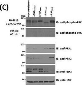
Western Blot: PKN3 Antibody [NBP1-30102] -
Western Blot: PKN3 Antibody [NBP1-30102] - Influence of TP Agonist on the Activation of PRK1, PRK2 & PRK3. Panel C. In order to identify the species of PRK subject to U46619-induced T-loop phosphorylation, PC-3 cells initially transfected for 72 hr with 30 nM siRNAPRK1, siRNAPRK2, siRNAPRK3 or, as controls, with a scrambled siRNAControl. Thereafter, cells serum starved & stimulated with U46619 for 60 min or with vehicle & then immunoblotted (20 μg/lane) with anti-phospho-PRK1Thr774/PRK2Thr816/PRK3Thr718 & with anti-PRK1, anti-PRK2, anti-PRK3 or, as loading controls, HDJ2 antisera, as indicated. Image collected & cropped by CiteAb from the following publication (https://www.oncotarget.com/lookup/doi/10.18632/oncotarget.4664), licensed under a CC-BY license. Not internally tested by Novus Biologicals.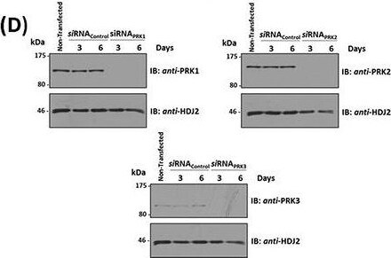
Western Blot: PKN3 Antibody [NBP1-30102] -
Western Blot: PKN3 Antibody [NBP1-30102] - Effect of siRNA-mediated disruption of PRK1, PRK2 & PRK3 expression on TP agonist-induced proliferation & migration of PC-3 cellsPanels A-D. PC-3 cells were transfected for 48 hr with 30 nM siRNAPRK1, siRNAPRK2, siRNAPRK3 or, as controls, with a scrambled siRNA (siRNAControl) prior to stimulation with U46619 (10 nM) or vehicle (0.0001% EtOH) for the indicated time specific to the assay, where non-transfected cells served as a reference. Panel A: For analysis of proliferation, 72 hr post-siRNA transfection PC-3 cells were stimulated for 48 hr with U46619 (10 nM) or vehicle (0.0001% EtOH). Panel B: For analysis of colony formation, 48 hr post-siRNA transfection PC-3 cells were stimulated with U46619 (10 nM) or vehicle (0.0001% EtOH) in soft agar & assessed 10 day post-treatment for colony formation. Panel C: For analysis of migration, 72 hr post-siRNA transfection PC-3 cells were stimulated for 8 hr with U46619 (10 nM) or vehicle (0.0001% EtOH). In Panels A-C, the bar charts show mean relative levels of PC-3 cell proliferation, colony formation & migration (± SEM, n ≥ 3), where levels in the vehicle-treated cells are assigned a value of 100%. The asterisks indicate where U46619-stimulation resulted in significant increases in proliferation, colony formation or migration by PC-3 cells compared to vehicle-treated cells, where ** & *** indicates p < 0.01 & p < 0.001, respectively. Panel D: Validation of the specificity & sustained siRNA-mediated disruption of PRK1, PRK2 & PRK3 expression was confirmed by immunoblot analysis of whole cell lysates (20 μg/lane) with the respective anti-PRK1/2/3 or, to validate protein loading, with anti-HDJ2 antisera where analysis was carried out 3 & 6 day post-transfection. Data n ≥ 3. Image collected & cropped by CiteAb from the following publication (https://www.oncotarget.com/lookup/doi/10.18632/oncotarget.4664), licensed under a CC-BY license. Not internally tested by Novus Biologicals.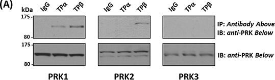
Western Blot: PKN3 Antibody [NBP1-30102] -
Western Blot: PKN3 Antibody [NBP1-30102] - Association of PRK1 & PRK2 with TP alpha & TP beta in prostate PC-3 cellsPanel A. PC-3 cells were immunoprecipitated with anti-TP alpha, anti-TP beta or, as controls, with the pre-immune (IgG) sera. Thereafter, immunoprecipitates (upper panels) or equivalent aliquots of whole cell lysates (20 μg/lane, lower panels) were immunoblotted (IB) with anti-PRK1, anti-PRK2 or anti-PRK3 antisera. The relative positions of the molecular size markers (kDa) are indicated to the left of the panels. Data shown are representative of at least three independent experiments (n ≥ 3). Panel B. PC-3 cells were incubated with U46619 (1 μM; 0–60 min) prior to immunoprecipitation with anti-TP alpha, anti-TP beta or, as controls, with the pre-immune (IgG) sera. Thereafter, immunoprecipitates (upper panels) or equivalent aliquots of whole cell lysates (20 μg/lane, lower panels) were IB with anti-PRK1, anti-PRK2 or anti-PRK3 antisera. Data n ≥ 3. Panel C. Bar charts show the mean relative levels of PRK1 or PRK2 associated with the anti-TP alpha or anti-TP beta immunoprecipitates, as determined by quantitative densitometry (± SEM), where levels associated with the respective immunoprecipitates in the absence of agonist are expressed as 1. The asterisks indicate where U46619 stimulation resulted in significant changes in complex-associated PRK1 or PRK2, where * & ** indicate p < 0.05 & p < 0.01, respectively. Image collected & cropped by CiteAb from the following publication (https://www.oncotarget.com/lookup/doi/10.18632/oncotarget.4664), licensed under a CC-BY license. Not internally tested by Novus Biologicals.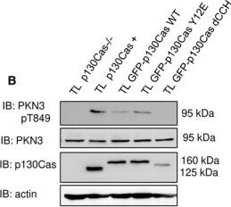
Western Blot: PKN3 Antibody [NBP1-30102] -
Western Blot: PKN3 Antibody [NBP1-30102] - PKN3 activity is important for stress fibers formation & is stimulated by the expression of p130Cas. (A) p130Cas−/−MEFs growing on FN‐coated cover slips were co‐transfected by GFP‐p130Cas & mCherry‐PKN3 fusion variant (WT, mPR, KD) or mCherry. After 48 h, cells were fixed & imaged by Leica TCS SP2 microscope (63×/1.45 oil objective). Stress fibers were visualized by Phalloidin (405) & focal adhesions by anti‐Paxillin staining (2nd 633). Representative images are shown. Scale bars represent 20 μm. (B) p130Cas−/−MEFs or p130Cas−/−MEFs re‐expressing p130Cas or transfected by GFP‐fused p130Cas variants (WT, YE, dCCH) were lysed in RIPA buffer, blotted to nitrocellulose membrane, & analyzed for endogenous PKN3 activity by antibody anti‐phosphoThr849 of PKN3 (pT849 PKN3). Expression of p130Cas mutants was verified by anti‐p130Cas antibody & loading by anti‐PKN3 & anti‐actin antibody. (C) Densitometric quantification of PKN3 activity (pT849 PKN3 phosphorylation). The effect of p130Cas re‐expression on PKN3 T849 phosphorylation was analyzed separately from the effect of transfected p130Cas mutants (indicated by a dotted line). Error bars indicate means ± SD from three independent experiments (four experiments for the left part). Statistical significance was evaluated by one‐way repeated ANOVA followed by Turkey's post hoc test (*P < 0.05; **P < 0.01). (D) Lysates or (E) immunoprecipitates (by Flag sepharose) from p130Cas−/−MEFs re‐expressing p130Cas & overexpressing PKN3 variants (WT, mPR, KD) were immunoblotted by anti‐PKN3, anti‐pT849 PKN3, & anti‐Akt antibodies (loading control). Image collected & cropped by CiteAb from the following publication (https://pubmed.ncbi.nlm.nih.gov/30422386), licensed under a CC-BY license. Not internally tested by Novus Biologicals.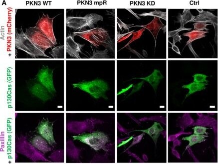
Immunocytochemistry/ Immunofluorescence: PKN3 Antibody [NBP1-30102] -
Immunocytochemistry/ Immunofluorescence: PKN3 Antibody [NBP1-30102] - PKN3 activity is important for stress fibers formation & is stimulated by the expression of p130Cas. (A) p130Cas−/−MEFs growing on FN‐coated cover slips were co‐transfected by GFP‐p130Cas & mCherry‐PKN3 fusion variant (WT, mPR, KD) or mCherry. After 48 h, cells were fixed & imaged by Leica TCS SP2 microscope (63×/1.45 oil objective). Stress fibers were visualized by Phalloidin (405) & focal adhesions by anti‐Paxillin staining (2nd 633). Representative images are shown. Scale bars represent 20 μm. (B) p130Cas−/−MEFs or p130Cas−/−MEFs re‐expressing p130Cas or transfected by GFP‐fused p130Cas variants (WT, YE, dCCH) were lysed in RIPA buffer, blotted to nitrocellulose membrane, & analyzed for endogenous PKN3 activity by antibody anti‐phosphoThr849 of PKN3 (pT849 PKN3). Expression of p130Cas mutants was verified by anti‐p130Cas antibody & loading by anti‐PKN3 & anti‐actin antibody. (C) Densitometric quantification of PKN3 activity (pT849 PKN3 phosphorylation). The effect of p130Cas re‐expression on PKN3 T849 phosphorylation was analyzed separately from the effect of transfected p130Cas mutants (indicated by a dotted line). Error bars indicate means ± SD from three independent experiments (four experiments for the left part). Statistical significance was evaluated by one‐way repeated ANOVA followed by Turkey's post hoc test (*P < 0.05; **P < 0.01). (D) Lysates or (E) immunoprecipitates (by Flag sepharose) from p130Cas−/−MEFs re‐expressing p130Cas & overexpressing PKN3 variants (WT, mPR, KD) were immunoblotted by anti‐PKN3, anti‐pT849 PKN3, & anti‐Akt antibodies (loading control). Image collected & cropped by CiteAb from the following publication (https://pubmed.ncbi.nlm.nih.gov/30422386), licensed under a CC-BY license. Not internally tested by Novus Biologicals.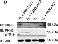
Western Blot: PKN3 Antibody [NBP1-30102] -
Western Blot: PKN3 Antibody [NBP1-30102] - PKN3 activity is important for stress fibers formation & is stimulated by the expression of p130Cas. (A) p130Cas−/−MEFs growing on FN‐coated cover slips were co‐transfected by GFP‐p130Cas & mCherry‐PKN3 fusion variant (WT, mPR, KD) or mCherry. After 48 h, cells were fixed & imaged by Leica TCS SP2 microscope (63×/1.45 oil objective). Stress fibers were visualized by Phalloidin (405) & focal adhesions by anti‐Paxillin staining (2nd 633). Representative images are shown. Scale bars represent 20 μm. (B) p130Cas−/−MEFs or p130Cas−/−MEFs re‐expressing p130Cas or transfected by GFP‐fused p130Cas variants (WT, YE, dCCH) were lysed in RIPA buffer, blotted to nitrocellulose membrane, & analyzed for endogenous PKN3 activity by antibody anti‐phosphoThr849 of PKN3 (pT849 PKN3). Expression of p130Cas mutants was verified by anti‐p130Cas antibody & loading by anti‐PKN3 & anti‐actin antibody. (C) Densitometric quantification of PKN3 activity (pT849 PKN3 phosphorylation). The effect of p130Cas re‐expression on PKN3 T849 phosphorylation was analyzed separately from the effect of transfected p130Cas mutants (indicated by a dotted line). Error bars indicate means ± SD from three independent experiments (four experiments for the left part). Statistical significance was evaluated by one‐way repeated ANOVA followed by Turkey's post hoc test (*P < 0.05; **P < 0.01). (D) Lysates or (E) immunoprecipitates (by Flag sepharose) from p130Cas−/−MEFs re‐expressing p130Cas & overexpressing PKN3 variants (WT, mPR, KD) were immunoblotted by anti‐PKN3, anti‐pT849 PKN3, & anti‐Akt antibodies (loading control). Image collected & cropped by CiteAb from the following publication (https://pubmed.ncbi.nlm.nih.gov/30422386), licensed under a CC-BY license. Not internally tested by Novus Biologicals.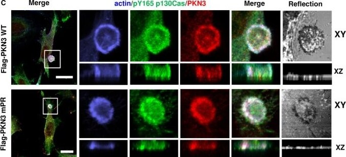
Immunocytochemistry/ Immunofluorescence: PKN3 Antibody [NBP1-30102] -
Immunocytochemistry/ Immunofluorescence: PKN3 Antibody [NBP1-30102] - PKN3 colocalizes with p130Cas in lamellipodia & podosome rosettes. Representative images are shown. (A) p130Cas−/−MEFs plated on fibronectin (FN) were transfected by GFP‐p130Cas, CFP‐LifeAct, & mCherry‐PKN3WT or mCherry‐PKN3 mPR & imaged live 24 h after transfection. White arrow indicates lamellipodia. Histogram of dotted straight line is shown. (B) Quantification of mCherry‐PKN3 WT, mCherry‐PKN3 mPR, & mCherry localization to lamellipodia (LifeAct as marker) was calculated as described in methods (values are mean ± SD from three independent experiments, n > 50 measurements – 3 per cell; ***P < 0.001, one‐way ANOVA on ranks followed by Dunn's post hoc test). (C) Src‐transformed p130Cas−/−MEFs co‐expressing p130Cas (SC) & mouse Flag tagged PKN3 WT or Flag‐PKN3 mPR are shown. Cells were grown on FN‐coated coverslips for 48 h, fixed, & stained for p130Cas by anti‐pTyr165 p130Cas antibody (pY165 p130Cas; 2nd 405), for actin by Phalloidin 488 & for Flag‐PKN3 by anti‐Flag antibody (2nd 633). Reflection (670 nm) indicates fibronectin degradation. All scale bars represent 20 μm. Cell were imaged by Leica TCS SP8 microscope system equipped with Leica 63×/1.45 oil objective. Image collected & cropped by CiteAb from the following publication (https://pubmed.ncbi.nlm.nih.gov/30422386), licensed under a CC-BY license. Not internally tested by Novus Biologicals.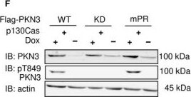
Western Blot: PKN3 Antibody [NBP1-30102] -
Western Blot: PKN3 Antibody [NBP1-30102] - PKN3 overexpression regulates growth of MEFs, & this effect requires PKN3–p130Cas interaction. (A) Immunoblotted lysates from MEFs p130Cas−/− or MEFs p130Cas−/− re‐expressing p130Cas (p130Cas+) treated by Doxycycline (Dox) to induce expression of mCherry‐PKN3 or mCherry alone. p130Cas presence detected by ani‐p130Cas antibody & mCherry epitope by anti‐mCherry antibody. (B) Dynamics of mCherry‐PKN3 expression after supplementation w/ Dox shown by immunoblot w/ anti‐mCherry antibody. (C–E) Effect of induced mCherry‐PKN3 expression on cell growth. Representative graphs showing growth of MEFs p130Cas−/− re‐expressing p130Cas (p130Cas+) (C) or MEFs p130Cas−/− (D) measured in real‐time using xCELLigence RTCA (real‐time cell analysis) system instrument. (E) Quantification of cell growth change induced by mCherry‐PKN3 expression (‘−’ indicates inducible mCherry expression used as negative control). Slope ratios reflecting cell growth calculated from log growth phase of cell growth (indicated by dotted lines; see C & D). (F) Immunoblotted lysates from MEFs p130Cas−/− re‐expressing p130Cas (p130Cas+) treated or not treated by Dox which induced expression of Flag‐fused PKN3 variants (WT, mPR, KD, empty vector). Stimulated overexpression of PKN3 detected by anti‐PKN3 antibody & its activity by antibody anti‐pT849 PKN3. (G) Quantification of cell growth change stimulated by Dox‐inducible expression of Flag‐fused PKN3 variants (WT, mPR, KD) in MEFs p130Cas−/− re‐expressing p130Cas (p130Cas+). All error bars indicate means ± SD calculated from 3 to 5 independent experiments (each in triplicates). Statistical significance always calculated between induced & noninduced cells & evaluated by one‐way repeated ANOVA followed by Turkey post hoc test (***P < 0.001). Image collected & cropped by CiteAb from following publication (https://pubmed.ncbi.nlm.nih.gov/30422386), licensed under a CC-BY license. Not internally tested by Novus Biologicals.Applications for PKN3 Antibody - BSA Free
Knockout Validated
Western Blot
Reviewed Applications
Read 2 reviews rated 4.5 using NBP1-30102 in the following applications:
Formulation, Preparation, and Storage
Purification
Formulation
Format
Preservative
Concentration
Shipping
Stability & Storage
Background: PKN3
Alternate Names
Entrez Gene IDs
Gene Symbol
UniProt
Additional PKN3 Products
Product Documents for PKN3 Antibody - BSA Free
Certificate of Analysis
To download a Certificate of Analysis, please enter a lot or batch number in the search box below.
Product Specific Notices for PKN3 Antibody - BSA Free
This product is for research use only and is not approved for use in humans or in clinical diagnosis. Primary Antibodies are guaranteed for 1 year from date of receipt.
Citations for PKN3 Antibody - BSA Free
 Powered by Bioz
Customer Reviews for PKN3 Antibody - BSA Free (2)
Have you used PKN3 Antibody - BSA Free?
Submit a review and receive an Amazon gift card!
$25/€18/£15/$25CAN/¥2500 Yen for a review with an image
$10/€7/£6/$10CAN/¥1110 Yen for a review without an image
Submit a review
Customer Images
-(01-ml)_NBP1-30102_9141.jpg)
-
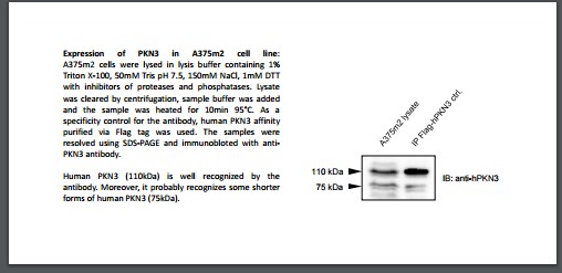
Application: Western BlotSample Tested: A375m2 cell lysate and ImmunoprecipitationSpecies: HumanVerified Customer | Posted 06/05/2017

-
Application: Western BlotSample Tested:Species: HumanVerified Customer | Posted 08/05/2014PKN3 Antibody Characterization
There are no reviews that match your criteria.
Protocols
Find general support by application which include: protocols, troubleshooting, illustrated assays, videos and webinars.
- Cellular Response to Hypoxia Protocols
- R&D Systems Quality Control Western Blot Protocol
- Troubleshooting Guide: Western Blot Figures
- Western Blot Conditions
- Western Blot Protocol
- Western Blot Protocol for Cell Lysates
- Western Blot Troubleshooting
- Western Blot Troubleshooting Guide
- View all Protocols, Troubleshooting, Illustrated assays and Webinars
FAQs for PKN3 Antibody - BSA Free
-
Q: A customer needs an antibody to PKN3 which is specific for this isoform and not cross reacting with the forms PKN1 and PKN2. Would this be suitable for NBP1-30102?
A: I ran an alignment of the immunogen sequence for NBP1-30102 against the sequences for PKN1 and PKN2 and the similarity is low. We haven't tested for cross reactivity, but the customer should feel confident that within these regions there is not many amino acids shared between PKN3 and PKN1 and PKN2.
-
Q: What I'm looking for is an antibody that recognizes preferably the N-terminal part of the human and mouse PKN3 kinase (since my protein of interest has been shown to bind to a region just upstream of the kinase domain - between aa 510 and 522). My intention is to use this antibody for Western blotting and IPs. I have found a PKN3 antibody (NBP1-30102) raised against an internal region of the human protein, that can recognize endogenous levels of the protein in HeLa lysates according to your datasheet (Western blot). This antibody however is not recommended for IPs. Does this mean that IPs have been tested and the antibody doesn't work for this application? Since I want to be able to (by Western blotting) detect mouse PKN3, do you know if this antibody is suitable for this?
A: NBP1-30102 recognizes a region from amino acids 225-275 which would be upstream of the kinase domain. Our antibodies are guaranteed for use in all species and applications listed, so for this antibody, it has been shown to work in Western Blots but looks to be unsuitable for IPs. The IP (-) means that we have tested for its use in IP and have been unsuccessful so we do not recommend if for IP. The region recognized shows only 75% sequence similarity in mouse so it may or may not cross-react with mouse but it is something we cannot guarantee. As far as an alternative antibody, our other PKN3 antibodies we currently sell are raised against full-length PKN3 and are not epitope mapped. One of these could be useful for IP and NBP1-30102 could be used to detect.
-
Q: A customer needs an antibody to PKN3 which is specific for this isoform and not cross reacting with the forms PKN1 and PKN2. Would this be suitable for NBP1-30102?
A: I ran an alignment of the immunogen sequence for NBP1-30102 against the sequences for PKN1 and PKN2 and the similarity is low. We haven't tested for cross reactivity, but the customer should feel confident that within these regions there is not many amino acids shared between PKN3 and PKN1 and PKN2.
-
Q: What I'm looking for is an antibody that recognizes preferably the N-terminal part of the human and mouse PKN3 kinase (since my protein of interest has been shown to bind to a region just upstream of the kinase domain - between aa 510 and 522). My intention is to use this antibody for Western blotting and IPs. I have found a PKN3 antibody (NBP1-30102) raised against an internal region of the human protein, that can recognize endogenous levels of the protein in HeLa lysates according to your datasheet (Western blot). This antibody however is not recommended for IPs. Does this mean that IPs have been tested and the antibody doesn't work for this application? Since I want to be able to (by Western blotting) detect mouse PKN3, do you know if this antibody is suitable for this?
A: NBP1-30102 recognizes a region from amino acids 225-275 which would be upstream of the kinase domain. Our antibodies are guaranteed for use in all species and applications listed, so for this antibody, it has been shown to work in Western Blots but looks to be unsuitable for IPs. The IP (-) means that we have tested for its use in IP and have been unsuccessful so we do not recommend if for IP. The region recognized shows only 75% sequence similarity in mouse so it may or may not cross-react with mouse but it is something we cannot guarantee. As far as an alternative antibody, our other PKN3 antibodies we currently sell are raised against full-length PKN3 and are not epitope mapped. One of these could be useful for IP and NBP1-30102 could be used to detect.