Plasma Cell Marker Antibody (SPM310)
Novus Biologicals | Catalog # NBP2-34359
Key Product Details
Species Reactivity
Human, Rat (Negative)
Applications
Validated:
Immunohistochemistry, Immunohistochemistry-Paraffin
Cited:
Immunohistochemistry-Paraffin
Label
Unconjugated
Antibody Source
Monoclonal Mouse IgG2a Kappa Clone # SPM310
Loading...
Product Specifications
Immunogen
Pancreatic cancer-related mucin
Reactivity Notes
Does not react with Rat.
Localization
Cytoplasmic
Marker
Plasma Cell Marker
Specificity
It recognizes an intra-cytoplasmic antigen, which shows a very high degree of specificity for plasma cells. This antigen is present in normal as well as neoplastic plasma cells. Plasma cells, which are large lymphocytes derived from an antigen-specific B cell, secrete antibodies and are responsible for humoral immunity. Plasma cells differentiate from B cells upon stimulation by CD4+ lymphocytes. The B cell acts as an antigen-presenting cell (APC), consuming an offending pathogen, which is taken up by the B cell by phagocytosis and broken down within proteosomes. Plasma cells contain basophilic cytoplasm; their nucleus contains heterochromatin organized in a characteristic cartwheel arrangement. This monoclonal antibody superbly recognizes normal and neoplastic plasma cells in routine formalin-fixed, paraffin-embedded tissue sections. It is of potential value in identifying myeloma or plasmacytoma in bone marrow or other tissues. It also helps differentiate lympho-plasmacytoid lymphoma from lymphocytic and follicular lymphoma. Note that this monoclonal antibody is not suitable for staining frozen tissues.
Clonality
Monoclonal
Host
Mouse
Isotype
IgG2a Kappa
Description
200ug/ml of antibody purified from Bioreactor Concentrate by Protein A or G. Prepared in 10 mM PBS with 0.05% BSA & 0.05% azide. Also available WITHOUT BSA & azide at 1.0 mg/ml. (NBP2-34407)
Antibody with azide - store at 2 to 8C. Antibody without azide - store at -20 to -80C.
Antibody with azide - store at 2 to 8C. Antibody without azide - store at -20 to -80C.
Scientific Data Images for Plasma Cell Marker Antibody (SPM310)
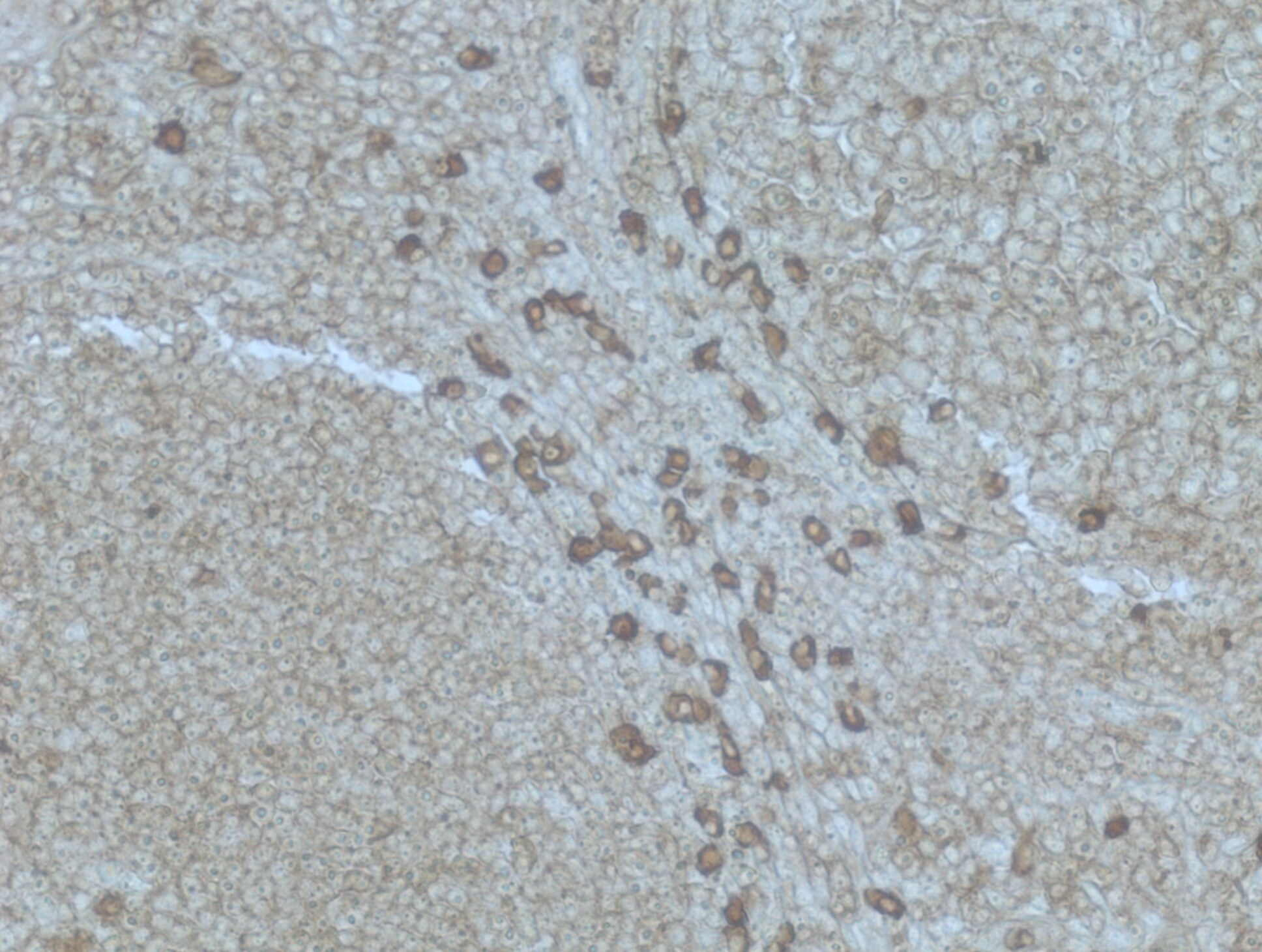
![Immunohistochemistry-Paraffin: Plasma Cell Marker Antibody (SPM310) [NBP2-34359]](https://resources.rndsystems.com/images/products/Plasma-Cell-Marker-Antibody-SPM310-Immunohistochemistry-Paraffin-NBP2-34359-img0002.jpg "Immunohistochemistry-Paraffin: Plasma Cell Marker Antibody (SPM310) [NBP2-34359]")
Immunohistochemistry-Paraffin: Plasma Cell Marker Antibody (SPM310) [NBP2-34359]
Immunohistochemistry-Paraffin: Plasma Cell Marker Antibody (SPM310) [NBP2-34359] - Human tonsil tissue. Heat induced antigen retrieval in citrate buffer pH 6 at 95C for 20 minutes. Image from verified customer review.![Immunohistochemistry-Paraffin: Plasma Cell Marker Antibody (SPM310) [NBP2-34359]](https://resources.rndsystems.com/images/products/Plasma-Cell-Marker-Antibody-SPM310-Immunohistochemistry-Paraffin-NBP2-34359-img0001.jpg "Immunohistochemistry-Paraffin: Plasma Cell Marker Antibody (SPM310) [NBP2-34359]")
Immunohistochemistry-Paraffin: Plasma Cell Marker Antibody (SPM310) [NBP2-34359]
Immunohistochemistry-Paraffin: Plasma Cell Marker Antibody (SPM310) [NBP2-34359] - Formalin-fixed paraffin-embedded human tonsil stained with plasma cell marker Monoclonal antibody (SPM310) [NBP2-34359] -")
Immunohistochemistry-Paraffin: Plasma Cell Marker Antibody (SPM310) [NBP2-34359] -
Immunohistochemistry-Paraffin: Plasma Cell Marker Antibody (SPM310) [NBP2-34359] - Formalin-paraffin human tonsil stained with Plasma Cell Marker MAb (SPM310). [NBP2-34359] -")
Immunohistochemistry: Plasma Cell Marker Antibody (SPM310) [NBP2-34359] -
The results of immunohistochemistry (IHC). In total of 29 patients, 15 were diagnosed with liposarcoma (Metastasis occurred in eight patients during follow-up) and 14 were diagnosed with leiomyosarcoma (Metastasis occurred in nine patients during follow-up). Proteins of PAX5 and LTB were shown to be significantly down-regulated in the tumor cells of primary sarcomas with metastasis while the markers of B cells (CD19 and CD 38) were not detected in almost all primary tumor (a). Although CD19 and CD 38 were found in lymph node, as the advice of a pathologist in our hospital, these were not surprising as B cells are abundant in lymph node. Therefore, the IHC results of CD19 and CD 38 did not prove or disprove the hypothesis. Furthermore, in the absence of a good CD19 and CD38 antibody, we considered CD138 (Syndean-1) and SPM310 plasma cell marker antibody (Novus NBP2-34359) as other proven plasma cell markers. However, only four of 29 sarcomas were found plasma cells in the HE staining section of the tumor and none of the four patients had metastases (b). This might be because tumor-infiltrating immune cells tended to be located around the tumor rather than within it, and sarcomas tended to be excised en bloc, so there were no paracancer tissues as a control. Fortunately, the results of Mann–Whitney U test suggested that PAX5 (P < 0.001), LTB (P < 0.001) in were all highly expressed in well differentiated primary sarcomas and primary sarcomas without metastasis (c, d) Image collected and cropped by CiteAb from the following open publication (https://pubmed.ncbi.nlm.nih.gov/33397394), licensed under a CC-BY license. Not internally tested by Novus Biologicals.Applications for Plasma Cell Marker Antibody (SPM310)
Application
Recommended Usage
Immunohistochemistry-Paraffin
0.1-0.2 ug/ml
Application Notes
Immunohistochemistry (Formalin-fixed): 0.1-0.2ug/ml for 30 minutes at RT. Staining of formalin-fixed tissues requires heating tissue sections in 10mM Tris with 1mM EDTA, pH 9.0, for 45 min at 95C followed by cooling at RT for 20 minutes.
Optimal dilution for a specific application should be determined.
Note that this antibody is not suitable for staining frozen tissues.
Optimal dilution for a specific application should be determined.
Note that this antibody is not suitable for staining frozen tissues.
Reviewed Applications
Read 1 review rated 4 using NBP2-34359 in the following applications:
Formulation, Preparation, and Storage
Purification
Protein A or G purified
Formulation
10 mM PBS with 0.05% BSA
Preservative
0.05% Sodium Azide
Concentration
0.2 mg/ml
Shipping
The product is shipped with polar packs. Upon receipt, store it immediately at the temperature recommended below.
Stability & Storage
Store at 4C.
Product Documents for Plasma Cell Marker Antibody (SPM310)
Certificate of Analysis
To download a Certificate of Analysis, please enter a lot or batch number in the search box below.
Product Specific Notices for Plasma Cell Marker Antibody (SPM310)
This product is for research use only and is not approved for use in humans or in clinical diagnosis. Primary Antibodies are guaranteed for 1 year from date of receipt.
Citations for Plasma Cell Marker Antibody (SPM310)
 Powered by Bioz
Customer Reviews for Plasma Cell Marker Antibody (SPM310) (1)
4 out of 5
1 Customer Rating
Have you used Plasma Cell Marker Antibody (SPM310)?
Submit a review and receive an Amazon gift card!
$25/€18/£15/$25CAN/¥2500 Yen for a review with an image
$10/€7/£6/$10CAN/¥1110 Yen for a review without an image
Submit a review
Customer Images
Showing
1
-
1 of
1 review
Showing All
Filter By:
-
Application: Immunohistochemistry-ParaffinSample Tested: Tonsil tissueSpecies: HumanVerified Customer | Posted 05/18/2018Heat induced antigen retrieval was performed in citrate buffer (pH 6) at 95C for 20 minutes.

There are no reviews that match your criteria.
Protocols
Find general support by application which include: protocols, troubleshooting, illustrated assays, videos and webinars.
- Antigen Retrieval Protocol (PIER)
- Antigen Retrieval for Frozen Sections Protocol
- Appropriate Fixation of IHC/ICC Samples
- Cellular Response to Hypoxia Protocols
- Chromogenic IHC Staining of Formalin-Fixed Paraffin-Embedded (FFPE) Tissue Protocol
- Chromogenic Immunohistochemistry Staining of Frozen Tissue
- ClariTSA™ Fluorophore Kits
- Detection & Visualization of Antibody Binding
- Fluorescent IHC Staining of Frozen Tissue Protocol
- Graphic Protocol for Heat-induced Epitope Retrieval
- Graphic Protocol for the Preparation and Fluorescent IHC Staining of Frozen Tissue Sections
- Graphic Protocol for the Preparation and Fluorescent IHC Staining of Paraffin-embedded Tissue Sections
- Graphic Protocol for the Preparation of Gelatin-coated Slides for Histological Tissue Sections
- IHC Sample Preparation (Frozen sections vs Paraffin)
- Immunofluorescent IHC Staining of Formalin-Fixed Paraffin-Embedded (FFPE) Tissue Protocol
- Immunohistochemistry (IHC) and Immunocytochemistry (ICC) Protocols
- Immunohistochemistry Frozen Troubleshooting
- Immunohistochemistry Paraffin Troubleshooting
- Preparing Samples for IHC/ICC Experiments
- Preventing Non-Specific Staining (Non-Specific Binding)
- Primary Antibody Selection & Optimization
- Protocol for Heat-Induced Epitope Retrieval (HIER)
- Protocol for Making a 4% Formaldehyde Solution in PBS
- Protocol for VisUCyte™ HRP Polymer Detection Reagent
- Protocol for the Preparation & Fixation of Cells on Coverslips
- Protocol for the Preparation and Chromogenic IHC Staining of Frozen Tissue Sections
- Protocol for the Preparation and Chromogenic IHC Staining of Frozen Tissue Sections - Graphic
- Protocol for the Preparation and Chromogenic IHC Staining of Paraffin-embedded Tissue Sections
- Protocol for the Preparation and Chromogenic IHC Staining of Paraffin-embedded Tissue Sections - Graphic
- Protocol for the Preparation and Fluorescent IHC Staining of Frozen Tissue Sections
- Protocol for the Preparation and Fluorescent IHC Staining of Paraffin-embedded Tissue Sections
- Protocol for the Preparation of Gelatin-coated Slides for Histological Tissue Sections
- TUNEL and Active Caspase-3 Detection by IHC/ICC Protocol
- The Importance of IHC/ICC Controls
- Troubleshooting Guide: Immunohistochemistry
- View all Protocols, Troubleshooting, Illustrated assays and Webinars
Loading...