PPP2R1A Antibody - BSA Free
Novus Biologicals | Catalog # NBP2-19907
![Western Blot: PPP2R1A Antibody [NBP2-19907]](https://resources.rndsystems.com/images/products/PPP2R1A-Antibody-Western-Blot-NBP2-19907-img0002.jpg "Western Blot: PPP2R1A Antibody [NBP2-19907]")
Loading...
Key Product Details
Species Reactivity
Validated:
Human, Mouse
Predicted:
Bovine (99%), Porcine (100%), Rat (100%), Rhesus Macaque (100%), Xenopus (95%). Backed by our 100% Guarantee.
Applications
Immunohistochemistry, Immunohistochemistry-Paraffin, Western Blot, Immunocytochemistry/ Immunofluorescence, Immunoprecipitation
Label
Unconjugated
Antibody Source
Polyclonal Rabbit IgG
Format
BSA Free
Loading...
Product Specifications
Immunogen
Recombinant protein encompassing a sequence within the center region of human PPP2R1A. The exact sequence is proprietary.
Reactivity Notes
Zebrafish (88%), Xenopus laevis (95%).
Clonality
Polyclonal
Host
Rabbit
Isotype
IgG
Theoretical MW
65 kDa.
Disclaimer note: The observed molecular weight of the protein may vary from the listed predicted molecular weight due to post translational modifications, post translation cleavages, relative charges, and other experimental factors.
Disclaimer note: The observed molecular weight of the protein may vary from the listed predicted molecular weight due to post translational modifications, post translation cleavages, relative charges, and other experimental factors.
Scientific Data Images for PPP2R1A Antibody - BSA Free
Western Blot: PPP2R1A Antibody [NBP2-19907]
Western Blot: PPP2R1A Antibody [NBP2-19907] - Sample (30 ug of whole cell lysate) A: NIH-3T3 B: JC C: BCL-1 7. 5% SDS PAGE gel, diluted at 1:5000.
Immunohistochemistry-Paraffin: PPP2R1A Antibody [NBP2-19907] -
Immunohistochemistry-Paraffin: PPP2R1A Antibody [NBP2-19907] - PPP2R1A antibody detects PPP2R1A protein at cytoplasm in mouse kidney by immunohistochemical analysis.Sample: Paraffin-embedded mouse kidney.
PPP2R1A antibody (NBP2-19907) diluted at 1:2500.
Antigen Retrieval: Citrate buffer, pH 6.0, 15 min

Western Blot: PPP2R1A Antibody [NBP2-19907] -
Whole cell extract (30 ug) was separated by 7.5% SDS-PAGE, and the membrane was blotted with PPP2R1A antibody (NBP2-19907) diluted at 1:1000.
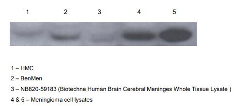
Western Blot: PPP2R1A Antibody - BSA Free [NBP2-19907] -
Protein expression levels of PPP2R5D and its interaction with the PPP2CA and PPP2R1A. (A) HA-tagged PPP2R5D WT/variants were transfected in 293T cells, protein expression was detected through Western blot. A 66 kDa band was detected in all missense variants. As for frameshift variant H436Mfs*3, a ~50 kDa band was detected and it was significantly weaker. (B) HA-tagged PPP2R5D WT/variants were purified through HA pull down, the interaction with endogenous PPP2CA and PPP2R1A were detected through Western blot. No band of PPP2CA and PPP2R1A was detected in W207R, D251H, D251Y and H463Mfs*3. A weaker band of PPP2CA and PPP2R1A was detected in E198K, E200K, L203P and Q211P. Image collected and cropped by CiteAb from the following open publication (https://www.mdpi.com/2227-9067/11/8/897), licensed under a CC-BY license. Not internally tested by Novus Biologicals.Applications for PPP2R1A Antibody - BSA Free
Application
Recommended Usage
Immunocytochemistry/ Immunofluorescence
1:100-1:1000
Immunohistochemistry
1:100-1:2500
Immunohistochemistry-Paraffin
1:100-1:2500
Western Blot
1:500-1:10000
Application Notes
Immunoprecipitation Assay-dependent dilution
Reviewed Applications
Read 1 review rated 4 using NBP2-19907 in the following applications:
Formulation, Preparation, and Storage
Purification
Antigen Affinity-purified
Formulation
PBS, 20% Glycerol
Format
BSA Free
Preservative
0.025% Proclin 300
Concentration
Concentrations vary lot to lot. See vial label for concentration. If unlisted please contact technical services.
Shipping
The product is shipped with polar packs. Upon receipt, store it immediately at the temperature recommended below.
Stability & Storage
Aliquot and store at -20C or -80C. Avoid freeze-thaw cycles.
Background: PPP2R1A
Alternate Names
Medium tumor antigen-associated 61 kDa protein, MGC786, PP2A subunit A isoform PR65-alpha, PP2A subunit A isoform R1-alpha, PP2AAALPHA, PP2A-Aalpha, PR65A, protein phosphatase 2 (formerly 2A), regulatory subunit A (PR 65), alphaisoform, protein phosphatase 2 (formerly 2A), regulatory subunit A, alpha isoform, protein phosphatase 2, regulatory subunit A, alpha, serine/threonine protein phosphatase 2A, 65 kDa regulatory subunit A, alphaisoform, serine/threonine-protein phosphatase 2A 65 kDa regulatory subunit A alphaisoform
Gene Symbol
PPP2R1A
UniProt
Additional PPP2R1A Products
Product Documents for PPP2R1A Antibody - BSA Free
Certificate of Analysis
To download a Certificate of Analysis, please enter a lot or batch number in the search box below.
Product Specific Notices for PPP2R1A Antibody - BSA Free
This product is for research use only and is not approved for use in humans or in clinical diagnosis. Primary Antibodies are guaranteed for 1 year from date of receipt.
Citations for PPP2R1A Antibody - BSA Free
 Powered by Bioz
Customer Reviews for PPP2R1A Antibody - BSA Free (1)
4 out of 5
1 Customer Rating
Have you used PPP2R1A Antibody - BSA Free?
Submit a review and receive an Amazon gift card!
$25/€18/£15/$25CAN/¥2500 Yen for a review with an image
$10/€7/£6/$10CAN/¥1110 Yen for a review without an image
Submit a review
Customer Images
Showing
1
-
1 of
1 review
Showing All
Filter By:
-
Application: Western BlotSample Tested: Cultured cancer cell lysates and Human Whole Cell lysateSpecies: HumanVerified Customer | Posted 09/30/2019These have been attached. The protein content of all samples were quantified prior to loading, and standardized at concentrations of 40ug.

There are no reviews that match your criteria.
Protocols
Find general support by application which include: protocols, troubleshooting, illustrated assays, videos and webinars.
- Antigen Retrieval Protocol (PIER)
- Antigen Retrieval for Frozen Sections Protocol
- Appropriate Fixation of IHC/ICC Samples
- Cellular Response to Hypoxia Protocols
- Chromogenic IHC Staining of Formalin-Fixed Paraffin-Embedded (FFPE) Tissue Protocol
- Chromogenic Immunohistochemistry Staining of Frozen Tissue
- ClariTSA™ Fluorophore Kits
- Detection & Visualization of Antibody Binding
- Fluorescent IHC Staining of Frozen Tissue Protocol
- Graphic Protocol for Heat-induced Epitope Retrieval
- Graphic Protocol for the Preparation and Fluorescent IHC Staining of Frozen Tissue Sections
- Graphic Protocol for the Preparation and Fluorescent IHC Staining of Paraffin-embedded Tissue Sections
- Graphic Protocol for the Preparation of Gelatin-coated Slides for Histological Tissue Sections
- ICC Cell Smear Protocol for Suspension Cells
- ICC Immunocytochemistry Protocol Videos
- ICC for Adherent Cells
- IHC Sample Preparation (Frozen sections vs Paraffin)
- Immunocytochemistry (ICC) Protocol
- Immunocytochemistry Troubleshooting
- Immunofluorescence of Organoids Embedded in Cultrex Basement Membrane Extract
- Immunofluorescent IHC Staining of Formalin-Fixed Paraffin-Embedded (FFPE) Tissue Protocol
- Immunohistochemistry (IHC) and Immunocytochemistry (ICC) Protocols
- Immunohistochemistry Frozen Troubleshooting
- Immunohistochemistry Paraffin Troubleshooting
- Immunoprecipitation Protocol
- Preparing Samples for IHC/ICC Experiments
- Preventing Non-Specific Staining (Non-Specific Binding)
- Primary Antibody Selection & Optimization
- Protocol for Heat-Induced Epitope Retrieval (HIER)
- Protocol for Making a 4% Formaldehyde Solution in PBS
- Protocol for VisUCyte™ HRP Polymer Detection Reagent
- Protocol for the Fluorescent ICC Staining of Cell Smears - Graphic
- Protocol for the Fluorescent ICC Staining of Cultured Cells on Coverslips - Graphic
- Protocol for the Preparation & Fixation of Cells on Coverslips
- Protocol for the Preparation and Chromogenic IHC Staining of Frozen Tissue Sections
- Protocol for the Preparation and Chromogenic IHC Staining of Frozen Tissue Sections - Graphic
- Protocol for the Preparation and Chromogenic IHC Staining of Paraffin-embedded Tissue Sections
- Protocol for the Preparation and Chromogenic IHC Staining of Paraffin-embedded Tissue Sections - Graphic
- Protocol for the Preparation and Fluorescent ICC Staining of Cells on Coverslips
- Protocol for the Preparation and Fluorescent ICC Staining of Non-adherent Cells
- Protocol for the Preparation and Fluorescent ICC Staining of Stem Cells on Coverslips
- Protocol for the Preparation and Fluorescent IHC Staining of Frozen Tissue Sections
- Protocol for the Preparation and Fluorescent IHC Staining of Paraffin-embedded Tissue Sections
- Protocol for the Preparation of Gelatin-coated Slides for Histological Tissue Sections
- Protocol for the Preparation of a Cell Smear for Non-adherent Cell ICC - Graphic
- R&D Systems Quality Control Western Blot Protocol
- TUNEL and Active Caspase-3 Detection by IHC/ICC Protocol
- The Importance of IHC/ICC Controls
- Troubleshooting Guide: Immunohistochemistry
- Troubleshooting Guide: Western Blot Figures
- Western Blot Conditions
- Western Blot Protocol
- Western Blot Protocol for Cell Lysates
- Western Blot Troubleshooting
- Western Blot Troubleshooting Guide
- View all Protocols, Troubleshooting, Illustrated assays and Webinars
Loading...