pSIVA Apoptosis Detection Kit
Novus Biologicals | Catalog # NBP2-29382
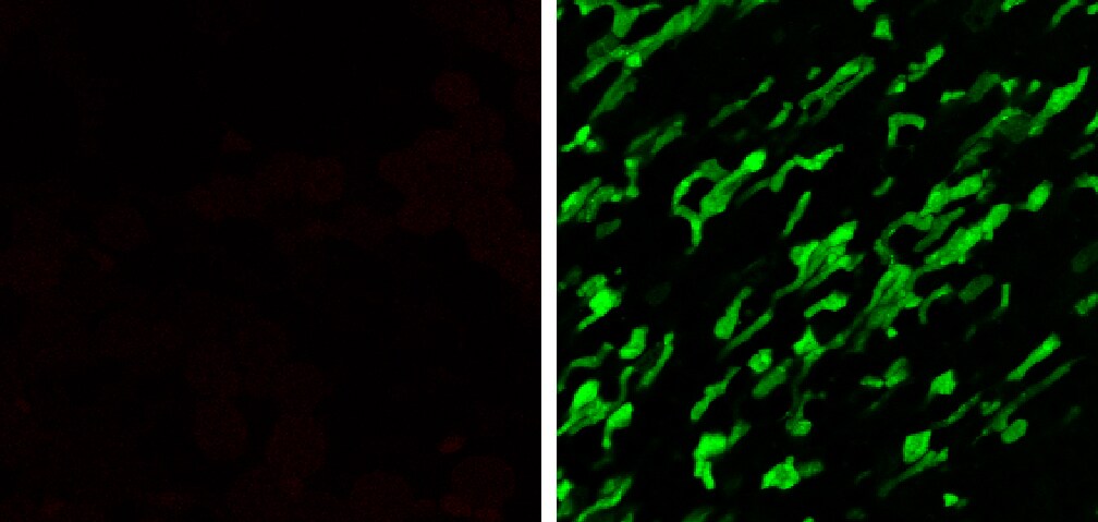
![Live Imaging Microscopy: pSIVA Apoptosis Detection Kit [NBP2-29382]](https://resources.rndsystems.com/images/products/Polarity-Sensitive-Indicator-of-Viability-Apoptosis-Microscopy-Kit-Live-Imaging--Microscopy-NBP2-29382-img0005.jpg "Live Imaging Microscopy: pSIVA Apoptosis Detection Kit [NBP2-29382]")
Key Product Details
Species
Applications
Kit Type
Conjugate
Product Summary for pSIVA Apoptosis Detection Kit
Product Specifications
Application Notes
Reactivity Notes
Scientific Data Images for pSIVA Apoptosis Detection Kit
Live Imaging Microscopy: pSIVA Apoptosis Detection Kit [NBP2-29382]
Live Imaging Microscopy: Polarity Sensitive Indicator of Viability Apoptosis Microscopy Kit [NBP2-29382] - Rescue of dying neurons: NGF withdrawal leads to cell death and pSIVA-IANBD(TM) fluorescence. Addition of NGF eventually rescues some neurons as shown by the loss of pSIVA-IANBD(TM) fluorescence (30h boxes).![Live Imaging Microscopy: pSIVA Apoptosis Detection Kit [NBP2-29382]](https://resources.rndsystems.com/images/products/Polarity-Sensitive-Indicator-of-Viability-Apoptosis-Microscopy-Kit-Live-Imaging--Microscopy-NBP2-29382-img0006.jpg "Live Imaging Microscopy: pSIVA Apoptosis Detection Kit [NBP2-29382]")
Live Imaging Microscopy: pSIVA Apoptosis Detection Kit [NBP2-29382]
Live Imaging Microscopy: Polarity Sensitive Indicator of Viability Apoptosis Microscopy Kit [NBP2-29382] - Rescue of dying neurons. NGF withdrawal leads to cell death and pSIVA-IANBD(TM) fluorescence. Correlation of fluorescence and phase microscopy shows that NGF rescued the neurons as shown by the loss of pSIVA-IANBD(TM) fluorescence at 30h.![Live Imaging Microscopy: pSIVA Apoptosis Detection Kit [NBP2-29382]](https://resources.rndsystems.com/images/products/Polarity-Sensitive-Indicator-of-Viability-Apoptosis-Microscopy-Kit-Live-Imaging--Microscopy-NBP2-29382-img0007.jpg "Live Imaging Microscopy: pSIVA Apoptosis Detection Kit [NBP2-29382]")
Live Imaging Microscopy: pSIVA Apoptosis Detection Kit [NBP2-29382]
Live Imaging Microscopy: Polarity Sensitive Indicator of Viability Apoptosis Microscopy Kit [NBP2-29382] - Quantitative analysis of pSIVA-IANBD™ fluorescence is rescued and NGF deprived neuronal cultures. pSIVA-IANBD(TM) fluorescence from 3 areas of the culture shows that NGF rescue (A) of dying cells was associated with loss of fluorescence whereas unrescued cells (B) continued to fluoresce.![Live Imaging Microscopy: pSIVA Apoptosis Detection Kit [NBP2-29382]](https://resources.rndsystems.com/images/products/Polarity-Sensitive-Indicator-of-Viability-Apoptosis-Microscopy-Kit-Live-Imaging--Microscopy-NBP2-29382-img0008.jpg "Live Imaging Microscopy: pSIVA Apoptosis Detection Kit [NBP2-29382]")
Live Imaging Microscopy: pSIVA Apoptosis Detection Kit [NBP2-29382]
Live Imaging Microscopy: Polarity Sensitive Indicator of Viability Apoptosis Microscopy Kit [NBP2-29382] - Rescue of dying photoreceptors in dystrophic rat neural retina. Image from verified customer review.Kit Contents for pSIVA Apoptosis Detection Kit
- Propidium Iodide Staining Solution (500 uL)
- pSIVA-IANBD (200 uL)
Formulation, Preparation, and Storage
Concentration
Shipping
Storage
Background: pSIVA Apoptosis Detection
Alternate Names
Additional pSIVA Apoptosis Detection Products
Product Documents for pSIVA Apoptosis Detection Kit
Certificate of Analysis
To download a Certificate of Analysis, please enter a lot or batch number in the search box below.
Product Specific Notices for pSIVA Apoptosis Detection Kit
pSIVA is protected under patent number: 8,541,549. This is the same kit which was sold as pSIVA(TM) - IANBD Apoptosis/Viability Microscopy Set [Catalog Number IMG-6701K] by Imgenex - now a part of Novus Biologicals, a Bio-Techne brand.
This product is for research use only and is not approved for use in humans or in clinical diagnosis. Kits are guaranteed for 6 months from date of receipt.
Citations for pSIVA Apoptosis Detection Kit
 Powered by Bioz
Customer Reviews for pSIVA Apoptosis Detection Kit (1)
Have you used pSIVA Apoptosis Detection Kit?
Submit a review and receive an Amazon gift card!
$25/€18/£15/$25CAN/¥2500 Yen for a review with an image
$10/€7/£6/$10CAN/¥1110 Yen for a review without an image
Submit a review
Customer Images
-
Application: ImmunofluorescenceSample Tested: dystrophic (RCS) rat neural retinaSpecies: RatVerified Customer | Posted 06/08/2016dying photoreceptors in dystrophic rat neural retina

There are no reviews that match your criteria.
Protocols
View specific protocols for pSIVA Apoptosis Detection Kit (NBP2-29382):
Hazard Information
Chemical Name: Propidium Iodide
CAS Number: 25535-16-4
Hazard Identification
Eye, skin
First Aid Measures
Eye Contact: Irrigate thoroughly with water for at least 15 minutes. Seek medical advice.
Skin Contact: Wash skin thoroughly with soap and water for at least 15 minutes. Remove contaminated clothing and wash before re-use. In severe cases, obtain medical attention.
Inhalation: Remove from exposure, rest and keep warm. In severe cases, seek medical advice.
Ingestion: Wash out mouth thoroughly with water and give plenty of water to drink. Seek medical advice.
Accidental Release Measures
Wear appropriate protective clothing. Inform others to keep a safe distance. Spread inert absorbent material liberally over spillage. If local regulations permit, mop up cautiously with plenty of water and run to waste, diluting greatly with running water. Otherwise transfer to container and arrange removal by disposal company. Wash site of spillage thoroughly with water.
Handling and Storage
Handling: No special handling required. Store at 4C or colder, protect from light.
Exposure Controls / Personal Protection
Ventilation: Use in an open, well-ventilated area
Gloves: Rubber or plastic
Eye Protection: Lab goggles or face shield
Physical and Chemical Properties
Form: Liquid
Color: Colorless
Odor: Odorless
Melting Point: 220-225C
Boiling Temperature: No data available
Density: No data available
Vapor Pressure: No data available
Solubility in Water: Very soluble
Flash Point: No data available
Explosion limits: No data available
Ignition Temperature: No data available
Stability and Reactivity: Stable at room temperature
Other Information
Transport: not classified as dangerous
Material Safety Data Sheet for IANBD
Hazard Information
Chemical Name: N-((2-(iodoacetoxy)ethyl)-N-methyl)amino-7-nitrobenz-2-oxa-1,3-diazole (IANBD ester)
Hazard Identification
The product contains no substances which at their given concentration are considered to be hazardous to health. NOVUS recommends handling all chemicals with caution.
Find general support by application which include: protocols, troubleshooting, illustrated assays, videos and webinars.
- Appropriate Fixation of IHC/ICC Samples
- Cellular Response to Hypoxia Protocols
- ClariTSA™ Fluorophore Kits
- Detection & Visualization of Antibody Binding
- ICC Cell Smear Protocol for Suspension Cells
- ICC Immunocytochemistry Protocol Videos
- ICC for Adherent Cells
- Immunocytochemistry (ICC) Protocol
- Immunocytochemistry Troubleshooting
- Immunofluorescence of Organoids Embedded in Cultrex Basement Membrane Extract
- Immunohistochemistry (IHC) and Immunocytochemistry (ICC) Protocols
- Preparing Samples for IHC/ICC Experiments
- Preventing Non-Specific Staining (Non-Specific Binding)
- Primary Antibody Selection & Optimization
- Protocol for VisUCyte™ HRP Polymer Detection Reagent
- Protocol for the Fluorescent ICC Staining of Cell Smears - Graphic
- Protocol for the Fluorescent ICC Staining of Cultured Cells on Coverslips - Graphic
- Protocol for the Preparation and Fluorescent ICC Staining of Cells on Coverslips
- Protocol for the Preparation and Fluorescent ICC Staining of Non-adherent Cells
- Protocol for the Preparation and Fluorescent ICC Staining of Stem Cells on Coverslips
- Protocol for the Preparation of a Cell Smear for Non-adherent Cell ICC - Graphic
- TUNEL and Active Caspase-3 Detection by IHC/ICC Protocol
- The Importance of IHC/ICC Controls
- View all Protocols, Troubleshooting, Illustrated assays and Webinars
FAQs for pSIVA Apoptosis Detection Kit
-
Q: How many tests can you perform with this kit?
A: 100 tests for the full sized kit and 25 tests for the 1 sample sized kit.
-
Q: Our end user is working on tissues (epithelium of imaginal disk from drosophila leg). The tissues are kept growing on a culture medium and they manage to follow apoptosis properly with orange acrylin but not with annexin V because of background. Did you ever test any of your pSIVA apoptosis kits (NBP2-29382, NBP2-29611) on tissue culture instead of cell culture ? Is there a chance the proble will work on such an application ? Would you be able to provide a sample for this application ? As for the other samples, I'm still waiting on customer feedback and I'll keep you posted.
A:
Yes, pSIVA has been used in tissue applications as denoted on the publication link. Specifically, I would encourage the research to look at the following publications from the link: 1. Diurnal, localized exposure of phosphatidylserine by rod outer segment tips in wild-type but not Itgb5-/- or Mfge8-/- mouse retina. Ruggiero L, MP Connor, J Chen, R Langen, SC Finnemann. PNAS 109:8145-4148 (2012). Live tissue imaging (mouse retina): Figs 4, 5. S4. pSIVA-IANBD was added to dissected live mouse retina and shown to label the tips of photoreceptor outer segments (POS). > The results suggested that phosphatidylserine (PS) exposure is specific to the POS surface. Furthermore, enhanced PS exposure preceded rod shedding and phagocytosis, suggesting that surface PS exposure promotes these processes. 2. Diurnal photoreceptor outer segment shedding: contributions of the RPE. Finnemann SC, L Ruggiero, MP Connor. Invest Opthalmol Vis Sci 53:4327 (2012). Live Imaging: mouse retina. pSIVA was used quantify the surface exposure of phosphotidylserine on the distal rod photoreceptor outer segments. 3. Characterization of cell cycle modifications induced by electric pulses in human corneal endothelium. Thi MH, Z He, N Campolmi, S Piselli, P Gain, M Peoc'H, JM Dumolllard, S Acquart, O Garraud, G Thuret. Acta Opthalmologica 90:s249 (2012). Live tissue imaging (human cornea organ cultures): pSIVA-IANBD was used to assess cell death following gene electrotransfer into corneal endothelial cells. > 3. Engineering a polarity-sensitive biosensor for time-lapse imaging of apoptotic processess and degeneration. Kim YE, J Chen, JR Chan, R Langen. Nature Methods 7:67-73 (2010a). Real-time live-cell imaging and time-lapse microscopy of apoptosis: Fig 2 (COS-7 cells), Fig 3 (rat neuronal degeneration), Fig 4 (rat axonal degeneration), Fig 5 (rescue of rat neuronal degeneration as visualized by pSIVA. (this was also used in a rat nerve injury tissue model) Please watch the pSIVA webinar on our website for additional information about pSIVA. The webinar is referred to March 25, 2014: Real-Time Tracking of Cell Death Webinar. We would certainly be pleased to offer any of your end user research customers a sample who is willing to provide a short explanation about what they are planning to do, their timeframe, and provide feedback about how it worked for them. For any samples, please specify if you
-
Q: How many tests can you perform with this kit?
A: 100 tests for the full sized kit and 25 tests for the 1 sample sized kit.
-
Q: Our end user is working on tissues (epithelium of imaginal disk from drosophila leg). The tissues are kept growing on a culture medium and they manage to follow apoptosis properly with orange acrylin but not with annexin V because of background. Did you ever test any of your pSIVA apoptosis kits (NBP2-29382, NBP2-29611) on tissue culture instead of cell culture ? Is there a chance the proble will work on such an application ? Would you be able to provide a sample for this application ? As for the other samples, I'm still waiting on customer feedback and I'll keep you posted.
A:
Yes, pSIVA has been used in tissue applications as denoted on the publication link. Specifically, I would encourage the research to look at the following publications from the link: 1. Diurnal, localized exposure of phosphatidylserine by rod outer segment tips in wild-type but not Itgb5-/- or Mfge8-/- mouse retina. Ruggiero L, MP Connor, J Chen, R Langen, SC Finnemann. PNAS 109:8145-4148 (2012). Live tissue imaging (mouse retina): Figs 4, 5. S4. pSIVA-IANBD was added to dissected live mouse retina and shown to label the tips of photoreceptor outer segments (POS). > The results suggested that phosphatidylserine (PS) exposure is specific to the POS surface. Furthermore, enhanced PS exposure preceded rod shedding and phagocytosis, suggesting that surface PS exposure promotes these processes. 2. Diurnal photoreceptor outer segment shedding: contributions of the RPE. Finnemann SC, L Ruggiero, MP Connor. Invest Opthalmol Vis Sci 53:4327 (2012). Live Imaging: mouse retina. pSIVA was used quantify the surface exposure of phosphotidylserine on the distal rod photoreceptor outer segments. 3. Characterization of cell cycle modifications induced by electric pulses in human corneal endothelium. Thi MH, Z He, N Campolmi, S Piselli, P Gain, M Peoc'H, JM Dumolllard, S Acquart, O Garraud, G Thuret. Acta Opthalmologica 90:s249 (2012). Live tissue imaging (human cornea organ cultures): pSIVA-IANBD was used to assess cell death following gene electrotransfer into corneal endothelial cells. > 3. Engineering a polarity-sensitive biosensor for time-lapse imaging of apoptotic processess and degeneration. Kim YE, J Chen, JR Chan, R Langen. Nature Methods 7:67-73 (2010a). Real-time live-cell imaging and time-lapse microscopy of apoptosis: Fig 2 (COS-7 cells), Fig 3 (rat neuronal degeneration), Fig 4 (rat axonal degeneration), Fig 5 (rescue of rat neuronal degeneration as visualized by pSIVA. (this was also used in a rat nerve injury tissue model) Please watch the pSIVA webinar on our website for additional information about pSIVA. The webinar is referred to March 25, 2014: Real-Time Tracking of Cell Death Webinar. We would certainly be pleased to offer any of your end user research customers a sample who is willing to provide a short explanation about what they are planning to do, their timeframe, and provide feedback about how it worked for them. For any samples, please specify if you