Key Product Details
Species Reactivity
Human, Mouse, Rat, Zebrafish
Applications
Immunohistochemistry, Immunohistochemistry-Paraffin, Western Blot, Immunocytochemistry/ Immunofluorescence
Label
Unconjugated
Antibody Source
Monoclonal Mouse IgG1 Clone # OTI5F2
Loading...
Product Specifications
Immunogen
Full length human recombinant protein of human RAB3IP (NP_071901) produced in HEK293T cell.
Reactivity Notes
Please note that this antibody is reactive to Mouse and derived from the same host, Mouse. Mouse-On-Mouse blocking reagent may be needed for IHC and ICC experiments to reduce high background signal. You can find these reagents under catalog numbers PK-2200-NB and MP-2400-NB. Please contact Technical Support if you have any questions.
Clonality
Monoclonal
Host
Mouse
Isotype
IgG1
Theoretical MW
51.1 kDa.
Disclaimer note: The observed molecular weight of the protein may vary from the listed predicted molecular weight due to post translational modifications, post translation cleavages, relative charges, and other experimental factors.
Disclaimer note: The observed molecular weight of the protein may vary from the listed predicted molecular weight due to post translational modifications, post translation cleavages, relative charges, and other experimental factors.
Scientific Data Images for RAB3IP Antibody (OTI5F2)
![Western Blot: RAB3IP Antibody (OTI5F2) [NBP2-45500]](https://resources.rndsystems.com/images/products/RAB3IP-Antibody-OTI5F2-Western-Blot-NBP2-45500-img0006.jpg "Western Blot: RAB3IP Antibody (OTI5F2) [NBP2-45500]")
Western Blot: RAB3IP Antibody (OTI5F2) [NBP2-45500]
Western Blot: RAB3IP Antibody (OTI5F2) [NBP2-45500] - HEK293T cells were transfected with the pCMV6-ENTRY control (Left lane) or pCMV6-ENTRY RAB3IP.![Immunocytochemistry/ Immunofluorescence: RAB3IP Antibody (OTI5F2) [NBP2-45500]](https://resources.rndsystems.com/images/products/RAB3IP-Antibody-OTI5F2-Immunocytochemistry-Immunofluorescence-NBP2-45500-img0001.jpg "Immunocytochemistry/ Immunofluorescence: RAB3IP Antibody (OTI5F2) [NBP2-45500]")
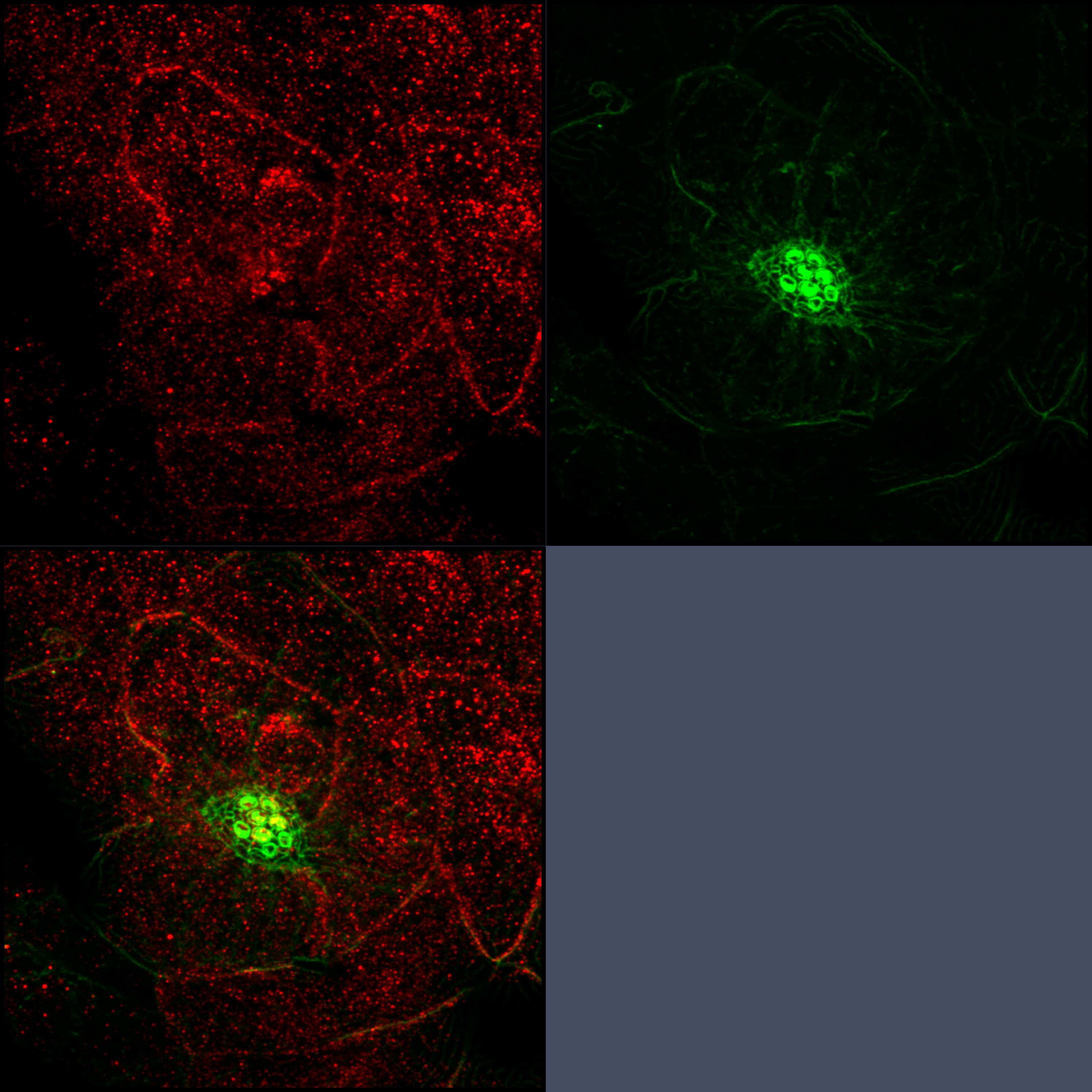
Immunocytochemistry/ Immunofluorescence: RAB3IP Antibody (OTI5F2) [NBP2-45500]
Immunocytochemistry/Immunofluorescence: RAB3IP Antibody (OTI5F2) [NBP2-45500] - RAB3IP Antibody (5F2) [NBP2-45500] - 3dpf zebrafish neuromast immunostained for Rab3IP (red) and counterstained with phalloidin (green). Image courtesy of customer.![Immunohistochemistry-Paraffin: RAB3IP Antibody (OTI5F2) [NBP2-45500]](https://resources.rndsystems.com/images/products/RAB3IP-Antibody-OTI5F2-Immunohistochemistry-Paraffin-NBP2-45500-img0005.jpg "Immunohistochemistry-Paraffin: RAB3IP Antibody (OTI5F2) [NBP2-45500]")
Immunohistochemistry-Paraffin: RAB3IP Antibody (OTI5F2) [NBP2-45500]
Immunohistochemistry-Paraffin: RAB3IP Antibody (OTI5F2) [NBP2-45500] - Staining of paraffin-embedded Human Kidney tissue within the normal limits using anti-RAB3IP mouse monoclonal antibody. (Heat-induced epitope retrieval by 1mM EDTA in 10mM Tris buffer (pH8.5) at 120C for 3min) (1:150)![Immunohistochemistry-Paraffin: RAB3IP Antibody (OTI5F2) [NBP2-45500]](https://resources.rndsystems.com/images/products/RAB3IP-Antibody-OTI5F2-Immunohistochemistry-Paraffin-NBP2-45500-img0002.jpg "Immunohistochemistry-Paraffin: RAB3IP Antibody (OTI5F2) [NBP2-45500]")
Immunohistochemistry-Paraffin: RAB3IP Antibody (OTI5F2) [NBP2-45500]
Immunohistochemistry-Paraffin: RAB3IP Antibody (OTI5F2) [NBP2-45500] - Staining of paraffin-embedded Carcinoma of Human liver tissue using anti-RAB3IP mouse monoclonal antibody. (Heat-induced epitope retrieval by 1mM EDTA in 10mM Tris buffer (pH8.5) at 120C for 3min) (1:150)![Immunohistochemistry-Paraffin: RAB3IP Antibody (OTI5F2) [NBP2-45500]](https://resources.rndsystems.com/images/products/RAB3IP-Antibody-OTI5F2-Immunohistochemistry-Paraffin-NBP2-45500-img0003.jpg "Immunohistochemistry-Paraffin: RAB3IP Antibody (OTI5F2) [NBP2-45500]")
Immunohistochemistry-Paraffin: RAB3IP Antibody (OTI5F2) [NBP2-45500]
Immunohistochemistry-Paraffin: RAB3IP Antibody (OTI5F2) [NBP2-45500] - Staining of paraffin-embedded Carcinoma of Human pancreas tissue using anti-RAB3IP mouse monoclonal antibody. (Heat-induced epitope retrieval by 1mM EDTA in 10mM Tris buffer (pH8.5) at 120C for 3min) (1:150)![Immunohistochemistry-Paraffin: RAB3IP Antibody (OTI5F2) [NBP2-45500]](https://resources.rndsystems.com/images/products/RAB3IP-Antibody-OTI5F2-Immunohistochemistry-Paraffin-NBP2-45500-img0004.jpg "Immunohistochemistry-Paraffin: RAB3IP Antibody (OTI5F2) [NBP2-45500]")
Immunohistochemistry-Paraffin: RAB3IP Antibody (OTI5F2) [NBP2-45500]
Immunohistochemistry-Paraffin: RAB3IP Antibody (OTI5F2) [NBP2-45500] - Staining of paraffin-embedded Human colon tissue within the normal limits using anti-RAB3IP mouse monoclonal antibody. (Heat-induced epitope retrieval by 1mM EDTA in 10mM Tris buffer (pH8.5) at 120C for 3min) (1:150)Applications for RAB3IP Antibody (OTI5F2)
Application
Recommended Usage
Immunocytochemistry/ Immunofluorescence
1:10-1:500
Immunohistochemistry
1:150
Western Blot
1:2000
Reviewed Applications
Read 1 review rated 3 using NBP2-45500 in the following applications:
Formulation, Preparation, and Storage
Purification
Immunogen affinity purified
Formulation
PBS (pH 7.3), 1.0% BSA and 50% Glycerol
Preservative
0.02% Sodium Azide
Concentration
1 mg/ml
Shipping
The product is shipped with polar packs. Upon receipt, store it immediately at the temperature recommended below.
Stability & Storage
Store at -20C. Avoid freeze-thaw cycles.
Background: RAB3IP
Alternate Names
FLJ14660, FLJ22548, MGC71495, RAB3A interacting protein (rabin3), rab-3A-interacting protein, Rab3A-interacting protein, RABIN3, Rabin-3, SSX2 interacting protein, SSX2-interacting protein
Gene Symbol
RAB3IP
Additional RAB3IP Products
Product Documents for RAB3IP Antibody (OTI5F2)
Certificate of Analysis
To download a Certificate of Analysis, please enter a lot or batch number in the search box below.
Product Specific Notices for RAB3IP Antibody (OTI5F2)
This product is for research use only and is not approved for use in humans or in clinical diagnosis. Primary Antibodies are guaranteed for 1 year from date of receipt.
Customer Reviews for RAB3IP Antibody (OTI5F2) (1)
3 out of 5
1 Customer Rating
Have you used RAB3IP Antibody (OTI5F2)?
Submit a review and receive an Amazon gift card!
$25/€18/£15/$25CAN/¥2500 Yen for a review with an image
$10/€7/£6/$10CAN/¥1110 Yen for a review without an image
Submit a review
Customer Images
Showing
1
-
1 of
1 review
Showing All
Filter By:
-
Application: ImmunofluorescenceSample Tested: whole mount zebrafishSpecies: ZebrafishVerified Customer | Posted 02/28/20173dpf zebrafish neuromast immunostained for Rab3IP (red) and counterstained with phalloidin (green)3-5dpf larvae were fixed at 4⁰C overnight with 4% PFA in PBS and then rinsed twice with PBS + Triton X-100 0.1%. Primary antibody was diluted in PBSTx 0.1% containing 2.5% BSA and 2.5% FBS at 1:200 dilution and samples incubated overnight with rocking at 4⁰C. After several washes larvae were incubated with AlexaFluor 594-mouse conjugated antibody and AlexaFluor 488-phalloidin.

There are no reviews that match your criteria.
Protocols
Find general support by application which include: protocols, troubleshooting, illustrated assays, videos and webinars.
- Antigen Retrieval Protocol (PIER)
- Antigen Retrieval for Frozen Sections Protocol
- Appropriate Fixation of IHC/ICC Samples
- Cellular Response to Hypoxia Protocols
- Chromogenic IHC Staining of Formalin-Fixed Paraffin-Embedded (FFPE) Tissue Protocol
- Chromogenic Immunohistochemistry Staining of Frozen Tissue
- ClariTSA™ Fluorophore Kits
- Detection & Visualization of Antibody Binding
- Fluorescent IHC Staining of Frozen Tissue Protocol
- Graphic Protocol for Heat-induced Epitope Retrieval
- Graphic Protocol for the Preparation and Fluorescent IHC Staining of Frozen Tissue Sections
- Graphic Protocol for the Preparation and Fluorescent IHC Staining of Paraffin-embedded Tissue Sections
- Graphic Protocol for the Preparation of Gelatin-coated Slides for Histological Tissue Sections
- ICC Cell Smear Protocol for Suspension Cells
- ICC Immunocytochemistry Protocol Videos
- ICC for Adherent Cells
- IHC Sample Preparation (Frozen sections vs Paraffin)
- Immunocytochemistry (ICC) Protocol
- Immunocytochemistry Troubleshooting
- Immunofluorescence of Organoids Embedded in Cultrex Basement Membrane Extract
- Immunofluorescent IHC Staining of Formalin-Fixed Paraffin-Embedded (FFPE) Tissue Protocol
- Immunohistochemistry (IHC) and Immunocytochemistry (ICC) Protocols
- Immunohistochemistry Frozen Troubleshooting
- Immunohistochemistry Paraffin Troubleshooting
- Preparing Samples for IHC/ICC Experiments
- Preventing Non-Specific Staining (Non-Specific Binding)
- Primary Antibody Selection & Optimization
- Protocol for Heat-Induced Epitope Retrieval (HIER)
- Protocol for Making a 4% Formaldehyde Solution in PBS
- Protocol for VisUCyte™ HRP Polymer Detection Reagent
- Protocol for the Fluorescent ICC Staining of Cell Smears - Graphic
- Protocol for the Fluorescent ICC Staining of Cultured Cells on Coverslips - Graphic
- Protocol for the Preparation & Fixation of Cells on Coverslips
- Protocol for the Preparation and Chromogenic IHC Staining of Frozen Tissue Sections
- Protocol for the Preparation and Chromogenic IHC Staining of Frozen Tissue Sections - Graphic
- Protocol for the Preparation and Chromogenic IHC Staining of Paraffin-embedded Tissue Sections
- Protocol for the Preparation and Chromogenic IHC Staining of Paraffin-embedded Tissue Sections - Graphic
- Protocol for the Preparation and Fluorescent ICC Staining of Cells on Coverslips
- Protocol for the Preparation and Fluorescent ICC Staining of Non-adherent Cells
- Protocol for the Preparation and Fluorescent ICC Staining of Stem Cells on Coverslips
- Protocol for the Preparation and Fluorescent IHC Staining of Frozen Tissue Sections
- Protocol for the Preparation and Fluorescent IHC Staining of Paraffin-embedded Tissue Sections
- Protocol for the Preparation of Gelatin-coated Slides for Histological Tissue Sections
- Protocol for the Preparation of a Cell Smear for Non-adherent Cell ICC - Graphic
- R&D Systems Quality Control Western Blot Protocol
- TUNEL and Active Caspase-3 Detection by IHC/ICC Protocol
- The Importance of IHC/ICC Controls
- Troubleshooting Guide: Immunohistochemistry
- Troubleshooting Guide: Western Blot Figures
- Western Blot Conditions
- Western Blot Protocol
- Western Blot Protocol for Cell Lysates
- Western Blot Troubleshooting
- Western Blot Troubleshooting Guide
- View all Protocols, Troubleshooting, Illustrated assays and Webinars
Loading...