SC35 Antibody - BSA Free
Novus Biologicals | Catalog # NBP2-47290
![Immunocytochemistry/ Immunofluorescence: SC35 Antibody [NBP2-47290]](https://resources.rndsystems.com/images/products/SC35-Antibody-Immunocytochemistry-Immunofluorescence-NBP2-47290-img0003.jpg "Immunocytochemistry/ Immunofluorescence: SC35 Antibody [NBP2-47290]")
Loading...
Key Product Details
Species Reactivity
Validated:
Human
Predicted:
Mouse (97%), Rat (97%). Backed by our 100% Guarantee.
Applications
Immunohistochemistry, Immunohistochemistry-Paraffin, Immunocytochemistry/ Immunofluorescence
Label
Unconjugated
Antibody Source
Polyclonal Rabbit IgG
Format
BSA Free
Loading...
Product Specifications
Immunogen
This antibody was developed against a recombinant protein corresponding to amino acids: LKVDNLTYRTSPDSLRRVFEKYGRVGDVYIPR
Reactivity Notes
Reactivity reported in scientific literature (PMID: 25800673)
Clonality
Polyclonal
Host
Rabbit
Isotype
IgG
Scientific Data Images for SC35 Antibody - BSA Free
Immunocytochemistry/ Immunofluorescence: SC35 Antibody [NBP2-47290]
Immunocytochemistry/Immunofluorescence: SC35 Antibody [NBP2-47290] - Staining of human cell line U-251 MG shows localization to nuclear speckles & cytosol. Antibody staining is shown in green.![Immunohistochemistry-Paraffin: SC35 Antibody [NBP2-47290]](https://resources.rndsystems.com/images/products/SC35-Antibody-Immunohistochemistry-Paraffin-NBP2-47290-img0009.jpg "Immunohistochemistry-Paraffin: SC35 Antibody [NBP2-47290]")
Immunohistochemistry-Paraffin: SC35 Antibody [NBP2-47290]
Immunohistochemistry-Paraffin: SC35 Antibody [NBP2-47290] - Staining of human skin shows moderate cytoplasmic positivity in epidermal cells.![Immunohistochemistry-Paraffin: SC35 Antibody [NBP2-47290]](https://resources.rndsystems.com/images/products/SC35-Antibody-Immunohistochemistry-Paraffin-NBP2-47290-img0006.jpg "Immunohistochemistry-Paraffin: SC35 Antibody [NBP2-47290]")
Immunohistochemistry-Paraffin: SC35 Antibody [NBP2-47290]
Immunohistochemistry-Paraffin: SC35 Antibody [NBP2-47290] - Staining of human cerebral cortex shows moderate to strong nuclear positivity in neurons.![Immunohistochemistry-Paraffin: SC35 Antibody [NBP2-47290]](https://resources.rndsystems.com/images/products/SC35-Antibody-Immunohistochemistry-Paraffin-NBP2-47290-img0007.jpg "Immunohistochemistry-Paraffin: SC35 Antibody [NBP2-47290]")
Immunohistochemistry-Paraffin: SC35 Antibody [NBP2-47290]
Immunohistochemistry-Paraffin: SC35 Antibody [NBP2-47290] - Staining of human duodenum shows moderate cytoplasmic positivity in glandular cells.![Immunohistochemistry-Paraffin: SC35 Antibody [NBP2-47290]](https://resources.rndsystems.com/images/products/SC35-Antibody-Immunohistochemistry-Paraffin-NBP2-47290-img0008.jpg "Immunohistochemistry-Paraffin: SC35 Antibody [NBP2-47290]")
Immunohistochemistry-Paraffin: SC35 Antibody [NBP2-47290]
Immunohistochemistry-Paraffin: SC35 Antibody [NBP2-47290] - Staining of human tonsil shows moderate to strong positivity in germinal center cells.Applications for SC35 Antibody - BSA Free
Application
Recommended Usage
Immunocytochemistry/ Immunofluorescence
0.25-2 ug/ml
Immunohistochemistry
1:50 - 1:200
Immunohistochemistry-Paraffin
1:50 - 1:200
Application Notes
For IHC-Paraffin, HIER pH 6 retrieval is recommended. ICC/IF Fixation Permeabilization: Use PFA/Triton X-100.
Reviewed Applications
Read 1 review rated 1 using NBP2-47290 in the following applications:
Formulation, Preparation, and Storage
Purification
Affinity purified
Formulation
PBS (pH 7.2) and 40% Glycerol
Format
BSA Free
Preservative
0.02% Sodium Azide
Concentration
Concentrations vary lot to lot. See vial label for concentration. If unlisted please contact technical services.
Shipping
The product is shipped with polar packs. Upon receipt, store it immediately at the temperature recommended below.
Stability & Storage
Store at 4C short term. Aliquot and store at -20C long term. Avoid freeze-thaw cycles.
Background: SC35
Alternate Names
PR264, SC35, SC-35, serine/arginine-rich splicing factor 2, SFRS2, SFRS2A, splicing component, 35 kDa, splicing factor SC35, splicing factor, arginine/serine-rich 2, SR splicing factor 2, SRp30b
Gene Symbol
SRSF2
Additional SC35 Products
Product Documents for SC35 Antibody - BSA Free
Certificate of Analysis
To download a Certificate of Analysis, please enter a lot or batch number in the search box below.
Product Specific Notices for SC35 Antibody - BSA Free
This product is for research use only and is not approved for use in humans or in clinical diagnosis. Primary Antibodies are guaranteed for 1 year from date of receipt.
Citations for SC35 Antibody - BSA Free
 Powered by Bioz
Customer Reviews for SC35 Antibody - BSA Free (1)
1 out of 5
1 Customer Rating
Have you used SC35 Antibody - BSA Free?
Submit a review and receive an Amazon gift card!
$25/€18/£15/$25CAN/¥2500 Yen for a review with an image
$10/€7/£6/$10CAN/¥1110 Yen for a review without an image
Submit a review
Customer Images
Showing
1
-
1 of
1 review
Showing All
Filter By:
-
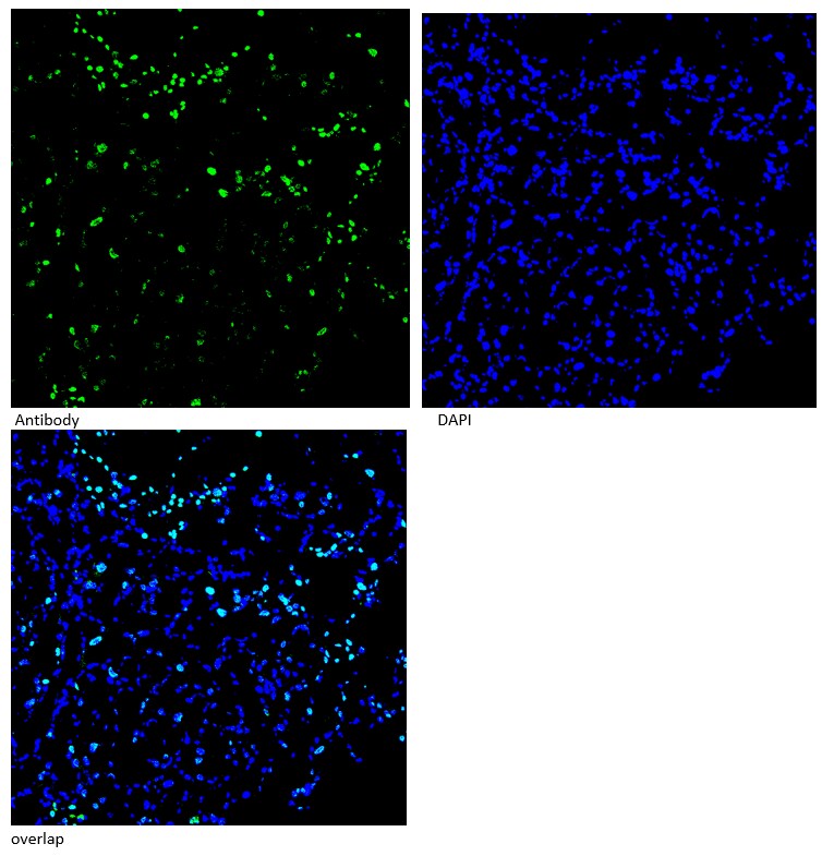
Application: Immunohistochemistry-FrozenSample Tested: LiverSpecies: MouseVerified Customer | Posted 12/20/2016This customer tested the product in an untested application (IHC-Fr) and earned the Innovator's Reward for this product. Antibody Storage Conditions: -20℃ Staining Expectations: positive signals should be within nucleus Fixative Composition: PFA Fixation Time & Temperature: 15 min RT Tissue Processing: as normal Blocking Solution: 5% goat serum in PBS Time & Temperature: 15min RT Primary Antibody: Dilution: 1:50 Diluent Buffer: 5% goat serum in PBS Time & Temperature: 15min RT Washing Conditions: Wash Buffer Composition: PBS Times/Washing: 5min x 3 times Repetitions: 3 Secondary Antibody Manufacturer and Catalog #: life technologies Ref:A11034 Secondary description: goat anti Rabit Dilution: 1:2000 Diluent Buffer: 5% goat serum in PBS Incubation Time & Temperature: 1 hour RT Post-Secondary Washing: PBS Detection Method: Detection: confocal Procedure: as olypus DAB-Incubation Time (if applicable): Click here to enter text. Controls: Positive Control: No Negative Control: no

There are no reviews that match your criteria.
Protocols
Find general support by application which include: protocols, troubleshooting, illustrated assays, videos and webinars.
- Antigen Retrieval Protocol (PIER)
- Antigen Retrieval for Frozen Sections Protocol
- Appropriate Fixation of IHC/ICC Samples
- Cellular Response to Hypoxia Protocols
- Chromogenic IHC Staining of Formalin-Fixed Paraffin-Embedded (FFPE) Tissue Protocol
- Chromogenic Immunohistochemistry Staining of Frozen Tissue
- ClariTSA™ Fluorophore Kits
- Detection & Visualization of Antibody Binding
- Fluorescent IHC Staining of Frozen Tissue Protocol
- Graphic Protocol for Heat-induced Epitope Retrieval
- Graphic Protocol for the Preparation and Fluorescent IHC Staining of Frozen Tissue Sections
- Graphic Protocol for the Preparation and Fluorescent IHC Staining of Paraffin-embedded Tissue Sections
- Graphic Protocol for the Preparation of Gelatin-coated Slides for Histological Tissue Sections
- ICC Cell Smear Protocol for Suspension Cells
- ICC Immunocytochemistry Protocol Videos
- ICC for Adherent Cells
- IHC Sample Preparation (Frozen sections vs Paraffin)
- Immunocytochemistry (ICC) Protocol
- Immunocytochemistry Troubleshooting
- Immunofluorescence of Organoids Embedded in Cultrex Basement Membrane Extract
- Immunofluorescent IHC Staining of Formalin-Fixed Paraffin-Embedded (FFPE) Tissue Protocol
- Immunohistochemistry (IHC) and Immunocytochemistry (ICC) Protocols
- Immunohistochemistry Frozen Troubleshooting
- Immunohistochemistry Paraffin Troubleshooting
- Preparing Samples for IHC/ICC Experiments
- Preventing Non-Specific Staining (Non-Specific Binding)
- Primary Antibody Selection & Optimization
- Protocol for Heat-Induced Epitope Retrieval (HIER)
- Protocol for Making a 4% Formaldehyde Solution in PBS
- Protocol for VisUCyte™ HRP Polymer Detection Reagent
- Protocol for the Fluorescent ICC Staining of Cell Smears - Graphic
- Protocol for the Fluorescent ICC Staining of Cultured Cells on Coverslips - Graphic
- Protocol for the Preparation & Fixation of Cells on Coverslips
- Protocol for the Preparation and Chromogenic IHC Staining of Frozen Tissue Sections
- Protocol for the Preparation and Chromogenic IHC Staining of Frozen Tissue Sections - Graphic
- Protocol for the Preparation and Chromogenic IHC Staining of Paraffin-embedded Tissue Sections
- Protocol for the Preparation and Chromogenic IHC Staining of Paraffin-embedded Tissue Sections - Graphic
- Protocol for the Preparation and Fluorescent ICC Staining of Cells on Coverslips
- Protocol for the Preparation and Fluorescent ICC Staining of Non-adherent Cells
- Protocol for the Preparation and Fluorescent ICC Staining of Stem Cells on Coverslips
- Protocol for the Preparation and Fluorescent IHC Staining of Frozen Tissue Sections
- Protocol for the Preparation and Fluorescent IHC Staining of Paraffin-embedded Tissue Sections
- Protocol for the Preparation of Gelatin-coated Slides for Histological Tissue Sections
- Protocol for the Preparation of a Cell Smear for Non-adherent Cell ICC - Graphic
- TUNEL and Active Caspase-3 Detection by IHC/ICC Protocol
- The Importance of IHC/ICC Controls
- Troubleshooting Guide: Immunohistochemistry
- View all Protocols, Troubleshooting, Illustrated assays and Webinars
Loading...