SCN3B Antibody (S396-29) - BSA Free
Novus Biologicals | Catalog # NBP2-59318
![Western Blot: SCN3B Antibody (S396-29) [NBP2-59318]](https://resources.rndsystems.com/images/products/SCN3B-Antibody-S396-29-Western-Blot-NBP2-59318-img0005.jpg "Western Blot: SCN3B Antibody (S396-29) [NBP2-59318]")
Key Product Details
Species Reactivity
Human, Mouse, Rat
Applications
Western Blot, Immunocytochemistry/ Immunofluorescence, Simple Western
Label
Unconjugated
Antibody Source
Monoclonal Mouse IgG2B Clone # S396-29
Format
BSA Free
Loading...
Product Specifications
Immunogen
Fusion protein amino acids 1-215 (full-length) of rat NavBeta3
Localization
Membrane
Specificity
Detects 40kDa. Does not cross-reat with NavBeta1, Navbeta2, or Navbeta4.
Clonality
Monoclonal
Host
Mouse
Isotype
IgG2B
Scientific Data Images for SCN3B Antibody (S396-29) - BSA Free
Western Blot: SCN3B Antibody (S396-29) [NBP2-59318]
Western Blot: SCN3B Antibody (S396-29) [NBP2-59318] - Western Blot analysis of Mouse Brain showing detection of ~40 kDa Nav Beta 3 protein using Mouse Anti-Nav Beta 3 Monoclonal Antibody, Clone S396-29 (NBP2-59318). Lane 1: MW Ladder. Lane 2: Mouse Brain. Load: 20 ug. Primary Antibody: Mouse Anti-Nav Beta 3 Monoclonal Antibody (NBP2-59318) at 1:1000 for 16 hours at 4C. Secondary Antibody: Goat Anti-Mouse IgG: HRP at 1:200 for 1 hour at RT. Predicted/Observed Size: ~40 kDa.![Immunocytochemistry/ Immunofluorescence: SCN3B Antibody (S396-29) [NBP2-59318]](https://resources.rndsystems.com/images/products/SCN3B-Antibody-S396-29-Immunocytochemistry-Immunofluorescence-NBP2-59318-img0006.jpg "Immunocytochemistry/ Immunofluorescence: SCN3B Antibody (S396-29) [NBP2-59318]")
Immunocytochemistry/ Immunofluorescence: SCN3B Antibody (S396-29) [NBP2-59318]
Immunocytochemistry/Immunofluorescence: SCN3B Antibody (S396-29) [NBP2-59318] - Immunocytochemistry/Immunofluorescence analysis using Mouse Anti-Nav beta3 Monoclonal Antibody, Clone S396-29 (NBP2-59318). Tissue: Neuroblastoma cells (SH-SY5Y). Species: Human. Fixation: 4% PFA for 15 min. Primary Antibody: Mouse Anti-Nav beta3 Monoclonal Antibody (NBP2-59318) at 1:200 for overnight at 4C with slow rocking. Secondary Antibody: AlexaFluor 488 at 1:1000 for 1 hour at RT. Counterstain: Phalloidin-iFluor 647 (red) F-Actin stain; Hoechst (blue) nuclear stain at 1:800, 1.6mM for 20 min at RT. (A) Hoechst (blue) nuclear stain. (B) Phalloidin-iFluor 647 (red) F-Actin stain. (C) Nav beta3 Antibody (D) Composite.![Immunocytochemistry/ Immunofluorescence: SCN3B Antibody (S396-29) [NBP2-59318]](https://resources.rndsystems.com/images/products/SCN3B-Antibody-S396-29-Immunocytochemistry-Immunofluorescence-NBP2-59318-img0002.jpg "Immunocytochemistry/ Immunofluorescence: SCN3B Antibody (S396-29) [NBP2-59318]")
Immunocytochemistry/ Immunofluorescence: SCN3B Antibody (S396-29) [NBP2-59318]
Immunocytochemistry/Immunofluorescence: SCN3B Antibody (S396-29) [NBP2-59318] - Tissue: Neuroblastoma cell line SK-N-BE. Species: Human. Fixation: 4% Formaldehyde for 15 min at RT. Primary Antibody: Mouse Anti-Nav beta 3 Monoclonal Antibody at 1:100 for 60 min at RT. Secondary Antibody: Goat Anti-Mouse ATTO 488 at 1:100 for 60 min at RT. Counterstain: Phalloidin Texas Red F-Actin stain; DAPI (blue) nuclear stain at 1:1000, 1:5000 for 60min RT, 5min RT. Localization: Cell Membrane, Nucleus. Magnification: 60X.Applications for SCN3B Antibody (S396-29) - BSA Free
Application
Recommended Usage
Immunocytochemistry/ Immunofluorescence
1:100
Western Blot
1:1000
Application Notes
This SCN3B Antibody (S396-29) has been validated for Simple Western from a verified customer review.
See Simple Western Antibody Database for Simple Western validation: separated by Size
See Simple Western Antibody Database for Simple Western validation: separated by Size
Reviewed Applications
Read 1 review rated 4 using NBP2-59318 in the following applications:
Formulation, Preparation, and Storage
Purification
Protein G purified
Formulation
PBS (pH 7.4), 50% Glycerol
Format
BSA Free
Preservative
0.1% Sodium Azide
Concentration
Please see the vial label for concentration. If unlisted please contact technical services.
Shipping
The product is shipped with polar packs. Upon receipt, store it immediately at the temperature recommended below.
Stability & Storage
Store at -20C.
Background: SCN3B
Alternate Names
HSA243396, KIAA1158, SCNB3, sodium channel subunit beta-3, sodium channel, voltage-gated, type III, beta, voltage-gated sodium channel beta-3 subunit
Gene Symbol
SCN3B
Additional SCN3B Products
Product Documents for SCN3B Antibody (S396-29) - BSA Free
Certificate of Analysis
To download a Certificate of Analysis, please enter a lot or batch number in the search box below.
Product Specific Notices for SCN3B Antibody (S396-29) - BSA Free
This product is for research use only and is not approved for use in humans or in clinical diagnosis. Primary Antibodies are guaranteed for 1 year from date of receipt.
Citations for SCN3B Antibody (S396-29) - BSA Free
 Powered by Bioz
Customer Reviews for SCN3B Antibody (S396-29) - BSA Free (1)
4 out of 5
1 Customer Rating
Have you used SCN3B Antibody (S396-29) - BSA Free?
Submit a review and receive an Amazon gift card!
$25/€18/£15/$25CAN/¥2500 Yen for a review with an image
$10/€7/£6/$10CAN/¥1110 Yen for a review without an image
Submit a review
Customer Images
Showing
1
-
1 of
1 review
Showing All
Filter By:
-
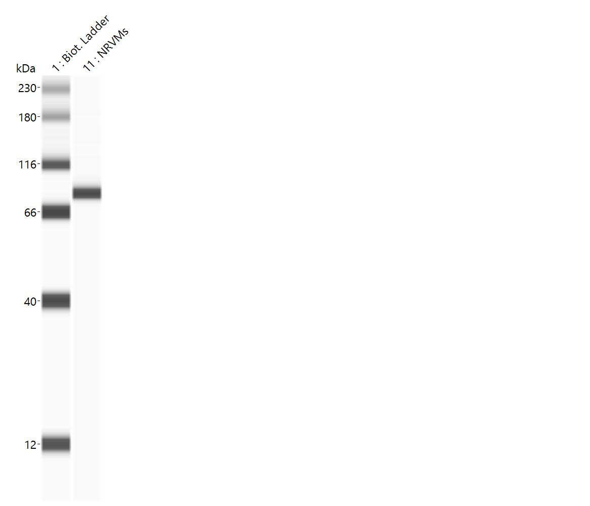
Application: Simple WesternSample Tested: primary neonatal ventricular cardiomyocytesSpecies: RatVerified Customer | Posted 02/17/2021Clear band without noise at 86kDa. Expected molecular weight should be around 30-40kDa. It can be the result of SCN3B dimers.Antibody used at a dilution of 1:50. Protein concentration of 0.8ug/uL.
 Bio-Techne ResponseThis review was submitted through the legacy Novus Innovators Program, reflecting a new species or application tested on a primary antibody.
There are no reviews that match your criteria.
Protocols
Find general support by application which include: protocols, troubleshooting, illustrated assays, videos and webinars.
- Appropriate Fixation of IHC/ICC Samples
- Cellular Response to Hypoxia Protocols
- ClariTSA™ Fluorophore Kits
- Detection & Visualization of Antibody Binding
- ICC Cell Smear Protocol for Suspension Cells
- ICC Immunocytochemistry Protocol Videos
- ICC for Adherent Cells
- Immunocytochemistry (ICC) Protocol
- Immunocytochemistry Troubleshooting
- Immunofluorescence of Organoids Embedded in Cultrex Basement Membrane Extract
- Immunohistochemistry (IHC) and Immunocytochemistry (ICC) Protocols
- Preparing Samples for IHC/ICC Experiments
- Preventing Non-Specific Staining (Non-Specific Binding)
- Primary Antibody Selection & Optimization
- Protocol for VisUCyte™ HRP Polymer Detection Reagent
- Protocol for the Fluorescent ICC Staining of Cell Smears - Graphic
- Protocol for the Fluorescent ICC Staining of Cultured Cells on Coverslips - Graphic
- Protocol for the Preparation and Fluorescent ICC Staining of Cells on Coverslips
- Protocol for the Preparation and Fluorescent ICC Staining of Non-adherent Cells
- Protocol for the Preparation and Fluorescent ICC Staining of Stem Cells on Coverslips
- Protocol for the Preparation of a Cell Smear for Non-adherent Cell ICC - Graphic
- R&D Systems Quality Control Western Blot Protocol
- TUNEL and Active Caspase-3 Detection by IHC/ICC Protocol
- The Importance of IHC/ICC Controls
- Troubleshooting Guide: Western Blot Figures
- Western Blot Conditions
- Western Blot Protocol
- Western Blot Protocol for Cell Lysates
- Western Blot Troubleshooting
- Western Blot Troubleshooting Guide
- View all Protocols, Troubleshooting, Illustrated assays and Webinars
Loading...