SUR1 Antibody (S289-16) - BSA Free
Novus Biologicals | Catalog # NBP2-59320
![Western Blot: SUR1 Antibody (S289-16) [NBP2-59320]](https://resources.rndsystems.com/images/products/SUR1-Antibody-S289-16-Western-Blot-NBP2-59320-img0007.jpg "Western Blot: SUR1 Antibody (S289-16) [NBP2-59320]")
Key Product Details
Species Reactivity
Validated:
Human, Mouse, Rat, Hamster
Cited:
Human, Mouse, Rat
Applications
Validated:
Immunohistochemistry, Western Blot, Immunocytochemistry/ Immunofluorescence
Cited:
IF/IHC
Label
Unconjugated
Antibody Source
Monoclonal Mouse IgG1 Clone # S289-16
Format
BSA Free
Loading...
Product Specifications
Immunogen
Fusion protein amino acids 1548-1582 (cytoplasmic C-terminus) of rat SUR1
Localization
Membrane
Specificity
Detects 160kDa. Does not cross-react with SUR2B.
Clonality
Monoclonal
Host
Mouse
Isotype
IgG1
Scientific Data Images for SUR1 Antibody (S289-16) - BSA Free
Western Blot: SUR1 Antibody (S289-16) [NBP2-59320]
Western Blot: SUR1 Antibody (S289-16) [NBP2-59320] - Western Blot analysis of Rat Brain Membrane showing detection of ~160 kDa SUR1 protein using Mouse Anti-SUR1 Monoclonal Antibody, Clone S289-16 (NBP2-59320). Lane 1: Molecular Weight Ladder. Lane 2: Rat Brain Membrane. Load: 15 ug. Block: 2% BSA and 2% Skim Milk in 1X TBST. Primary Antibody: Mouse Anti-SUR1 Monoclonal Antibody (NBP2-59320) at 1:200 for 16 hours at 4C. Secondary Antibody: Goat Anti-Mouse IgG: HRP at 1:1000 for 1 hour RT. Color Development: ECL solution for 6 min in RT. Predicted/Observed Size: ~160 kDa.![Immunocytochemistry/ Immunofluorescence: SUR1 Antibody (S289-16) [NBP2-59320]](https://resources.rndsystems.com/images/products/SUR1-Antibody-S289-16-Immunocytochemistry-Immunofluorescence-NBP2-59320-img0008.jpg "Immunocytochemistry/ Immunofluorescence: SUR1 Antibody (S289-16) [NBP2-59320]")
Immunocytochemistry/ Immunofluorescence: SUR1 Antibody (S289-16) [NBP2-59320]
Immunocytochemistry/Immunofluorescence: SUR1 Antibody (S289-16) [NBP2-59320] - Immunocytochemistry/Immunofluorescence analysis using Mouse Anti-SUR1 Monoclonal Antibody, Clone S289-16 (NBP2-59320). Tissue: Neuroblastoma cells (SH-SY5Y). Species: Human. Fixation: 4% PFA for 15 min. Primary Antibody: Mouse Anti-SUR1 Monoclonal Antibody (NBP2-59320) at 1:50 for overnight at 4C with slow rocking. Secondary Antibody: AlexaFluor 488 at 1:1000 for 1 hour at RT. Counterstain: Phalloidin-iFluor 647 (red) F-Actin stain; Hoechst (blue) nuclear stain at 1:800, 1.6mM for 20 min at RT. (A) Hoechst (blue) nuclear stain. (B) Phalloidin-iFluor 647 (red) F-Actin stain. (C) SUR1 Antibody (D) Composite.![Western Blot: SUR1 Antibody (S289-16) [NBP2-59320]](https://resources.rndsystems.com/images/products/SUR1-Antibody-S289-16-Western-Blot-NBP2-59320-img0005.jpg "Western Blot: SUR1 Antibody (S289-16) [NBP2-59320]")
Western Blot: SUR1 Antibody (S289-16) [NBP2-59320]
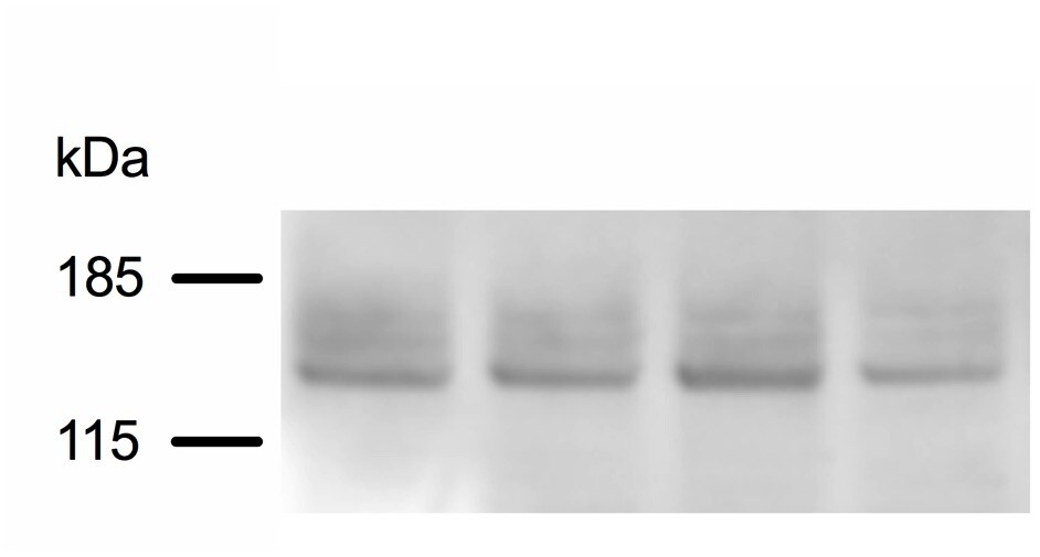
Western Blot: SUR1 Antibody (S289-16) [NBP2-59320] - Analysis of mouse pancreatic islets. Lane 1: Molecular Weight Ladder. Lanes 2-5: Samples from pooled pancreatic islets. Load: ~10ug. Primary Antibody: Anti-SUR1 Monoclonal Antibody at 1:200 for ~16hrs at 4C. Secondary Antibody: Goat Anti-Mouse IgG: HRP at 1:1000 for 1hr RT. Image from verified customer review.![Immunocytochemistry/ Immunofluorescence: SUR1 Antibody (S289-16) [NBP2-59320]](https://resources.rndsystems.com/images/products/SUR1-Antibody-S289-16-Immunocytochemistry-Immunofluorescence-NBP2-59320-img0002.jpg "Immunocytochemistry/ Immunofluorescence: SUR1 Antibody (S289-16) [NBP2-59320]")
Immunocytochemistry/ Immunofluorescence: SUR1 Antibody (S289-16) [NBP2-59320]
Immunocytochemistry/Immunofluorescence: SUR1 Antibody (S289-16) [NBP2-59320] - Analysis using Mouse Anti-SUR1 Monoclonal Antibody, Clone S289-16. Tissue: Neuroblastoma cell line SK-N-BE. Species: Human. Fixation: 4% Formaldehyde for 15 min at RT. Primary Antibody: Mouse Anti-SUR1 Monoclonal Antibody at 1:100 for 60 min at RT. Secondary Antibody: Goat Anti-Mouse ATTO 488 at 1:100 for 60 min at RT. Counterstain: Phalloidin Texas Red F-Actin stain; DAPI (blue) nuclear stain at 1:1000, 1:5000 for 60min RT, 5min RT. Localization: Cytoplasm, Nucleus. Magnification: 60X. (A) DAPI (blue) nuclear stain (B) Phalloidin Texas Red F-Actin stain (C) SUR1 Antibody (D) Composite.![Immunocytochemistry/ Immunofluorescence: SUR1 Antibody (S289-16) [NBP2-59320]](https://resources.rndsystems.com/images/products/SUR1-Antibody-S289-16-Immunocytochemistry-Immunofluorescence-NBP2-59320-img0006.jpg "Immunocytochemistry/ Immunofluorescence: SUR1 Antibody (S289-16) [NBP2-59320]")
Immunocytochemistry/ Immunofluorescence: SUR1 Antibody (S289-16) [NBP2-59320]
SUR1-Antibody-S289-16-Immunocytochemistry-Immunofluorescence-NBP2-59320-img0006.jpg [NBP2-59320] -")
Immunocytochemistry/ Immunofluorescence: SUR1 Antibody (S289-16) [NBP2-59320] -
Immunocytochemistry/ Immunofluorescence: SUR1 Antibody (S289-16) [NBP2-59320] - Glial fibrillary acidic protein (GFAP)–positive specimens from human contusion- traumatic brain injury (TBI) exhibit KIR6.2 expression in astrocytes. (A) Immunolabeling for KIR6.2 showed sparse immunoreactivity in the control specimen (CTR) vs. widespread expression in a GFAP-positive specimen from contusion-TBI. (B) Double immunolabeling for GFAP (red) & KIR6.2 (green) showed astrocyte expression of KIR6.2; merged images confirm co-localization (yellow); in situ hybridization of the same tissue section for Kcnj11 messenger RNA showed positive signal co-localized with GFAP-positive, KIR6.2-expressing astrocytes; arrowheads point to cells with all three signals. (C) Double immunolabeling showed that KIR6.2 (red) & sulfonylurea receptor 1 (SUR1; green) were co-localized (yellow) in astrocytes. (D) ImmunoFRET for SUR1 (magenta) & KIR6.2 (red) showed co-assembly of SUR1-KIR6.2 heteromers (yellow pseudocolor) in astrocytes. (E) Double immunolabeling showed that KIR6.2 (green) & transient receptor potential cation channel subfamily M member 4 (TRPM4) (red) were co-localized in astrocytes. The findings illustrated are representative of all GFAP-positive specimens from eight cases of human contusion-TBI. (case #2, 11 days post-TBI; case #5, 1 day post-TBI) Image collected & cropped by CiteAb from the following publication (https://pubmed.ncbi.nlm.nih.gov/30160201), licensed under a CC-BY license. Not internally tested by Novus Biologicals.Applications for SUR1 Antibody (S289-16) - BSA Free
Application
Recommended Usage
Immunocytochemistry/ Immunofluorescence
1:100
Immunohistochemistry
1:1000
Western Blot
1:1000
Reviewed Applications
Read 2 reviews rated 4 using NBP2-59320 in the following applications:
Formulation, Preparation, and Storage
Purification
Protein G purified
Formulation
PBS (pH 7.4), 50% Glycerol
Format
BSA Free
Preservative
0.09% Sodium Azide
Concentration
Please see the vial label for concentration. If unlisted please contact technical services.
Shipping
The product is shipped with polar packs. Upon receipt, store it immediately at the temperature recommended below.
Stability & Storage
Store at -20C.
Background: SUR1
Long Name
ATP-binding cassette sub-family C member 8
Alternate Names
ABCC8, HRINS, SUR
Gene Symbol
ABCC8
Additional SUR1 Products
Product Documents for SUR1 Antibody (S289-16) - BSA Free
Certificate of Analysis
To download a Certificate of Analysis, please enter a lot or batch number in the search box below.
Product Specific Notices for SUR1 Antibody (S289-16) - BSA Free
This product is for research use only and is not approved for use in humans or in clinical diagnosis. Primary Antibodies are guaranteed for 1 year from date of receipt.
Citations for SUR1 Antibody (S289-16) - BSA Free
 Powered by Bioz
Customer Reviews for SUR1 Antibody (S289-16) - BSA Free (2)
4 out of 5
2 Customer Ratings
Have you used SUR1 Antibody (S289-16) - BSA Free?
Submit a review and receive an Amazon gift card!
$25/€18/£15/$25CAN/¥2500 Yen for a review with an image
$10/€7/£6/$10CAN/¥1110 Yen for a review without an image
Submit a review
Customer Images
Showing
1
-
2 of
2 reviews
Showing All
Filter By:
-
Application: Western BlotSample Tested: Mouse pancreatic isletsSpecies: MouseVerified Customer | Posted 02/25/2019Western Blot analysis of mouse pancreatic islets using Mouse Anti-SUR1 S289-16. Lane 1: Molecular Weight Ladder. Lanes 2-5: Samples from pooled pancreatic islets. Load: ~10 ug. Primary Antibody: Anti-SUR1 Monoclonal Antibody at 1:200 for ~16 hours at 4 degrees celsius. Secondary Antibody: Goat Anti-Mouse IgG: HRP at 1:1000 for 1 hour RT.

-
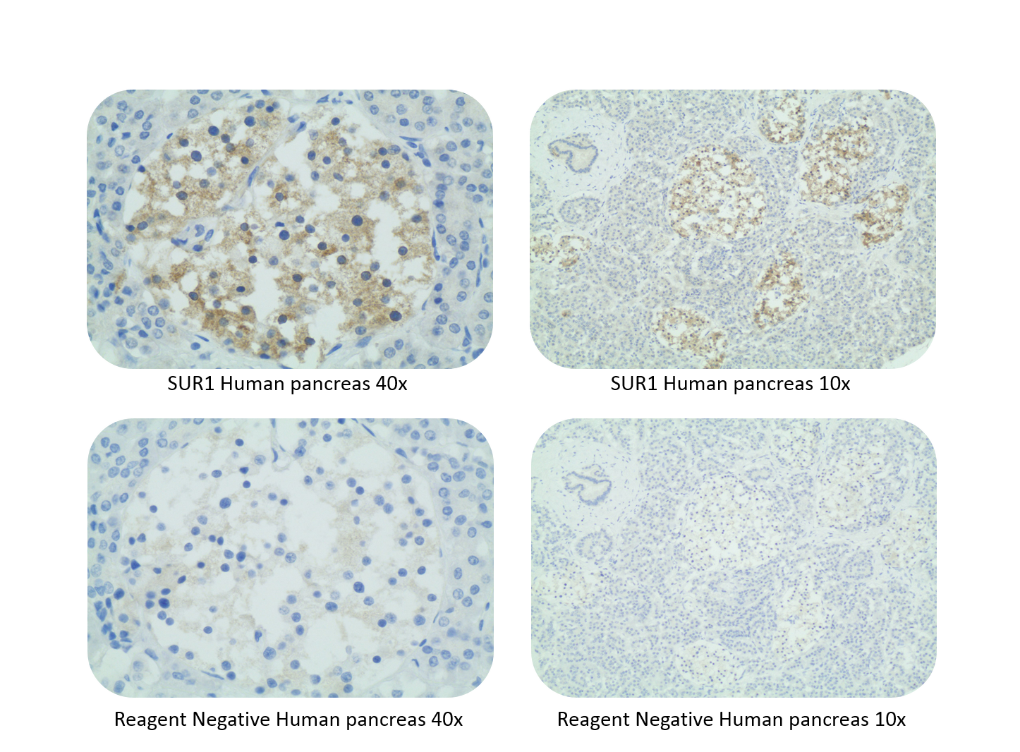
Application: Immunohistochemistry-ParaffinSample Tested: Human Tissue Sections - PancreasSpecies: HumanVerified Customer | Posted 07/07/2017Immunohistochemistry-Paraffin: SUR1 Antibody (S289-16) [NBP2-59320]SUR1 was detected in immersion fixed paraffin-embedded sections of normal human pancreas using citrate pH6 pretreatment and incubating at 1:50 dilution for 30 minutes, with LabVision Quanto Detection.
There are no reviews that match your criteria.
Protocols
Find general support by application which include: protocols, troubleshooting, illustrated assays, videos and webinars.
- Antigen Retrieval Protocol (PIER)
- Antigen Retrieval for Frozen Sections Protocol
- Appropriate Fixation of IHC/ICC Samples
- Cellular Response to Hypoxia Protocols
- Chromogenic IHC Staining of Formalin-Fixed Paraffin-Embedded (FFPE) Tissue Protocol
- Chromogenic Immunohistochemistry Staining of Frozen Tissue
- ClariTSA™ Fluorophore Kits
- Detection & Visualization of Antibody Binding
- Fluorescent IHC Staining of Frozen Tissue Protocol
- Graphic Protocol for Heat-induced Epitope Retrieval
- Graphic Protocol for the Preparation and Fluorescent IHC Staining of Frozen Tissue Sections
- Graphic Protocol for the Preparation and Fluorescent IHC Staining of Paraffin-embedded Tissue Sections
- Graphic Protocol for the Preparation of Gelatin-coated Slides for Histological Tissue Sections
- ICC Cell Smear Protocol for Suspension Cells
- ICC Immunocytochemistry Protocol Videos
- ICC for Adherent Cells
- IHC Sample Preparation (Frozen sections vs Paraffin)
- Immunocytochemistry (ICC) Protocol
- Immunocytochemistry Troubleshooting
- Immunofluorescence of Organoids Embedded in Cultrex Basement Membrane Extract
- Immunofluorescent IHC Staining of Formalin-Fixed Paraffin-Embedded (FFPE) Tissue Protocol
- Immunohistochemistry (IHC) and Immunocytochemistry (ICC) Protocols
- Immunohistochemistry Frozen Troubleshooting
- Immunohistochemistry Paraffin Troubleshooting
- Preparing Samples for IHC/ICC Experiments
- Preventing Non-Specific Staining (Non-Specific Binding)
- Primary Antibody Selection & Optimization
- Protocol for Heat-Induced Epitope Retrieval (HIER)
- Protocol for Making a 4% Formaldehyde Solution in PBS
- Protocol for VisUCyte™ HRP Polymer Detection Reagent
- Protocol for the Fluorescent ICC Staining of Cell Smears - Graphic
- Protocol for the Fluorescent ICC Staining of Cultured Cells on Coverslips - Graphic
- Protocol for the Preparation & Fixation of Cells on Coverslips
- Protocol for the Preparation and Chromogenic IHC Staining of Frozen Tissue Sections
- Protocol for the Preparation and Chromogenic IHC Staining of Frozen Tissue Sections - Graphic
- Protocol for the Preparation and Chromogenic IHC Staining of Paraffin-embedded Tissue Sections
- Protocol for the Preparation and Chromogenic IHC Staining of Paraffin-embedded Tissue Sections - Graphic
- Protocol for the Preparation and Fluorescent ICC Staining of Cells on Coverslips
- Protocol for the Preparation and Fluorescent ICC Staining of Non-adherent Cells
- Protocol for the Preparation and Fluorescent ICC Staining of Stem Cells on Coverslips
- Protocol for the Preparation and Fluorescent IHC Staining of Frozen Tissue Sections
- Protocol for the Preparation and Fluorescent IHC Staining of Paraffin-embedded Tissue Sections
- Protocol for the Preparation of Gelatin-coated Slides for Histological Tissue Sections
- Protocol for the Preparation of a Cell Smear for Non-adherent Cell ICC - Graphic
- R&D Systems Quality Control Western Blot Protocol
- TUNEL and Active Caspase-3 Detection by IHC/ICC Protocol
- The Importance of IHC/ICC Controls
- Troubleshooting Guide: Immunohistochemistry
- Troubleshooting Guide: Western Blot Figures
- Western Blot Conditions
- Western Blot Protocol
- Western Blot Protocol for Cell Lysates
- Western Blot Troubleshooting
- Western Blot Troubleshooting Guide
- View all Protocols, Troubleshooting, Illustrated assays and Webinars
Loading...