Thyroglobulin Antibody (2H11 + 6E1)
Novus Biologicals | Catalog # NBP2-34294
Key Product Details
Species Reactivity
Validated:
Human, Mouse, Rat
Cited:
Mouse
Applications
Validated:
Immunohistochemistry, Immunohistochemistry-Paraffin, Flow Cytometry
Cited:
Immunohistochemistry-Paraffin
Label
Unconjugated
Antibody Source
Monoclonal Mouse IgG1 Kappa/IgG1 Kappa Clone # 2H11 + 6E1
Loading...
Product Specifications
Immunogen
Human thyroid follicular cells
Localization
Cytoplasmic and secreted
Marker
Thyroidal Cell Marker
Specificity
Thyroglobulin is a 660kDa dimeric pre-protein with multiple glycosylation sites. It is produced by and processed within the thyroid gland to produce the hormone thyroxine and triiodothyronine. Prior to forming dimers, thyroglobulin monomers undergo conformational maturation in the endoplasmic reticulation. The vast majority of follicular carcinomas of the thyroid will give positive immunoreactivity for anti-thyroglobulin even though sometimes only focally. Poorly differentiated carcinomas of the thyroid are frequently anti-thyroglobulin negative. Adenocarcinomas of other-than-thyroid origin do not react with this antibody. This antibody is useful in identification of thyroid carcinoma of the papillary and follicular types. Presence of thyroglobulin in metastatic lesions establishes the thyroid origin of tumor. Anti-thyroglobulin, combined with anti-calcitonin, can identify medullary carcinomas of the thyroid. Furthermore, anti-thyroglobulin, combined with anti-TTF1, can be a reliable marker to differentiate between primary thyroid and lung neoplasms.
Clonality
Monoclonal
Host
Mouse
Isotype
IgG1 Kappa/IgG1 Kappa
Theoretical MW
660 kDa.
Disclaimer note: The observed molecular weight of the protein may vary from the listed predicted molecular weight due to post translational modifications, post translation cleavages, relative charges, and other experimental factors.
Disclaimer note: The observed molecular weight of the protein may vary from the listed predicted molecular weight due to post translational modifications, post translation cleavages, relative charges, and other experimental factors.
Description
200ug/ml of antibody purified from Bioreactor Concentrate by Protein A or G. Prepared in 10 mM PBS with 0.05% BSA & 0.05% azide. Also available WITHOUT BSA & azide at 1.0 mg/ml. (NBP2-34530)
Antibody with azide - store at 2 to 8C. Antibody without azide - store at -20 to -80C.
Antibody with azide - store at 2 to 8C. Antibody without azide - store at -20 to -80C.
Scientific Data Images for Thyroglobulin Antibody (2H11 + 6E1)
![Immunohistochemistry-Paraffin: Thyroglobulin Antibody (2H11 + 6E1) [NBP2-34294]](https://resources.rndsystems.com/images/products/Thyroglobulin-Antibody-2H11-+-6E1-Immunohistochemistry-Paraffin-NBP2-34294-img0006.jpg "Immunohistochemistry-Paraffin: Thyroglobulin Antibody (2H11 + 6E1) [NBP2-34294]")
Immunohistochemistry-Paraffin: Thyroglobulin Antibody (2H11 + 6E1) [NBP2-34294]
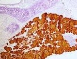
Immunohistochemistry-Paraffin: Thyroglobulin Antibody (2H11 + 6E1) [NBP2-34294] - Mouse thyroid tissue section stained with Thyroglobulin Antibody (2H11 + 6E1). IHC-P image submitted by a verified customer review.![Immunohistochemistry-Paraffin: Thyroglobulin Antibody (2H11 + 6E1) [NBP2-34294]](https://resources.rndsystems.com/images/products/Thyroglobulin-Antibody-2H11-+-6E1-Immunohistochemistry-Paraffin-NBP2-34294-img0003.jpg "Immunohistochemistry-Paraffin: Thyroglobulin Antibody (2H11 + 6E1) [NBP2-34294]")
Immunohistochemistry-Paraffin: Thyroglobulin Antibody (2H11 + 6E1) [NBP2-34294]
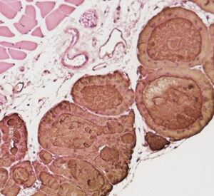
Immunohistochemistry-Paraffin: Thyroglobulin Antibody (2H11 + 6E1) [NBP2-34294] - Analysis using Azide/BSA FREE version of NBP2-34294. Human Thyroid stained with Thyroglobulin Ab (2H11 + 6E1).![Immunohistochemistry: Thyroglobulin Antibody (2H11 + 6E1) [NBP2-34294]](https://resources.rndsystems.com/images/products/Thyroglobulin-Antibody-2H11-+-6E1-Immunohistochemistry-NBP2-34294-img0004.jpg "Immunohistochemistry: Thyroglobulin Antibody (2H11 + 6E1) [NBP2-34294]")
Immunohistochemistry: Thyroglobulin Antibody (2H11 + 6E1) [NBP2-34294]
Immunohistochemistry: Thyroglobulin Antibody (2H11 + 6E1) [NBP2-34294] - Imaging of Mouse stomach tissue. Positive staining can only be observed inside the thyroid cells and the follicle in thyroid tissue. This image was submitted via customer Review.![Immunohistochemistry-Paraffin: Thyroglobulin Antibody (2H11 + 6E1) [NBP2-34294]](https://resources.rndsystems.com/images/products/Thyroglobulin-Antibody-2H11-+-6E1-Immunohistochemistry-Paraffin-NBP2-34294-img0005.jpg "Immunohistochemistry-Paraffin: Thyroglobulin Antibody (2H11 + 6E1) [NBP2-34294]")
Immunohistochemistry-Paraffin: Thyroglobulin Antibody (2H11 + 6E1) [NBP2-34294]
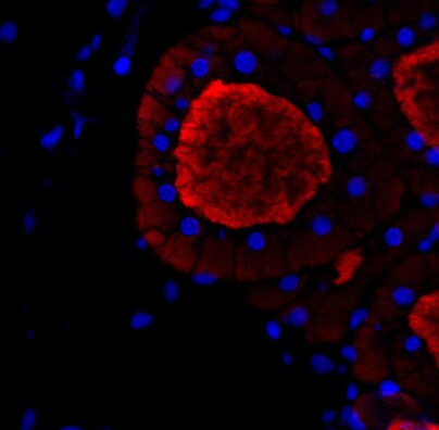
Immunohistochemistry-Paraffin: Thyroglobulin Antibody (2H11 + 6E1) [NBP2-34294] - Thyroglobulin staining in thyroid tissue of normal C57 mouse (Red). Dilution is 1:100. IHC-P image submitted by a verified customer review.Applications for Thyroglobulin Antibody (2H11 + 6E1)
Application
Recommended Usage
Flow Cytometry
0.5-1.0 ug/million cells
Immunohistochemistry-Paraffin
0.1-0.2 ug/ml
Application Notes
Immunohistochemistry (Formalin-fixed): 0.1-0.2ug/ml for 30 min at RT. Staining of formalin-fixed tissues requires heating tissue sections in 10mM Tris with 1mM EDTA, pH 9.0, for 45 min at 95C followed by cooling at RT for 20 minutes.
Optimal dilution for a specific application should be determined.
Optimal dilution for a specific application should be determined.
Reviewed Applications
Read 3 reviews rated 5 using NBP2-34294 in the following applications:
Flow Cytometry Panel Builder
Bio-Techne Knows Flow Cytometry
Save time and reduce costly mistakes by quickly finding compatible reagents using the Panel Builder Tool.
Advanced Features
- Spectra Viewer - Custom analysis of spectra from multiple fluorochromes
- Spillover Popups - Visualize the spectra of individual fluorochromes
- Antigen Density Selector - Match fluorochrome brightness with antigen density

Formulation, Preparation, and Storage
Purification
Protein A or G purified
Formulation
10 mM PBS with 0.05% BSA
Preservative
0.05% Sodium Azide
Concentration
0.2 mg/ml
Shipping
The product is shipped with polar packs. Upon receipt, store it immediately at the temperature recommended below.
Stability & Storage
Store at 4C.
Background: Thyroglobulin
Additional Thyroglobulin Products
Product Documents for Thyroglobulin Antibody (2H11 + 6E1)
Certificate of Analysis
To download a Certificate of Analysis, please enter a lot or batch number in the search box below.
Product Specific Notices for Thyroglobulin Antibody (2H11 + 6E1)
This product is for research use only and is not approved for use in humans or in clinical diagnosis. Primary Antibodies are guaranteed for 1 year from date of receipt.
Related Research Areas
Citations for Thyroglobulin Antibody (2H11 + 6E1)
 Powered by Bioz
Customer Reviews for Thyroglobulin Antibody (2H11 + 6E1) (3)
5 out of 5
3 Customer Ratings
Have you used Thyroglobulin Antibody (2H11 + 6E1)?
Submit a review and receive an Amazon gift card!
$25/€18/£15/$25CAN/¥2500 Yen for a review with an image
$10/€7/£6/$10CAN/¥1110 Yen for a review without an image
Submit a review
Customer Images
Showing
1
-
3 of
3 reviews
Showing All
Filter By:
-
Application: Immunohistochemistry-ParaffinSample Tested: Thyroid tissueSpecies: MouseVerified Customer | Posted 10/21/2021Thyroid tissue staining

-
Application: ImmunohistochemistrySample Tested: Stomach tissueSpecies: MouseVerified Customer | Posted 06/20/2017Very specific antibody. Positive staining can only be observed inside the thyroid cells and the follicle in thyroid tissue.
-
Application: Immunohistochemistry-ParaffinSample Tested: Thyroid tissueSpecies: MouseVerified Customer | Posted 06/09/2017Thyroglobulin staining in thyroid tissue of normal C57 mouse (Red). Dilution is 1:100Immunofluorescence using paraffin-embedded mouse thyroid tissue section. Antigen retrieval was by heat mediation. Samples were incubated with primary antibody (1/100 in antibody diluent reagent) for 12 hours at 4°C.
There are no reviews that match your criteria.
Protocols
Find general support by application which include: protocols, troubleshooting, illustrated assays, videos and webinars.
- 7-Amino Actinomycin D (7-AAD) Cell Viability Flow Cytometry Protocol
- Antigen Retrieval Protocol (PIER)
- Antigen Retrieval for Frozen Sections Protocol
- Appropriate Fixation of IHC/ICC Samples
- Cellular Response to Hypoxia Protocols
- Chromogenic IHC Staining of Formalin-Fixed Paraffin-Embedded (FFPE) Tissue Protocol
- Chromogenic Immunohistochemistry Staining of Frozen Tissue
- ClariTSA™ Fluorophore Kits
- Detection & Visualization of Antibody Binding
- Extracellular Membrane Flow Cytometry Protocol
- Flow Cytometry Protocol for Cell Surface Markers
- Flow Cytometry Protocol for Staining Membrane Associated Proteins
- Flow Cytometry Staining Protocols
- Flow Cytometry Troubleshooting Guide
- Fluorescent IHC Staining of Frozen Tissue Protocol
- Graphic Protocol for Heat-induced Epitope Retrieval
- Graphic Protocol for the Preparation and Fluorescent IHC Staining of Frozen Tissue Sections
- Graphic Protocol for the Preparation and Fluorescent IHC Staining of Paraffin-embedded Tissue Sections
- Graphic Protocol for the Preparation of Gelatin-coated Slides for Histological Tissue Sections
- IHC Sample Preparation (Frozen sections vs Paraffin)
- Immunofluorescent IHC Staining of Formalin-Fixed Paraffin-Embedded (FFPE) Tissue Protocol
- Immunohistochemistry (IHC) and Immunocytochemistry (ICC) Protocols
- Immunohistochemistry Frozen Troubleshooting
- Immunohistochemistry Paraffin Troubleshooting
- Intracellular Flow Cytometry Protocol Using Alcohol (Methanol)
- Intracellular Flow Cytometry Protocol Using Detergents
- Intracellular Nuclear Staining Flow Cytometry Protocol Using Detergents
- Intracellular Staining Flow Cytometry Protocol Using Alcohol Permeabilization
- Intracellular Staining Flow Cytometry Protocol Using Detergents to Permeabilize Cells
- Preparing Samples for IHC/ICC Experiments
- Preventing Non-Specific Staining (Non-Specific Binding)
- Primary Antibody Selection & Optimization
- Propidium Iodide Cell Viability Flow Cytometry Protocol
- Protocol for Heat-Induced Epitope Retrieval (HIER)
- Protocol for Liperfluo
- Protocol for Making a 4% Formaldehyde Solution in PBS
- Protocol for VisUCyte™ HRP Polymer Detection Reagent
- Protocol for the Characterization of Human Th22 Cells
- Protocol for the Characterization of Human Th9 Cells
- Protocol for the Preparation & Fixation of Cells on Coverslips
- Protocol for the Preparation and Chromogenic IHC Staining of Frozen Tissue Sections
- Protocol for the Preparation and Chromogenic IHC Staining of Frozen Tissue Sections - Graphic
- Protocol for the Preparation and Chromogenic IHC Staining of Paraffin-embedded Tissue Sections
- Protocol for the Preparation and Chromogenic IHC Staining of Paraffin-embedded Tissue Sections - Graphic
- Protocol for the Preparation and Fluorescent IHC Staining of Frozen Tissue Sections
- Protocol for the Preparation and Fluorescent IHC Staining of Paraffin-embedded Tissue Sections
- Protocol for the Preparation of Gelatin-coated Slides for Histological Tissue Sections
- Protocol: Annexin V and PI Staining by Flow Cytometry
- Protocol: Annexin V and PI Staining for Apoptosis by Flow Cytometry
- TUNEL and Active Caspase-3 Detection by IHC/ICC Protocol
- The Importance of IHC/ICC Controls
- Troubleshooting Guide: Fluorokine Flow Cytometry Kits
- Troubleshooting Guide: Immunohistochemistry
- View all Protocols, Troubleshooting, Illustrated assays and Webinars
Loading...