TM4SF18 also known as AAB5 and L6D, was cloned from human umbilical vein endothelial cells. It shares approximately 40 - 50% amino acid sequence identity with TM4SF1/L6, TM4SF4/IL-TMP, and TM4SF5, a group of proteins with limited homology to the tetraspanin superfamily.
![Immunohistochemistry-Paraffin: TM4SF18 Antibody [NBP2-57543]](https://resources.rndsystems.com/images/products/TM4SF18-Antibody-Immunohistochemistry-Paraffin-NBP2-57543-img0001.jpg "Immunohistochemistry-Paraffin: TM4SF18 Antibody [NBP2-57543]")
Loading...
Key Product Details
Species Reactivity
Human
Applications
Immunohistochemistry, Immunohistochemistry-Paraffin
Label
Unconjugated
Antibody Source
Polyclonal Rabbit IgG
Format
BSA Free
Loading...
Product Specifications
Immunogen
This antibody was developed against a recombinant protein corresponding to the following amino acid sequence: NGQTSYASSNKLTNYVWYFE
Clonality
Polyclonal
Host
Rabbit
Isotype
IgG
Scientific Data Images for TM4SF18 Antibody - BSA Free
Immunohistochemistry-Paraffin: TM4SF18 Antibody [NBP2-57543]
Immunohistochemistry-Paraffin: TM4SF18 Antibody [NBP2-57543] - Immunohistochemical staining of human kidney shows strong cytoplasmic positivity in cells of proximal tubules.Applications for TM4SF18 Antibody - BSA Free
Application
Recommended Usage
Immunohistochemistry
1:20 - 1:50
Immunohistochemistry-Paraffin
1:20 - 1:50
Application Notes
For IHC-Paraffin, HIER pH 6 retrieval is recommended.
Reviewed Applications
Read 1 review rated 1 using NBP2-57543 in the following applications:
Formulation, Preparation, and Storage
Purification
Affinity purified
Formulation
PBS (pH 7.2) and 40% Glycerol
Format
BSA Free
Preservative
0.02% Sodium Azide
Concentration
Concentrations vary lot to lot. See vial label for concentration. If unlisted please contact technical services.
Shipping
The product is shipped with polar packs. Upon receipt, store it immediately at the temperature recommended below.
Stability & Storage
Store at 4C short term. Aliquot and store at -20C long term. Avoid freeze-thaw cycles.
Background: TM4SF18
Long Name
Transmembrane 4 L Six Family Member 18
Alternate Names
AAB5, L6D
Gene Symbol
TM4SF18
Additional TM4SF18 Products
Product Documents for TM4SF18 Antibody - BSA Free
Certificate of Analysis
To download a Certificate of Analysis, please enter a lot or batch number in the search box below.
Product Specific Notices for TM4SF18 Antibody - BSA Free
This product is for research use only and is not approved for use in humans or in clinical diagnosis. Primary Antibodies are guaranteed for 1 year from date of receipt.
Customer Reviews for TM4SF18 Antibody - BSA Free (1)
1 out of 5
1 Customer Rating
Have you used TM4SF18 Antibody - BSA Free?
Submit a review and receive an Amazon gift card!
$25/€18/£15/$25CAN/¥2500 Yen for a review with an image
$10/€7/£6/$10CAN/¥1110 Yen for a review without an image
Submit a review
Customer Images
Showing
1
-
1 of
1 review
Showing All
Filter By:
-
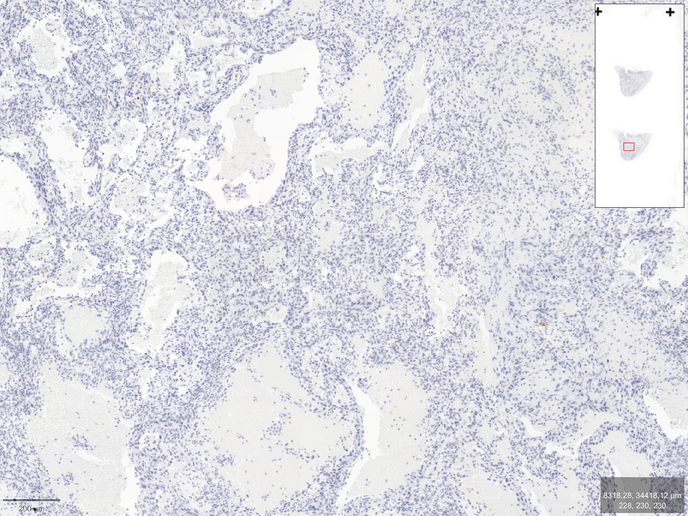
Application: Immunohistochemistry-ParaffinSample Tested: Multiple tissuesSpecies: Human, Canine and MouseVerified Customer | Posted 03/13/2025Negative TM4SF18 staining in a canine splenic hemangiosarcoma tumor expressing TM4SF18 on snRNAseq. IHC staining was also negative in multiple human and mouse tissues: kidneys, liver, spleen, lungs...Tissue slides were loaded into an automated research stainer, dewaxed and pretreated with EDTA-based epitope retrieval ER2 solution for 20 mins at 100°C. After a peroxide incubation for 5 min, the rabbit antibody against TM4SF18 (NBP2-57543-25UL) (1:50) was incubated for 60 mins at room temperature followed by Bond Polymer (anti-rabbit HRP) incubation for 8 min at room temperature. Mixed DAB reagent (Polymer Refine Detection Kit) was incubated for 10 mins, and Hematoxylin (Refine Detection Kit) counterstaining for 10 mins. After staining, sample slides were washed in water, dehydrated using ethanol gradient (70%, 90%, 100%), washed three times, and mounted in mounting medium.
 Bio-Techne ResponseThank you for reviewing our product. We are sorry to hear that this product did not perform as expected. We have been in touch with the customer to resolve this issue according to our Product Guarantee and to the customer’s satisfaction.
There are no reviews that match your criteria.
Protocols
Find general support by application which include: protocols, troubleshooting, illustrated assays, videos and webinars.
- Antigen Retrieval Protocol (PIER)
- Antigen Retrieval for Frozen Sections Protocol
- Appropriate Fixation of IHC/ICC Samples
- Cellular Response to Hypoxia Protocols
- Chromogenic IHC Staining of Formalin-Fixed Paraffin-Embedded (FFPE) Tissue Protocol
- Chromogenic Immunohistochemistry Staining of Frozen Tissue
- ClariTSA™ Fluorophore Kits
- Detection & Visualization of Antibody Binding
- Fluorescent IHC Staining of Frozen Tissue Protocol
- Graphic Protocol for Heat-induced Epitope Retrieval
- Graphic Protocol for the Preparation and Fluorescent IHC Staining of Frozen Tissue Sections
- Graphic Protocol for the Preparation and Fluorescent IHC Staining of Paraffin-embedded Tissue Sections
- Graphic Protocol for the Preparation of Gelatin-coated Slides for Histological Tissue Sections
- IHC Sample Preparation (Frozen sections vs Paraffin)
- Immunofluorescent IHC Staining of Formalin-Fixed Paraffin-Embedded (FFPE) Tissue Protocol
- Immunohistochemistry (IHC) and Immunocytochemistry (ICC) Protocols
- Immunohistochemistry Frozen Troubleshooting
- Immunohistochemistry Paraffin Troubleshooting
- Preparing Samples for IHC/ICC Experiments
- Preventing Non-Specific Staining (Non-Specific Binding)
- Primary Antibody Selection & Optimization
- Protocol for Heat-Induced Epitope Retrieval (HIER)
- Protocol for Making a 4% Formaldehyde Solution in PBS
- Protocol for VisUCyte™ HRP Polymer Detection Reagent
- Protocol for the Preparation & Fixation of Cells on Coverslips
- Protocol for the Preparation and Chromogenic IHC Staining of Frozen Tissue Sections
- Protocol for the Preparation and Chromogenic IHC Staining of Frozen Tissue Sections - Graphic
- Protocol for the Preparation and Chromogenic IHC Staining of Paraffin-embedded Tissue Sections
- Protocol for the Preparation and Chromogenic IHC Staining of Paraffin-embedded Tissue Sections - Graphic
- Protocol for the Preparation and Fluorescent IHC Staining of Frozen Tissue Sections
- Protocol for the Preparation and Fluorescent IHC Staining of Paraffin-embedded Tissue Sections
- Protocol for the Preparation of Gelatin-coated Slides for Histological Tissue Sections
- TUNEL and Active Caspase-3 Detection by IHC/ICC Protocol
- The Importance of IHC/ICC Controls
- Troubleshooting Guide: Immunohistochemistry
- View all Protocols, Troubleshooting, Illustrated assays and Webinars
Loading...