Human Various Tissue MicroArray (Cancer)
Novus Biologicals | Catalog # NBP2-30331
Loading...
Key Product Details
Species
Human
Applications
Immunohistochemistry
Product Summary for Human Various Tissue MicroArray (Cancer)
Microarray Panel:
Various cancers, high density (3)
No. of samples: 146
No. of patients: 146
Core diameter: 1.0 mm
Format: Formalin Fixed
Section thickness: 4 micrometer
Please see manual for tissue information and location.
All tissues are fixed in 10% neutral buffered formalin for 12 to 24 hours, dehydrated with gradient ethanol, cleared with xylene, and embedded in paraffin. Then each slide is tested for immunohistochemistry on multiple antibodies including the p53 protein. Quality control is ensured as every tissue block is collected and arranged by certified pathologists, who used an identical process for their own research.
About 100,000 cells are included in each sample with 2 mm core diameter. Some slides may have less than 60 cores due to sample loss when obtaining multiple sections. Maximum number of missing cores in a slide is less than 10%.
Loading...
Product Specifications
Type
Tissue
Tissue Condition
Cancer
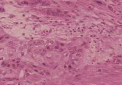
Scientific Data Images for Human Various Tissue MicroArray (Cancer)
Hematoxylin & Eosin Stain: Human Various Tissue MicroArray (Cancer) [NBP2-30331] - Urinary Bladder, Transitional Cell Carcinoma
Formulation, Preparation, and Storage
Concentration
Concentration is not relevant for this product. Please see the protocols for proper use of this product.
Shipping
The product is shipped with polar packs. Upon receipt, store it immediately at the temperature recommended below.
Storage
Store at 4C. Do not freeze.
Product Documents for Human Various Tissue MicroArray (Cancer)
Certificate of Analysis
To download a Certificate of Analysis, please enter a lot or batch number in the search box below.
Product Specific Notices for Human Various Tissue MicroArray (Cancer)
This product is for research use only and is not approved for use in humans or in clinical diagnosis. Tissue Micro Arrays are guaranteed for 1 year from date of receipt.
Customer Reviews for Human Various Tissue MicroArray (Cancer)
There are currently no reviews for this product. Be the first to review Human Various Tissue MicroArray (Cancer) and earn rewards!
Have you used Human Various Tissue MicroArray (Cancer)?
Submit a review and receive an Amazon gift card!
$25/€18/£15/$25CAN/¥2500 Yen for a review with an image
$10/€7/£6/$10CAN/¥1110 Yen for a review without an image
Submit a review
Protocols
Find general support by application which include: protocols, troubleshooting, illustrated assays, videos and webinars.
- Antigen Retrieval Protocol (PIER)
- Antigen Retrieval for Frozen Sections Protocol
- Appropriate Fixation of IHC/ICC Samples
- Cellular Response to Hypoxia Protocols
- Chromogenic IHC Staining of Formalin-Fixed Paraffin-Embedded (FFPE) Tissue Protocol
- Chromogenic Immunohistochemistry Staining of Frozen Tissue
- ClariTSA™ Fluorophore Kits
- Detection & Visualization of Antibody Binding
- Fluorescent IHC Staining of Frozen Tissue Protocol
- Graphic Protocol for Heat-induced Epitope Retrieval
- Graphic Protocol for the Preparation and Fluorescent IHC Staining of Frozen Tissue Sections
- Graphic Protocol for the Preparation and Fluorescent IHC Staining of Paraffin-embedded Tissue Sections
- Graphic Protocol for the Preparation of Gelatin-coated Slides for Histological Tissue Sections
- IHC Sample Preparation (Frozen sections vs Paraffin)
- Immunofluorescent IHC Staining of Formalin-Fixed Paraffin-Embedded (FFPE) Tissue Protocol
- Immunohistochemistry (IHC) and Immunocytochemistry (ICC) Protocols
- Immunohistochemistry Frozen Troubleshooting
- Immunohistochemistry Paraffin Troubleshooting
- Preparing Samples for IHC/ICC Experiments
- Preventing Non-Specific Staining (Non-Specific Binding)
- Primary Antibody Selection & Optimization
- Protocol for Heat-Induced Epitope Retrieval (HIER)
- Protocol for Making a 4% Formaldehyde Solution in PBS
- Protocol for VisUCyte™ HRP Polymer Detection Reagent
- Protocol for the Preparation & Fixation of Cells on Coverslips
- Protocol for the Preparation and Chromogenic IHC Staining of Frozen Tissue Sections
- Protocol for the Preparation and Chromogenic IHC Staining of Frozen Tissue Sections - Graphic
- Protocol for the Preparation and Chromogenic IHC Staining of Paraffin-embedded Tissue Sections
- Protocol for the Preparation and Chromogenic IHC Staining of Paraffin-embedded Tissue Sections - Graphic
- Protocol for the Preparation and Fluorescent IHC Staining of Frozen Tissue Sections
- Protocol for the Preparation and Fluorescent IHC Staining of Paraffin-embedded Tissue Sections
- Protocol for the Preparation of Gelatin-coated Slides for Histological Tissue Sections
- TUNEL and Active Caspase-3 Detection by IHC/ICC Protocol
- The Importance of IHC/ICC Controls
- Troubleshooting Guide: Immunohistochemistry
- View all Protocols, Troubleshooting, Illustrated assays and Webinars
Loading...