Fluorescent Probes and Stains
Fluorescent probes target specific cellular and sub-cellular components, or they may selectively target a single biomolecule. Alternatively, they might provide a functional read-out, such as live versus dead cell staining. They enable researchers to detect particular components of complex biomolecular assemblies, such as microtubules, with high sensitivity and selectivity allowing the exploration of cell structure and function. R&D Systems portfolio offers a wide selection of fluorescent probes, covering organelle probes and cell viability stains.
MitoBrilliant™ Dyes
MitoBrilliant are next-generation fluorescent stains for the localization and tracking of mitochondria in both live and fixed cells. Our range harnesses Janelia Fluor® dye technology, conferring some of the properties that make these widely used dyes, into mitochondrial stains. The MitoBrilliant Live dyes accumulate in the mitochondria of live cells. Two dyes are available: MitoBrilliant™ Live 646 and MitoBrilliant™ Live 549. A universal stain for mitochondria in both live and fixed cells is also available: MitoBrilliant™ 646.
MitoBrilliant™ 646
- Suitable for Live and Fixed Cell Applications
- Improved Performance and Staining Fidelity Post-Fixation
Product Name | Core Dye Structure | Abs/Em (nm) | Δψm Dependent | Live/Fixed Cell Use? | Stains Pre-Fixed Cells? | Image Without Wash Step? | Demonstrated Applications |
MitoBrilliant™ 646 Cat. No. 7700 |
Janelia Fluor® technology |
655/668 |
No* |
Suitable for live and fixed cell work |
Yes |
Yes, but replacing media recommended | Fixed-cell imaging, Live-cell imaging Flow cytometry, IHC/ICC, Super-resolution, Microscopy – STED, High-content screening |
MitoBrilliant™ Live 646 Cat. No. 7417 |
Janelia Fluor® technology |
648/662 |
Yes |
Live-cell work only |
No |
Yes, but replacing media recommended | Live-cell imaging, Flow cytometry, High-content screening |
MitoBrilliant™ Live 549 Cat. No. 7693 |
Janelia Fluor® technology |
550/568 |
Yes |
Live-cell work only |
No |
Yes, but replacing media recommended | Live-cell imaging, Flow cytometry, High-content screening |
* In live-cell staining, Δψm drives initial recruitment of the dye into mitochondria. After staining, localization of the dyes becomes insensitive to Δψm changes. In pre-fixed cell staining, MitoBrilliant™ 646 can localize and stay in mitochondria without Δψm.
MitoBriliant CTA
Find Out More About MitoBrilliant DyesFluorescent Lysosome Probes
- Our range of fluorescent lysosome probes include HB-2-30, a fluorescent transglutaminase-2 probe for imaging endocytose in vitro and in vivo. Suitable for use with confocal microscopy
- Discover also tri-GalNAc-C5-AF647, a fluorescent asialoglycoprotein receptor (ASGPR) ligand for lysosomal uptake imaging in live cells
Image kindly provided by Fu-Chen Yang and Harrison Besser, University or Stanford.


Fluorescent Cell Metabolism Probes
- Discover our SCOTfluor probes range for real-time tracking of essential metabolites in live cells and in vivo
- SCOTfluor probes are suitable for use with flow cytometry and super-resolution microscopy
- They can be multiplexed with blue and green fluorescent proteins (BFP/GFP)
Fluorescent Microtubule Probes
- Our range of fluorescent microtubule probes include Flutax and Taxol Janelia Fluor probes, both Taxol based molecules
- Taxol Janelia Fluor probes enable ‘no-wash’ protocols, are bright, photostable, and well suited for confocal microscopy and super-resolution microscopy techniques

Fluorescent Probes and Stains Portfolio
Flow Cytometry
Bio-Techne has a large catalog of antibodies, reagents and tools to streamline your flow cytometry experiments.
Fluorescent Dyes
Bio-Techne through the Tocris brand, offers a wide range of fluorescent dyes including bright and photostable Janelia fluor dyes.
Fluorokines™: Fluorescently Labeled Proteins
Easily measure transduction efficiency of CAR T or CAR NK cell preparations or determine B cell specificity after COVID-19 infection or vaccination.
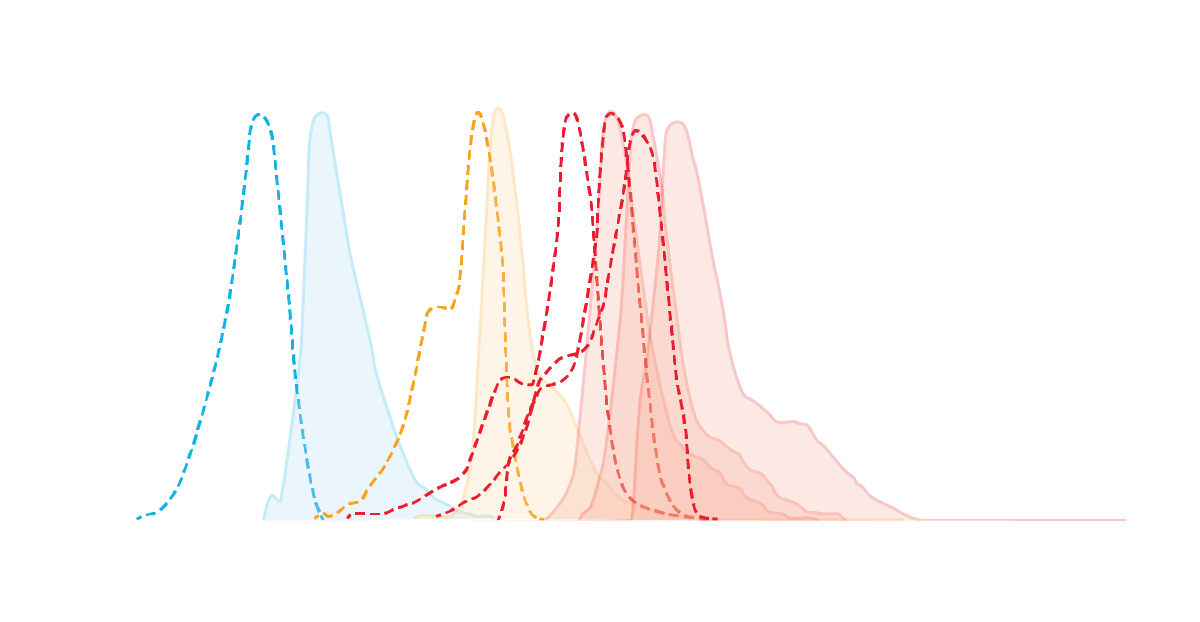
Spectra Viewer
Use our spectra viewer to interactively plan your experiments, assessing multiplexing options. View the excitation and emission spectra for our fluorescent dye range and other commonly used dyes.
MitoBrilliant Guide

This product guide provides a background, protocols and data from use in different research applications for our Mitobrilliant fluorescent mitochondrial probes.
Fluorescent Dyes and Probes Brochure
Our brochure showcases our range and gives background on the use of Fluorescent Dyes and Probes.
Flow Cytometry Handbook
Explore a Step-By-Step Guide to Flow Cytometry.