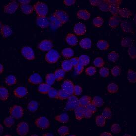
FGF R3 in U937 Human Cell Line.
Fibroblast Growth Factor Receptor 3 (FGF R3) was detected in immersion fixed U937 human histiocytic lymphoma cell line using Human FGF R3 (IIIc) Monoclonal Antibody (Catalog # MAB7662) at 10 µg/mL for 3 hours at room temperature. Cells were stained using the NorthernLights™ 557-conjugated Anti-Mouse IgG Secondary Antibody (red; Catalog # NL007) and counterstained with DAPI (blue). View our protocol for Fluorescent ICC Staining of Non-adherent Cells.
Secondary Antibodies
R&D Systems offers a wide range of biotinylated, HRP-conjugated, fluorochrome-labeled, and unlabeled species-specific secondary antibodies. Our NorthernLights™ fluorescent secondary antibodies are available with three distinct excitation and emission maxima, making them ideal for multi-color fluorescence microscopy.
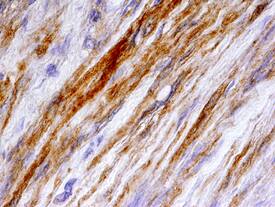
FGF R3 (IIIc) in Human Bladder Cancer Tissue.
Fibroblast Growth Factor Receptor 3 (FGF R3) was detected in immersion fixed paraffin-embedded sections of human bladder cancer tissue using Human FGF R3 (IIIc) Monoclonal Antibody (Catalog # MAB7662) at 25 µg/mL overnight at 4 °C. Tissue was stained using the Anti-Mouse HRP-DAB Cell & Tissue Staining Kit (brown; Catalog # CTS002) and counterstained with hematoxylin (blue). Specific staining was localized to smooth muscle cells. View our protocol for Chromogenic IHC Staining of Paraffin-embedded Tissue Sections.