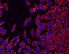
FGF Receptor 4 in MIA PaCa-2 Human Cell Line.
Fibroblast Growth Factor Receptor 4 (FGF R4) was detected in immersion fixed MIA PaCa‑2 human pancreatic cancer cell line using Human FGF R4 Monoclonal Antibody (Catalog # MAB6852) at 10 µg/mL for 3 hours at room temperature. Cells were stained using the NorthernLights™ 557-conjugated Anti-Rat IgG Secondary Antibody (red; Catalog # NL013) and counterstained with DAPI (blue). View our protocol for Fluorescent ICC Staining of Cells on Coverslips.
This antibody specifically recognizes human FGF R4. In Western blot experiments, this antibody shows less than 5% cross-reactivity with recombinant mouse FGF R4 and recombinant human FGF R5 and no cross-reactivity with any isoform of recombinant human FGF R1, FGF R2, or FGF R3.
Secondary Antibodies
R&D Systems offers a wide range of biotinylated, HRP-conjugated, fluorochrome-labeled, and unlabeled species-specific secondary antibodies. Our NorthernLights™ fluorescent secondary antibodies are available with three distinct excitation and emission maxima, making them ideal for multi-color fluorescence microscopy.
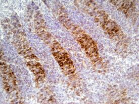
FGF R4 in Mouse Embryo.
Fibroblast Growth Factor Receptor 4 (FGF R4) was detected in immersion fixed frozen sections of mouse embryo (11.5 d.p.c.) using Mouse FGF R4 Antigen Affinity-purified Polyclonal Antibody (Catalog # AF2265) at 5 µg/mL overnight at 4 °C. Tissue was stained using the Anti-Goat HRP-DAB Cell & Tissue Staining Kit (brown; Catalog # CTS008) and counterstained with hematoxylin (blue). Specific staining was localized to cartilage. View our protocol for Chromogenic IHC Staining of Frozen Tissue Sections.
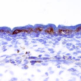
FGF R4 in Mouse Embryo.
Fibroblast Growth Factor Receptor 4 (FGF R4) was detected in immersion fixed frozen sections of mouse embryo (E11.5) using Mouse FGF R4 Biotinylated Antigen Affinity-purified Polyclonal Antibody (Catalog # BAF2265) at 15 µg/mL overnight at 4 °C. Tissue was stained using the Anti-Goat HRP-DAB Cell & Tissue Staining Kit (brown; Catalog # CTS008) and counterstained with hematoxylin (blue). Specific staining was localized to the epidermis. View our protocol for Chromogenic IHC Staining of Frozen Tissue Sections.
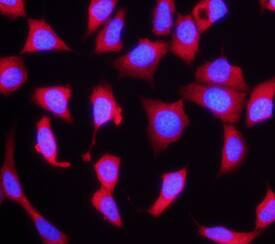
FGF R4 in MCF‑7 Human Cell Line.
Fibroblast Growth Factor Receptor 4 (FGF R4) was detected in immersion fixed MCF‑7 human breast cancer cell line using Rat Anti-Human FGF R4 Monoclonal Antibody (Catalog # MAB6852) at 10 µg/mL for 3 hours at room temperature. Cells were stained using the NorthernLights™ 557-conjugated Anti-Rat IgG Secondary Antibody (red; Catalog # NL013) and counterstained with DAPI (blue). Specific staining was localized to cytoplasm. View our protocol for Fluorescent ICC Staining of Cells on Coverslips.
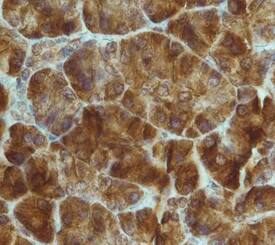
FGF R4 in Human Pancreas.
Fibroblast Growth Factor Receptor 4 (FGF R4) was detected in immersion fixed paraffin-embedded sections of human pancreas using Rat Anti-Human FGF R4 Monoclonal Antibody (Catalog # MAB6852) at 15 µg/mL overnight at 4 °C. Tissue was stained using the Anti-Rat HRP-DAB Cell & Tissue Staining Kit (brown; Catalog # CTS017) and counterstained with hematoxylin (blue). Specific staining was localized to nuclei and cytoplasm. View our protocol for Chromogenic IHC Staining of Paraffin-embedded Tissue Sections.