
Key Product Details
Species Reactivity
Validated:
Human
Cited:
Human, Mouse, Rat, Porcine, Bovine
Applications
Validated:
Immunohistochemistry, Western Blot, Neutralization, Immunocytochemistry, Simple Western
Cited:
Immunohistochemistry, Immunohistochemistry-Paraffin, Immunohistochemistry-Frozen, Western Blot, Neutralization, Immunocytochemistry, Immunoprecipitation, ELISA Development, ELISA Development (Detection), Neutralizing
Label
Unconjugated
Antibody Source
Polyclonal Goat IgG
Loading...
Product Specifications
Immunogen
E. coli-derived recombinant human IGF-I/IGF-1
Specificity
Detects human IGF-I/IGF-1 in direct ELISAs and Western blots. In direct ELISAs, approximately 50% cross-reactivity with recombinant mouse IGF-I/IGF-1 and recombinant rat IGF-I/IGF-1 is observed.
Clonality
Polyclonal
Host
Goat
Isotype
IgG
Endotoxin Level
<0.10 EU per 1 μg of the antibody by the LAL method.
Scientific Data Images for Human IGF-I/IGF-1 Antibody
Cell Proliferation Induced by IGF-I/IGF-1 and Neutralization by Human IGF-I/IGF-1 Antibody.
Recombinant Human IGF-I/IGF-1 (291-G1) stimulates proliferation in the MCF-7 human breast cancer cell line in a dose-dependent manner (orange line). Proliferation elicited by Recombinant Human IGF-I/IGF-1 (6 ng/mL) is neutralized (green line) by increasing concentrations of Goat Anti-Human IGF-I/IGF-1 Antigen Affinity-purified Polyclonal Antibody (Catalog # AF-291-NA). The ND50 is typically 3-12 µg/mL.
IGF-I/IGF-1 in MDA‑MB‑231 Human Cell Line.
IGF-I/IGF-1 was detected in immersion fixed MDA-MB-231 human breast cancer cell line using Goat Anti-Human IGF-I/IGF-1 Antigen Affinity-purified Polyclonal Antibody (Catalog # AF-291-NA) at 10 µg/mL for 3 hours at room temperature. Cells were stained using the NorthernLights™ 557-conjugated Anti-Goat IgG Secondary Antibody (yellow; NL001) and counterstained with DAPI (blue). View our protocol for Fluorescent ICC Staining of Cells on Coverslips.
IGF-I/IGF-1 in Human Placenta.
IGF-I/IGF-1 was detected in immersion fixed paraffin-embedded sections of human placenta using Goat Anti-Human IGF-I/IGF-1 Antigen Affinity-purified Polyclonal Antibody (Catalog # AF-291-NA) at 5 µg/mL overnight at 4 °C. Tissue was stained using the Anti-Goat HRP-DAB Cell & Tissue Staining Kit (brown; CTS008) and counterstained with hematoxylin (blue). Specific staining was localized to syncytiotrophoblasts. View our protocol for Chromogenic IHC Staining of Paraffin-embedded Tissue Sections.
Detection of Human IGF-I/IGF-1 by Simple WesternTM.
Simple Western lane view shows lysates of Recombinant Human IGF-1, loaded at 0.2 mg/mL. A specific band was detected for IGF-I/IGF-1 at approximately 6 kDa (as indicated) using 50 µg/mL of Goat Anti-Human IGF-I/IGF-1 Antigen Affinity-purified Polyclonal Antibody (Catalog # AF-291-NA). This experiment was conducted under reducing conditions and using the 2-40kDa separation system.Applications for Human IGF-I/IGF-1 Antibody
Application
Recommended Usage
Immunocytochemistry
5-15 µg/mL
Sample: Immersion fixed MDA-MB-231 human breast cancer cell line
Sample: Immersion fixed MDA-MB-231 human breast cancer cell line
Immunohistochemistry
5-15 µg/mL
Sample: Immersion fixed paraffin-embedded sections of human placenta (chorionic villi) subjected to Antigen Retrieval Reagent-Basic (Catalog # CTS013)
Sample: Immersion fixed paraffin-embedded sections of human placenta (chorionic villi) subjected to Antigen Retrieval Reagent-Basic (Catalog # CTS013)
Simple Western
50 µg/mL
Sample: Recombinant Human IGF-1
Sample: Recombinant Human IGF-1
Western Blot
0.1 µg/mL
Sample: Recombinant Human IGF-I/IGF-1 (Catalog # 291-G1)
Sample: Recombinant Human IGF-I/IGF-1 (Catalog # 291-G1)
Neutralization
Measured by its ability to neutralize IGF-I/IGF-1-induced proliferation in the MCF‑7 human breast cancer cell line. Karey, K.P. et al. (1988) Cancer Research 48:4083. The Neutralization Dose (ND50) is typically 3-12 µg/mL in the presence of 6 ng/mL Recombinant Human IGF-I/IGF-1.
Reviewed Applications
Read 4 reviews rated 4.5 using AF-291-NA in the following applications:
Formulation, Preparation, and Storage
Purification
Antigen Affinity-purified
Reconstitution
Reconstitute at 0.2 mg/mL in sterile PBS. For liquid material, refer to CoA for concentration.
Loading...
Formulation
Lyophilized from a 0.2 μm filtered solution in PBS with Trehalose. *Small pack size (SP) is supplied either lyophilized or as a 0.2 µm filtered solution in PBS.
Shipping
Lyophilized product is shipped at ambient temperature. Liquid small pack size (-SP) is shipped with polar packs. Upon receipt, store immediately at the temperature recommended below.
Stability & Storage
Use a manual defrost freezer and avoid repeated freeze-thaw cycles.
- 12 months from date of receipt, -20 to -70 °C as supplied.
- 1 month, 2 to 8 °C under sterile conditions after reconstitution.
- 6 months, -20 to -70 °C under sterile conditions after reconstitution.
Calculators
Background: IGF-I/IGF-1
Long Name
Insulin-like Growth Factor I/Insulin-like Growth Factor 1
Alternate Names
IGF-1, IGF1, IGFI, Somatomedin A, Somatomedin C
Gene Symbol
IGF1
Additional IGF-I/IGF-1 Products
Product Documents for Human IGF-I/IGF-1 Antibody
Certificate of Analysis
To download a Certificate of Analysis, please enter a lot or batch number in the search box below.
Note: Certificate of Analysis not available for kit components.
Product Specific Notices for Human IGF-I/IGF-1 Antibody
For research use only
Citations for Human IGF-I/IGF-1 Antibody
 Powered by Bioz
Customer Reviews for Human IGF-I/IGF-1 Antibody (4)
4.5 out of 5
4 Customer Ratings
Have you used Human IGF-I/IGF-1 Antibody?
Submit a review and receive an Amazon gift card!
$25/€18/£15/$25CAN/¥2500 Yen for a review with an image
$10/€7/£6/$10CAN/¥1110 Yen for a review without an image
Submit a review
Customer Images
Showing
1
-
4 of
4 reviews
Showing All
Filter By:
-
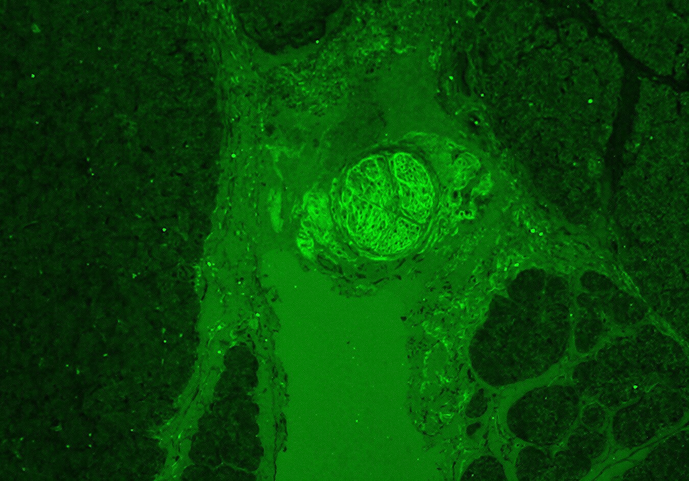
Application: Immunocytochemistry/ImmunofluorescenceSample Tested: Pancreas tissueSpecies: CanineVerified Customer | Posted 03/08/2022Used at 1:50 concentration in Casein in PBS overnight at 4 C. HIER was performed for two hours at 75 C in a citrate buffer.

-
Application: Siteclcik antibody biotinylationSample Tested: B16-F1 mouse melanoma cell lineSpecies: MouseVerified Customer | Posted 10/27/2017
-
Application: Western BlotSample Tested: See PMID 24036101Species: HumanVerified Customer | Posted 01/07/2015
-
Application: Western BlotSample Tested: See PMID 23424195Species: HumanVerified Customer | Posted 01/07/2015
There are no reviews that match your criteria.
Protocols
Find general support by application which include: protocols, troubleshooting, illustrated assays, videos and webinars.
- Antigen Retrieval Protocol (PIER)
- Antigen Retrieval for Frozen Sections Protocol
- Appropriate Fixation of IHC/ICC Samples
- Cellular Response to Hypoxia Protocols
- Chromogenic IHC Staining of Formalin-Fixed Paraffin-Embedded (FFPE) Tissue Protocol
- Chromogenic Immunohistochemistry Staining of Frozen Tissue
- ClariTSA™ Fluorophore Kits
- Detection & Visualization of Antibody Binding
- Fluorescent IHC Staining of Frozen Tissue Protocol
- Graphic Protocol for Heat-induced Epitope Retrieval
- Graphic Protocol for the Preparation and Fluorescent IHC Staining of Frozen Tissue Sections
- Graphic Protocol for the Preparation and Fluorescent IHC Staining of Paraffin-embedded Tissue Sections
- Graphic Protocol for the Preparation of Gelatin-coated Slides for Histological Tissue Sections
- ICC Cell Smear Protocol for Suspension Cells
- ICC Immunocytochemistry Protocol Videos
- ICC for Adherent Cells
- IHC Sample Preparation (Frozen sections vs Paraffin)
- Immunocytochemistry (ICC) Protocol
- Immunocytochemistry Troubleshooting
- Immunofluorescence of Organoids Embedded in Cultrex Basement Membrane Extract
- Immunofluorescent IHC Staining of Formalin-Fixed Paraffin-Embedded (FFPE) Tissue Protocol
- Immunohistochemistry (IHC) and Immunocytochemistry (ICC) Protocols
- Immunohistochemistry Frozen Troubleshooting
- Immunohistochemistry Paraffin Troubleshooting
- Preparing Samples for IHC/ICC Experiments
- Preventing Non-Specific Staining (Non-Specific Binding)
- Primary Antibody Selection & Optimization
- Protocol for Heat-Induced Epitope Retrieval (HIER)
- Protocol for Making a 4% Formaldehyde Solution in PBS
- Protocol for VisUCyte™ HRP Polymer Detection Reagent
- Protocol for the Fluorescent ICC Staining of Cell Smears - Graphic
- Protocol for the Fluorescent ICC Staining of Cultured Cells on Coverslips - Graphic
- Protocol for the Preparation & Fixation of Cells on Coverslips
- Protocol for the Preparation and Chromogenic IHC Staining of Frozen Tissue Sections
- Protocol for the Preparation and Chromogenic IHC Staining of Frozen Tissue Sections - Graphic
- Protocol for the Preparation and Chromogenic IHC Staining of Paraffin-embedded Tissue Sections
- Protocol for the Preparation and Chromogenic IHC Staining of Paraffin-embedded Tissue Sections - Graphic
- Protocol for the Preparation and Fluorescent ICC Staining of Cells on Coverslips
- Protocol for the Preparation and Fluorescent ICC Staining of Non-adherent Cells
- Protocol for the Preparation and Fluorescent ICC Staining of Stem Cells on Coverslips
- Protocol for the Preparation and Fluorescent IHC Staining of Frozen Tissue Sections
- Protocol for the Preparation and Fluorescent IHC Staining of Paraffin-embedded Tissue Sections
- Protocol for the Preparation of Gelatin-coated Slides for Histological Tissue Sections
- Protocol for the Preparation of a Cell Smear for Non-adherent Cell ICC - Graphic
- R&D Systems Quality Control Western Blot Protocol
- TUNEL and Active Caspase-3 Detection by IHC/ICC Protocol
- The Importance of IHC/ICC Controls
- Troubleshooting Guide: Immunohistochemistry
- Troubleshooting Guide: Western Blot Figures
- Western Blot Conditions
- Western Blot Protocol
- Western Blot Protocol for Cell Lysates
- Western Blot Troubleshooting
- Western Blot Troubleshooting Guide
- View all Protocols, Troubleshooting, Illustrated assays and Webinars


