
MKI67/Ki-67 in A549 Human Cell Line.
MKI67/Ki-67 was detected in immersion fixed A549 human lung carcinoma cell line using Sheep Anti-Human MKI67/Ki-67 Antigen Affinity-purified Polyclonal Antibody (Catalog # AF7617) at 5 µg/mL for 3 hours at room temperature. Cells were stained using the NorthernLights™ 557-conjugated Anti-Sheep IgG Secondary Antibody (red; Catalog # NL010) and counterstained with DAPI (blue). Specific staining was localized to nuclei and nucleoli. View our protocol for Fluorescent ICC Staining of Cells on Coverslips.
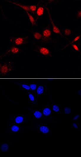
MKI67/Ki-67 in NIH‑3T3 Mouse Cell Line.
MKI67/Ki-67 was detected in immersion fixed NIH‑3T3 mouse embryonic fibroblast cell line using Sheep Anti-Mouse MKI67/Ki-67 Antigen Affinity-purified Polyclonal Antibody (Catalog # AF7649) at 10 µg/mL for 3 hours at room temperature. Cells were stained using the NorthernLights™ 557-conjugated Anti-Sheep IgG Secondary Antibody (red, upper panel; Catalog # NL010) and counterstained with DAPI (blue, lower panel). Specific staining was localized to nuclei and nucleoli. View our protocol for Fluorescent ICC Staining of Cells on Coverslips.
Secondary Antibodies
R&D Systems offers a wide range of biotinylated, HRP-conjugated, fluorochrome-labeled, and unlabeled species-specific secondary antibodies. Our NorthernLights™ fluorescent secondary antibodies are available with three distinct excitation and emission maxima, making them ideal for multi-color fluorescence microscopy.