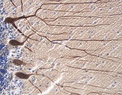
RAGE in Human Alzheimer's Disease Brain.
Receptor for Advanced Glycation End products (RAGE) was detected in immersion fixed paraffin-embedded sections of human Alzheimer's disease brain (cerebellum) using Human RAGE Antigen Affinity-purified Polyclonal Antibody (Catalog # AF1145) at 15 µg/mL overnight at 4 °C. Tissue was stained using the Anti-Goat HRP-DAB Cell & Tissue Staining Kit (brown; Catalog # CTS008) and counterstained with hematoxylin (blue). View our protocol for Chromogenic IHC Staining of Paraffin-embedded Tissue Sections.
This antibody specifically recognizes human RAGE. Reactivity with RAGE from other species has not been determined.
Secondary Antibodies
R&D Systems offers a wide range of biotinylated, HRP-conjugated, fluorochrome-labeled, and unlabeled species-specific secondary antibodies. Our NorthernLights™ fluorescent secondary antibodies are available with three distinct excitation and emission maxima, making them ideal for multi-color fluorescence microscopy.
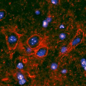
RAGE in Mouse Spinal Cord.
RAGE was detected in perfusion fixed frozen sections of mouse spinal cord using Rat Anti-Mouse RAGE Monoclonal Antibody (Catalog # MAB11794) at 25 µg/mL overnight at 4 °C. Tissue was stained using the NorthernLights™ 557-conjugated Anti-Rat IgG Secondary Antibody (red; Catalog # NL013) and counterstained with DAPI (blue). Specific staining was localized to the plasma membranes of motor neurons. View our protocol for Fluorescent IHC Staining of Frozen Tissue Sections.