
Aconitase 2 Antibody - BSA Free
Novus Biologicals | Catalog # NBP1-90264


Scientific images may be resized, cropped, or adjusted for brightness or contrast for presentation purposes. These updates are limited to presentation and do not affect the underlying scientific interpretation of the data. Where available, additional source data may be provided upon request.
Key Product Details
Validated by
Species Reactivity
Validated:
Cited:
Applications
Validated:
Cited:
Label
Antibody Source
Format
Product Specifications
Immunogen
Clonality
Host
Isotype
Theoretical MW
Disclaimer note: The observed molecular weight of the protein may vary from the listed predicted molecular weight due to post translational modifications, post translation cleavages, relative charges, and other experimental factors.
Scientific Data Images for Aconitase 2 Antibody - BSA Free
Immunocytochemistry/ Immunofluorescence: Aconitase 2 Antibody [NBP1-90264]
Immunocytochemistry/Immunofluorescence: Aconitase 2 Antibody [NBP1-90264] - Immunofluorescent staining of human cell line U-2 OS shows localization to mitochondria.

Immunohistochemistry-Paraffin: Aconitase 2 Antibody [NBP1-90264]
Immunohistochemistry-Paraffin: Aconitase 2 Antibody [NBP1-90264] - Staining of human heart muscle shows strong granular cytoplasmic positivity in cardiomyocytes.
Immunohistochemistry-Paraffin: Aconitase 2 Antibody [NBP1-90264]
Immunohistochemistry-Paraffin: Aconitase 2 Antibody [NBP1-90264] - Staining of human liver shows very weak positivity in hepatocytes as expected.
Immunohistochemistry-Paraffin: Aconitase 2 Antibody [NBP1-90264]
Immunohistochemistry-Paraffin: Aconitase 2 Antibody [NBP1-90264] - Staining of human duodenum shows strong strong granular cytoplasmic positivity in glandular cells.
Immunohistochemistry-Paraffin: Aconitase 2 Antibody [NBP1-90264]
Immunohistochemistry-Paraffin: Aconitase 2 Antibody [NBP1-90264] - Staining of human kidney shows strong strong granular cytoplasmic positivity in cells in tubules.
Western Blot: Aconitase 2 Antibody - BSA Free [NBP1-90264]
Analysis in human cell line HL-60.
Western Blot: Aconitase 2 Antibody - BSA Free [NBP1-90264]
Analysis in mouse cell line NIH-3T3, rat cell line NBT-II and rat cell line pC12.
Immunohistochemistry: Aconitase 2 Antibody [NBP1-90264]
Aconitase-2-Antibody-Immunohistochemistry-NBP1-90264-img0013.jpg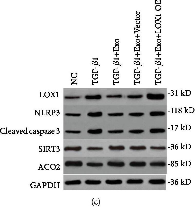
Western Blot: Aconitase 2 Antibody [NBP1-90264] -
Western Blot: Aconitase 2 Antibody [NBP1-90264] - The regulation mechanism of LOX1 on alveolar epithelial cell apoptosis & fibrosis. (a) Cells were transfected with a LOX1 overexpression plasmid & control vector. ROS levels were determined by DCFH-DA assay in MLE-12 cells treated by TGF-beta 1, TGF-beta 1 plus exosome, TGF-beta 1 plus exosome & vector, & TGF-beta 1 plus exosome & LOX1 overexpression plasmid (n = 3). (b) Detection of cellular mtDNA/18sRNA in each of the above cell groups (n = 3). (c) The expression of LOX1, caspase 3, mtDNA damage markers SIRT3 & ACO2, & NLRP3 was measured by western blotting in each of the above cell groups. (d) The expression of the same signal cascades including LOX1, caspase 3, SIRT3, ACO2, & NLRP3 was confirmed in an animal model with pulmonary fibrosis by western blot assay. Data is shown as the means ± SD, n ≥ 3. ∗∗p < 0.01. Image collected & cropped by CiteAb from the following publication (https://pubmed.ncbi.nlm.nih.gov/31949877), licensed under a CC-BY license. Not internally tested by Novus Biologicals.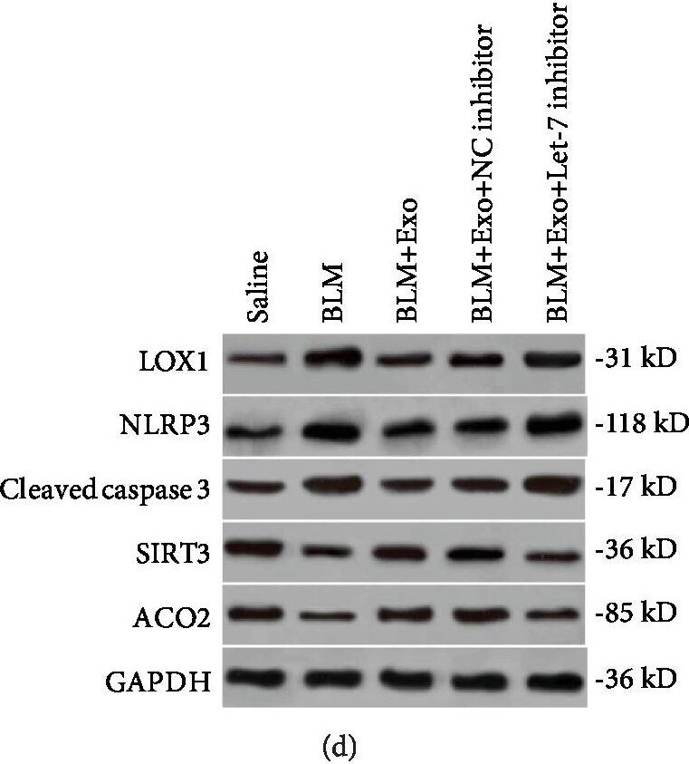
Western Blot: Aconitase 2 Antibody [NBP1-90264] -
Western Blot: Aconitase 2 Antibody [NBP1-90264] - The regulation mechanism of LOX1 on alveolar epithelial cell apoptosis & fibrosis. (a) Cells were transfected with a LOX1 overexpression plasmid & control vector. ROS levels were determined by DCFH-DA assay in MLE-12 cells treated by TGF-beta 1, TGF-beta 1 plus exosome, TGF-beta 1 plus exosome & vector, & TGF-beta 1 plus exosome & LOX1 overexpression plasmid (n = 3). (b) Detection of cellular mtDNA/18sRNA in each of the above cell groups (n = 3). (c) The expression of LOX1, caspase 3, mtDNA damage markers SIRT3 & ACO2, & NLRP3 was measured by western blotting in each of the above cell groups. (d) The expression of the same signal cascades including LOX1, caspase 3, SIRT3, ACO2, & NLRP3 was confirmed in an animal model with pulmonary fibrosis by western blot assay. Data is shown as the means ± SD, n ≥ 3. ∗∗p < 0.01. Image collected & cropped by CiteAb from the following publication (https://pubmed.ncbi.nlm.nih.gov/31949877), licensed under a CC-BY license. Not internally tested by Novus Biologicals.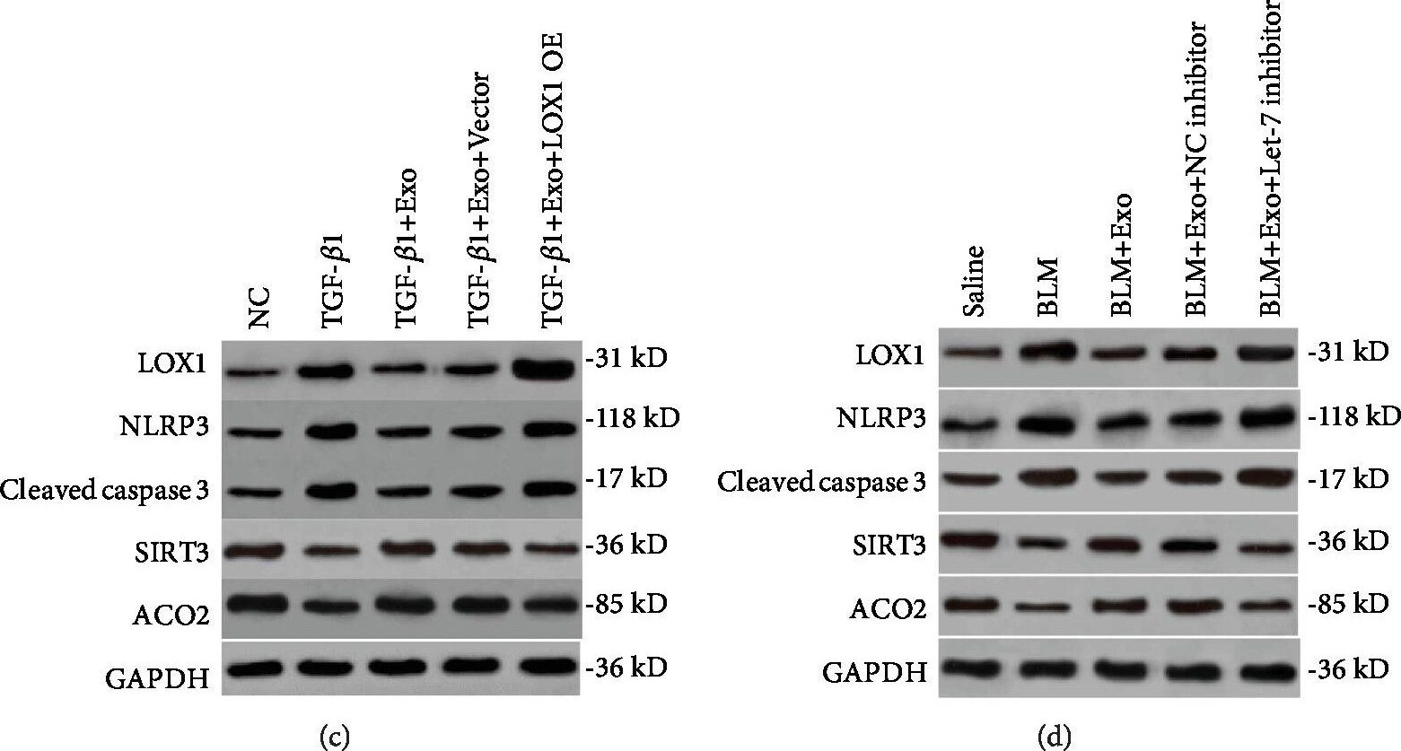
Western Blot: Aconitase 2 Antibody [NBP1-90264] -
Western Blot: Aconitase 2 Antibody [NBP1-90264] - The regulation mechanism of LOX1 on alveolar epithelial cell apoptosis & fibrosis. (a) Cells were transfected with a LOX1 overexpression plasmid & control vector. ROS levels were determined by DCFH-DA assay in MLE-12 cells treated by TGF-beta 1, TGF-beta 1 plus exosome, TGF-beta 1 plus exosome & vector, & TGF-beta 1 plus exosome & LOX1 overexpression plasmid (n = 3). (b) Detection of cellular mtDNA/18sRNA in each of the above cell groups (n = 3). (c) The expression of LOX1, caspase 3, mtDNA damage markers SIRT3 & ACO2, & NLRP3 was measured by western blotting in each of the above cell groups. (d) The expression of the same signal cascades including LOX1, caspase 3, SIRT3, ACO2, & NLRP3 was confirmed in an animal model with pulmonary fibrosis by western blot assay. Data is shown as the means ± SD, n ≥ 3. ∗∗p < 0.01. Image collected & cropped by CiteAb from the following publication (https://pubmed.ncbi.nlm.nih.gov/31949877), licensed under a CC-BY license. Not internally tested by Novus Biologicals.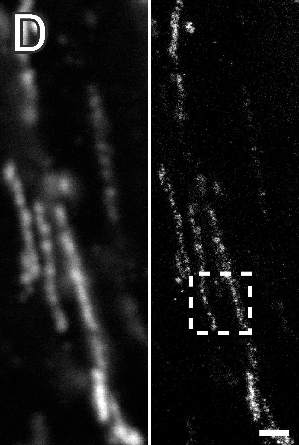
Immunohistochemistry: Aconitase 2 Antibody - BSA Free [NBP1-90264] -
STED super-resolution microscopy of mitochondria in the rectal Muscularis externa demonstrates high structural preservation of the stored paraffin-embedded tissue.STED recordings were performed on 2 um thick dewaxed sections cut along the longitudinal axis of the rectum. (A) Left: STED overview image of a region of the inner circular layer of the rectal Muscularis externa decorated with an antiserum against Tom20. Right: Magnifications of the areas in the indicated dashed squares showing the distribution of TOM clusters within the mitochondria. (B–E) STED images of tissue sections decorated with antisera against Tom20 (B), Mic60 (mitofilin) (C), aconitase (D), and cyclophilin D (E). In each panel the confocal (top, left) and the corresponding STED image (top, right) is displayed. Bottom: Magnification of the STED image as indicated by a dashed square. Note the different distributions of the four proteins within the mitochondria. Scale bars: 20 um (A, left); 1 um (A, right) and (B–E, top); 200 nm (B–E, bottom). Image collected and cropped by CiteAb from the following open publication (https://pubmed.ncbi.nlm.nih.gov/25025184), licensed under a CC-BY license. Not internally tested by Novus Biologicals.Applications for Aconitase 2 Antibody - BSA Free
Immunohistochemistry
Immunohistochemistry-Paraffin
Western Blot
Formulation, Preparation, and Storage
Purification
Formulation
Format
Preservative
Concentration
Shipping
Stability & Storage
Background: Aconitase 2
Alternate Names
Gene Symbol
UniProt
Additional Aconitase 2 Products
Product Documents for Aconitase 2 Antibody - BSA Free
Certificate of Analysis
To download a Certificate of Analysis, please enter a lot or batch number in the search box below.
Product Specific Notices for Aconitase 2 Antibody - BSA Free
This product is for research use only and is not approved for use in humans or in clinical diagnosis. Primary Antibodies are guaranteed for 1 year from date of receipt.
Citations for Aconitase 2 Antibody - BSA Free
 Powered by Bioz
Customer Reviews for Aconitase 2 Antibody - BSA Free
There are currently no reviews for this product. Be the first to review Aconitase 2 Antibody - BSA Free and earn rewards!
Have you used Aconitase 2 Antibody - BSA Free?
Submit a review and receive an Amazon gift card!
$25/€18/£15/$25CAN/¥2500 for a review with an image
$10/€7/£6/$10CAN/¥1110 for a review without an image
Submit a review
Protocols
Find general support by application which include: protocols, troubleshooting, illustrated assays, videos and webinars.
- Antigen Retrieval Protocol (PIER)
- Antigen Retrieval for Frozen Sections Protocol
- Appropriate Fixation of IHC/ICC Samples
- Cellular Response to Hypoxia Protocols
- Chromogenic IHC Staining of Formalin-Fixed Paraffin-Embedded (FFPE) Tissue Protocol
- Chromogenic Immunohistochemistry Staining of Frozen Tissue
- ClariTSA™ Fluorophore Kits
- Detection & Visualization of Antibody Binding
- Fluorescent IHC Staining of Frozen Tissue Protocol
- Graphic Protocol for Heat-induced Epitope Retrieval
- Graphic Protocol for the Preparation and Fluorescent IHC Staining of Frozen Tissue Sections
- Graphic Protocol for the Preparation and Fluorescent IHC Staining of Paraffin-embedded Tissue Sections
- Graphic Protocol for the Preparation of Gelatin-coated Slides for Histological Tissue Sections
- IHC Sample Preparation (Frozen sections vs Paraffin)
- Immunofluorescent IHC Staining of Formalin-Fixed Paraffin-Embedded (FFPE) Tissue Protocol
- Immunohistochemistry (IHC) and Immunocytochemistry (ICC) Protocols
- Immunohistochemistry Frozen Troubleshooting
- Immunohistochemistry Paraffin Troubleshooting
- Preparing Samples for IHC/ICC Experiments
- Preventing Non-Specific Staining (Non-Specific Binding)
- Primary Antibody Selection & Optimization
- Protocol for Heat-Induced Epitope Retrieval (HIER)
- Protocol for Making a 4% Formaldehyde Solution in PBS
- Protocol for VisUCyte™ HRP Polymer Detection Reagent
- Protocol for the Preparation & Fixation of Cells on Coverslips
- Protocol for the Preparation and Chromogenic IHC Staining of Frozen Tissue Sections
- Protocol for the Preparation and Chromogenic IHC Staining of Frozen Tissue Sections - Graphic
- Protocol for the Preparation and Chromogenic IHC Staining of Paraffin-embedded Tissue Sections
- Protocol for the Preparation and Chromogenic IHC Staining of Paraffin-embedded Tissue Sections - Graphic
- Protocol for the Preparation and Fluorescent IHC Staining of Frozen Tissue Sections
- Protocol for the Preparation and Fluorescent IHC Staining of Paraffin-embedded Tissue Sections
- Protocol for the Preparation of Gelatin-coated Slides for Histological Tissue Sections
- R&D Systems Quality Control Western Blot Protocol
- TUNEL and Active Caspase-3 Detection by IHC/ICC Protocol
- The Importance of IHC/ICC Controls
- Troubleshooting Guide: Immunohistochemistry
- Troubleshooting Guide: Western Blot Figures
- Western Blot Conditions
- Western Blot Protocol
- Western Blot Protocol for Cell Lysates
- Western Blot Troubleshooting
- Western Blot Troubleshooting Guide
- View all Protocols, Troubleshooting, Illustrated assays and Webinars