![Western Blot: BDNF Antibody [NB100-98682]](https://resources.rndsystems.com/images/products/BDNF-Antibody-Western-Blot-NB100-98682-img0010.jpg "Western Blot: BDNF Antibody [NB100-98682]")
Loading...
Key Product Details
Validated by
Biological Validation
Species Reactivity
Validated:
Human, Mouse, Rat, Zebrafish
Cited:
Human, Mouse, Rat, Fish - Danio rerio (Zebrafish)
Applications
Validated:
Immunohistochemistry, Immunohistochemistry-Paraffin, Western Blot, Immunocytochemistry/ Immunofluorescence
Cited:
Immunohistochemistry-Paraffin, Immunohistochemistry-Frozen, Western Blot, Immunocytochemistry/ Immunofluorescence, IF/IHC
Label
Unconjugated
Antibody Source
Polyclonal Rabbit IgG
Loading...
Product Specifications
Immunogen
A synthetic peptide from n-terminal region of human mature BDNF conjugated to blue carrier protein was used as the antigen. The peptide is homologous in many other species including rat, mouse, zebra fish and xenopus.
Reactivity Notes
Zebrafish reactivity reported in scientific literature (PMID: 30222997).
Specificity
Specific for mature BDNF.
Clonality
Polyclonal
Host
Rabbit
Isotype
IgG
Theoretical MW
28 kDa.
Disclaimer note: The observed molecular weight of the protein may vary from the listed predicted molecular weight due to post translational modifications, post translation cleavages, relative charges, and other experimental factors.
Disclaimer note: The observed molecular weight of the protein may vary from the listed predicted molecular weight due to post translational modifications, post translation cleavages, relative charges, and other experimental factors.
Scientific Data Images for BDNF Antibody
Western Blot: BDNF Antibody [NB100-98682]
Western Blot: BDNF Antibody [NB100-98682] - BDNF lowered the mechanical withdrawal threshold further and promoted activation of astrocytes and microglia, and enhanced the p38/JNK pathway to aggravate the release of IL-1beta and TNF-alpha in the SDH of CYP-induced cystitis. Western blots showing the expression of BDNF was further upregulated when compared with the CYP + PBS group. Data of mechanical withdrawal threshold were analyzed using a two-way analysis of variance (ANOVA) followed by the Sidak's multiple comparisons test. All data were calculated as mean +/- SEM (n = 10 per group). **p < 0.01, ***p < 0.001 vs. the control group. #p < 0.05, ##p < 0.01, ###p < 0.001 vs. the CYP + rBDNF group. $p < 0.05, $$p < 0.01, $$$p < 0.001 vs. the CYP + PBS group.![Immunohistochemistry: BDNF Antibody [NB100-98682]](https://resources.rndsystems.com/images/products/BDNF-Antibody-Immunohistochemistry-NB100-98682-img0007.jpg "Immunohistochemistry: BDNF Antibody [NB100-98682]")
![Western Blot: BDNF Antibody [NB100-98682]](https://resources.rndsystems.com/images/products/BDNF-Antibody-Western-Blot-NB100-98682-img0006.jpg "Western Blot: BDNF Antibody [NB100-98682]")
Western Blot: BDNF Antibody [NB100-98682]
Western Blot: BDNF Antibody [NB100-98682] - Human platelet lysate and recombinant mature BDNF. Blocking: 1% LFDM for 30 min at RT; primary antibody (1:1000) incubated at 4C overnight.![Immunohistochemistry-Paraffin: BDNF Antibody [NB100-98682]](https://resources.rndsystems.com/images/products/BDNF-Antibody-Immunohistochemistry-Paraffin-NB100-98682-img0001.jpg "Immunohistochemistry-Paraffin: BDNF Antibody [NB100-98682]")
Immunohistochemistry-Paraffin: BDNF Antibody [NB100-98682]
Immunohistochemistry-Paraffin: BDNF Antibody [NB100-98682] - Mouse hippocampus. The animal was perfused at a pressure of 130 mmHg with 300 ml 4% FA before being processed for paraffin embedding. HIER: Tris-EDTA, pH 9 for 20 min. Blocking: 0.2% LFDM in TBST filtered thru 0.2 um. Detection was done using Novolink HRP polymer from Leica following manufacturers instructions; DAB chromogen: Candela DAB chromogen. Primary antibody: dilution 1: 1000, incubated 30 min at RT. Sections were counterstained with Harris Hematoxylin.![Immunohistochemistry-Paraffin: BDNF Antibody [NB100-98682]](https://resources.rndsystems.com/images/products/BDNF-Antibody-Immunohistochemistry-Paraffin-NB100-98682-img0003.jpg "Immunohistochemistry-Paraffin: BDNF Antibody [NB100-98682]")
Immunohistochemistry-Paraffin: BDNF Antibody [NB100-98682]
Immunohistochemistry-Paraffin: BDNF Antibody [NB100-98682] - Mouse hippocampus. The animal was perfused at a pressure of 130 mmHg with 300 ml 4% FA before being processed for paraffin embedding. HIER: Tris-EDTA, pH 9 for 20 min. Blocking: 0.2% LFDM in TBST filtered thru 0.2 um. Detection was done using Novolink HRP polymer from Leica following manufacturers instructions; DAB chromogen: Candela DAB chromogen. Primary antibody: dilution 1: 1000, incubated 30 min at RT. Sections were counterstained with Harris Hematoxylin.![Immunohistochemistry-Paraffin: BDNF Antibody [NB100-98682]](https://resources.rndsystems.com/images/products/BDNF-Antibody-Immunohistochemistry-Paraffin-NB100-98682-img0005.jpg "Immunohistochemistry-Paraffin: BDNF Antibody [NB100-98682]")
Immunohistochemistry-Paraffin: BDNF Antibody [NB100-98682]
Immunohistochemistry-Paraffin: BDNF Antibody [NB100-98682] - Mouse hippocampus. The animal was perfused at a pressure of 130 mmHg with 300 ml 4% FA before being processed for paraffin embedding. HIER: Tris-EDTA, pH 9 for 20 min. Blocking: 0.2% LFDM in TBST filtered thru 0.2 um. Detection was done using Novolink HRP polymer from Leica following manufacturers instructions; DAB chromogen: Candela DAB chromogen. Primary antibody: dilution 1: 1000, incubated 30 min at RT. Sections were counterstained with Harris Hematoxylin.
Western Blot: BDNF Antibody [NB100-98682] -
Western Blot: BDNF Antibody [NB100-98682] - Effect of treatment with FX extract on CREB/BDNF signaling. (a-c) Differences in the phosphorylation of CREB & expression of BDNF between groups in the PFC & hippocampus (n = 3-4). (d, e) pCREB & BDNF immunofluorescence was assessed in the PFC & hippocampus (n = 3-4). Mean ± SD. #P < 0.05 versus normal group; ∗P < 0.05 & ∗∗P < 0.01 versus control group. Scale bar = 200 μm. Image collected & cropped by CiteAb from the following publication (https://pubmed.ncbi.nlm.nih.gov/30065945), licensed under a CC-BY license. Not internally tested by Novus Biologicals.
Western Blot: BDNF Antibody [NB100-98682] -
Western Blot: BDNF Antibody [NB100-98682] - Antagonized TrkB could restrain the activation of astrocytes & microglia & supress the p38/JNK pathway to alleviate the release of IL-1 beta & TNF-alpha in the SDH of CYP-induced cystitis. Western blots showed that the overexpression of a BDNF, b TrkB, cp-TrkB, d Iba1, e GFAP, fp-p38, gp-JNK, h TNF-alpha, & i IL-1 beta were downregulated in comparison to the CYP + DMSO group after ANA-12 treatment. All data were calculated as mean ± SEM (n = 5 per group). **p < 0.01, ***p < 0.001 vs. control group. ##p < 0.01, ###p < 0.001 vs. CYP + ANA-12 group Image collected & cropped by CiteAb from the following publication (https://pubmed.ncbi.nlm.nih.gov/31931832), licensed under a CC-BY license. Not internally tested by Novus Biologicals.
Western Blot: BDNF Antibody [NB100-98682] -
Western Blot: BDNF Antibody [NB100-98682] - BDNF lowered the mechanical withdrawal threshold further & promoted activation of astrocytes & microglia, & enhanced the p38/JNK pathway to aggravate the release of IL-1 beta & TNF-alpha in the SDH of CYP-induced cystitis. a BDNF treated every other day after CYP injection could further lower the mechanical withdrawal threshold & suppress the retrieval of mechanical threshold when compared with the CYP + PBS group. After the exogenous BDNF injection, Western blots showing the expression of b BDNF, c TrkB, dp-TrkB, e Iba1, f GFAP, gp-p38, hp-JNK, i TNF-alpha, & j IL-1 beta were all further upregulated when compared with the CYP + PBS group. Data of mechanical withdrawal threshold were analyzed using a two-way analysis of variance (ANOVA) followed by the Sidak's multiple comparisons test. All data were calculated as mean ± SEM (n = 10 per group). **p < 0.01, ***p < 0.001 vs. the control group. #p < 0.05, ##p < 0.01, ###p < 0.001 vs. the CYP + rBDNF group. $p < 0.05, $$p < 0.01, $$$p < 0.001 vs. the CYP + PBS group Image collected & cropped by CiteAb from the following publication (https://pubmed.ncbi.nlm.nih.gov/31931832), licensed under a CC-BY license. Not internally tested by Novus Biologicals.
Immunohistochemistry: BDNF Antibody [NB100-98682] -
Immunohistochemistry: BDNF Antibody [NB100-98682] - Effects of mBMSC & EA treatment on the expression of trophic factors. (A) Photomicrographs & (B‒D) histograms for mBDNF, NT4, & VEGF expression (n = 4–5). mBDNF expression was markedly increased by treatment with mBMSC and/or EA compared to vehicle treatment. mBDNF expression levels were higher in the EA-treated MCAO group than in the mBMSC-treated MCAO group. NT4 expression in the SVZ & hippocampus was significantly higher in the combined mBMSC & EA treatment group than in the other groups. VEGF expression in the hippocampus was markedly higher in the mBMSC treatment group than in the vehicle-treated MCAO group. #p < 0.05, ##p < 0.01, & ###p < 0.001 versus control group; *p < 0.05, **p < 0.01, & ***p < 0.001 versus vehicle-treated MCAO group; &p < 0.05, &&p < 0.01, & &&&p < 0.001 versus mBMSC-treated MCAO group; Scale bar = 1 mm. Image collected & cropped by CiteAb from the following publication (https://pubmed.ncbi.nlm.nih.gov/29391466), licensed under a CC-BY license. Not internally tested by Novus Biologicals.
Western Blot: BDNF Antibody [NB100-98682] -
Western Blot: BDNF Antibody [NB100-98682] - The expression of BDNF-TrkB signaling in the SDH of CYP-induced cystitis. a Changes of the mechanical threshold in CYP-induced cystitis model. Compared to that in the control group, the mechanical threshold of the cystitis group decreased significantly after the CYP injection & remained low until day 17, & the minimum threshold value was reached on day 12. The expression of b BDNF, c TrkB, & dp-TrkB were evaluated by western blots. Compared to the control group, they were upregulated on days 8, 12, & 17. e Immunofluorescence double staining assay of BDNF & p-TrkB in the SDH. BDNF & p-TrkB (red), NeuN, GFAP, & OX-42( green), co-localization (yellow). BDNF was mainly colocalized in neurons which mainly located in Laminate II to IV. & TrkB receptors expressed in neurons, microglia, & astrocytes. The white dotted lines in picture “BDNF/NeuN” showed the laminate of the SDH according to Rexed & Steiner. Scale bar = 100 μm. All data were calculated as mean ± SEM (n = 5 per group). *p < 0.05, ** p < 0.01, *** p < 0.001 vs. the control group Image collected & cropped by CiteAb from the following publication (https://pubmed.ncbi.nlm.nih.gov/31931832), licensed under a CC-BY license. Not internally tested by Novus Biologicals.Applications for BDNF Antibody
Application
Recommended Usage
Immunocytochemistry/ Immunofluorescence
1:10-1:500
Immunohistochemistry
1:1000
Immunohistochemistry-Paraffin
1:1000
Western Blot
1:1000
Application Notes
Use in IHC-P reported in scientific literature (PMID: 30222997). ICC/IF reported in scientific literature (PMID: 27914953).
Reviewed Applications
Read 1 review rated 5 using NB100-98682 in the following applications:
Formulation, Preparation, and Storage
Purification
Unpurified
Reconstitution
Reconstitute in 0.1 ml of sterile water. Centrifuge to remove any insoluble material. Glycerol may be added (1:1) for additional stability. Please note the sample size is provided in reconstituted format.
Formulation
Lyophilized from whole antisera
Preservative
No Preservative
Concentration
This product is unpurified. The exact concentration of antibody is not quantifiable.
Shipping
The product is shipped with polar packs. Upon receipt, store it immediately at the temperature recommended below.
Stability & Storage
Store at 4C short term. Aliquot and store at -20C long term. Avoid freeze-thaw cycles.
Calculators
Background: BDNF
Long Name
Brain-derived Neurotrophic Factor
Alternate Names
Abrineurin, ANON2, BULN2, Neurotrophin
Gene Symbol
BDNF
UniProt
Additional BDNF Products
Product Documents for BDNF Antibody
Certificate of Analysis
To download a Certificate of Analysis, please enter a lot or batch number in the search box below.
Product Specific Notices for BDNF Antibody
This product is for research use only and is not approved for use in humans or in clinical diagnosis. Primary Antibodies are guaranteed for 1 year from date of receipt.
Related Research Areas
Citations for BDNF Antibody
 Powered by Bioz
Customer Reviews for BDNF Antibody (1)
5 out of 5
1 Customer Rating
Have you used BDNF Antibody?
Submit a review and receive an Amazon gift card!
$25/€18/£15/$25CAN/¥2500 Yen for a review with an image
$10/€7/£6/$10CAN/¥1110 Yen for a review without an image
Submit a review
Customer Images
Showing
1
-
1 的
1 review
Showing All
Filter By:
-
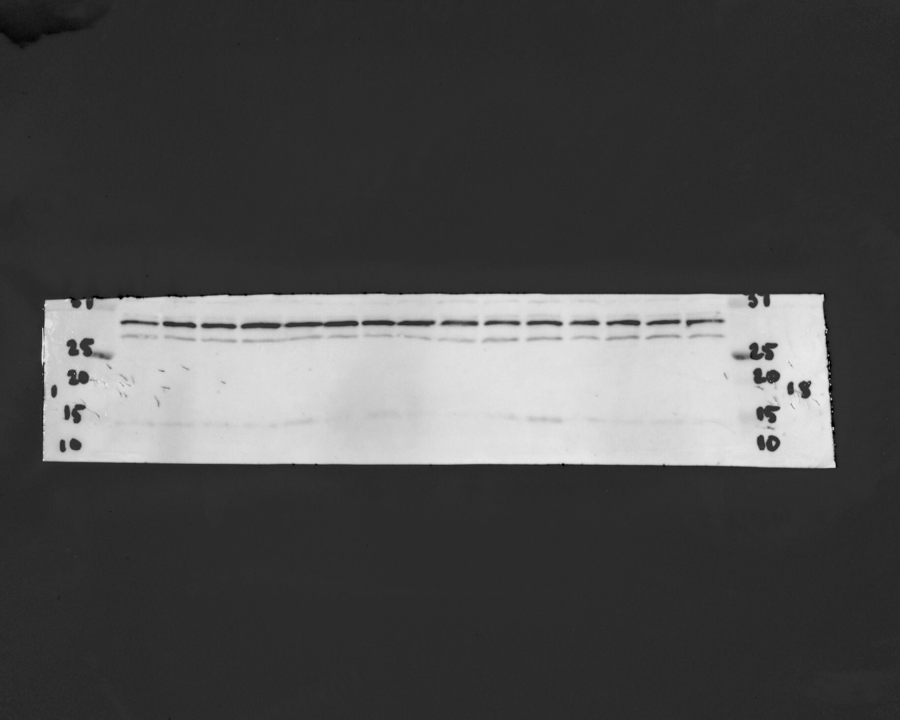
Application: Western BlotSample Tested: Rat hippocampusSpecies: RatVerified Customer | Posted 04/23/2025BDNF Bands in Rat Hippocampal SamplesThere are three aging groups on this blot: rats fed a Mediterranean diet, rats fed a western diet, and rats fed a chow diet. Animals were perfused with PBS and brains were micro dissected and frozen at -80C until needed. Dilution was 1:1,000. Two bands are clearly noticeable at ~32 and 30 kDa, while one band at ~13 kDa was less noticeable.

There are no reviews that match your criteria.
Protocols
Find general support by application which include: protocols, troubleshooting, illustrated assays, videos and webinars.
- Antigen Retrieval Protocol (PIER)
- Antigen Retrieval for Frozen Sections Protocol
- Appropriate Fixation of IHC/ICC Samples
- Cellular Response to Hypoxia Protocols
- Chromogenic IHC Staining of Formalin-Fixed Paraffin-Embedded (FFPE) Tissue Protocol
- Chromogenic Immunohistochemistry Staining of Frozen Tissue
- ClariTSA™ Fluorophore Kits
- Detection & Visualization of Antibody Binding
- Fluorescent IHC Staining of Frozen Tissue Protocol
- Graphic Protocol for Heat-induced Epitope Retrieval
- Graphic Protocol for the Preparation and Fluorescent IHC Staining of Frozen Tissue Sections
- Graphic Protocol for the Preparation and Fluorescent IHC Staining of Paraffin-embedded Tissue Sections
- Graphic Protocol for the Preparation of Gelatin-coated Slides for Histological Tissue Sections
- ICC Cell Smear Protocol for Suspension Cells
- ICC Immunocytochemistry Protocol Videos
- ICC for Adherent Cells
- IHC Sample Preparation (Frozen sections vs Paraffin)
- Immunocytochemistry (ICC) Protocol
- Immunocytochemistry Troubleshooting
- Immunofluorescence of Organoids Embedded in Cultrex Basement Membrane Extract
- Immunofluorescent IHC Staining of Formalin-Fixed Paraffin-Embedded (FFPE) Tissue Protocol
- Immunohistochemistry (IHC) and Immunocytochemistry (ICC) Protocols
- Immunohistochemistry Frozen Troubleshooting
- Immunohistochemistry Paraffin Troubleshooting
- Preparing Samples for IHC/ICC Experiments
- Preventing Non-Specific Staining (Non-Specific Binding)
- Primary Antibody Selection & Optimization
- Protocol for Heat-Induced Epitope Retrieval (HIER)
- Protocol for Making a 4% Formaldehyde Solution in PBS
- Protocol for VisUCyte™ HRP Polymer Detection Reagent
- Protocol for the Fluorescent ICC Staining of Cell Smears - Graphic
- Protocol for the Fluorescent ICC Staining of Cultured Cells on Coverslips - Graphic
- Protocol for the Preparation & Fixation of Cells on Coverslips
- Protocol for the Preparation and Chromogenic IHC Staining of Frozen Tissue Sections
- Protocol for the Preparation and Chromogenic IHC Staining of Frozen Tissue Sections - Graphic
- Protocol for the Preparation and Chromogenic IHC Staining of Paraffin-embedded Tissue Sections
- Protocol for the Preparation and Chromogenic IHC Staining of Paraffin-embedded Tissue Sections - Graphic
- Protocol for the Preparation and Fluorescent ICC Staining of Cells on Coverslips
- Protocol for the Preparation and Fluorescent ICC Staining of Non-adherent Cells
- Protocol for the Preparation and Fluorescent ICC Staining of Stem Cells on Coverslips
- Protocol for the Preparation and Fluorescent IHC Staining of Frozen Tissue Sections
- Protocol for the Preparation and Fluorescent IHC Staining of Paraffin-embedded Tissue Sections
- Protocol for the Preparation of Gelatin-coated Slides for Histological Tissue Sections
- Protocol for the Preparation of a Cell Smear for Non-adherent Cell ICC - Graphic
- R&D Systems Quality Control Western Blot Protocol
- TUNEL and Active Caspase-3 Detection by IHC/ICC Protocol
- The Importance of IHC/ICC Controls
- Troubleshooting Guide: Immunohistochemistry
- Troubleshooting Guide: Western Blot Figures
- Western Blot Conditions
- Western Blot Protocol
- Western Blot Protocol for Cell Lysates
- Western Blot Troubleshooting
- Western Blot Troubleshooting Guide
- View all Protocols, Troubleshooting, Illustrated assays and Webinars

