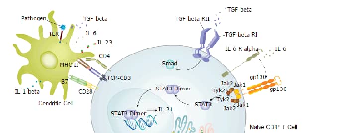
CD3 Antibody (MSVA-003R)
Novus Biologicals | Catalog # NBP3-57213
Recombinant Monoclonal Antibody

Scientific images may be resized, cropped, or adjusted for brightness or contrast for presentation purposes. These updates are limited to presentation and do not affect the underlying scientific interpretation of the data. Where available, additional source data may be provided upon request.
Key Product Details
Species Reactivity
Human
Applications
Immunohistochemistry, Immunohistochemistry-Paraffin
Label
Unconjugated
Antibody Source
Monoclonal Rabbit IgG Clone # MSVA-003R
Loading...
Product Specifications
Immunogen
A synthetic peptide from human CD3e protein (around aa 20-40) (exact sequence is proprietary) Swissprot P07766
Localization
Cell surface. Cytoplasm
Clonality
Monoclonal
Host
Rabbit
Isotype
IgG
Description
This antibody is highly specific and optimized for use in Immunohistochemistry.
It has been validated on TMAs covering 76 normal tissues and numerous cancer tissues.
The antibody concentration is optimized for dilution within dilution range using commercially available antibody diluent for immunohistochemistry.
Positive Control
Postive control:Tonsil: A moderate to strong membranous staining should be seen in all T-cells in the interfollicular T-zones and in the germinal centres (few cells)..
Negative Control
Negitve control:Tonsil: All epithelial structures and B-cells should not show any CD3 immunostaining.
Storage
Antibody can be stored at 4degC. For extended storage, aliquot and store at -20degC. Avoid repeated freeze-thaw cycles.
Scientific Data Images for CD3 Antibody (MSVA-003R)
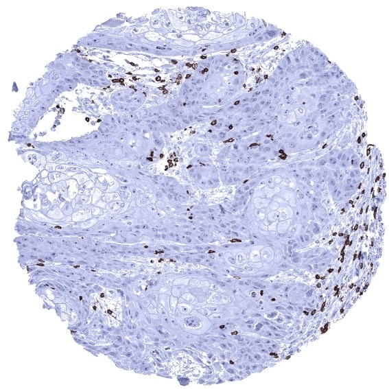
Immunohistochemistry-Paraffin:CD3 Antibody (MSVA-003R)NBP3-57213
Squamous cell carcinoma containing numerous CD3 positive T lymphocytes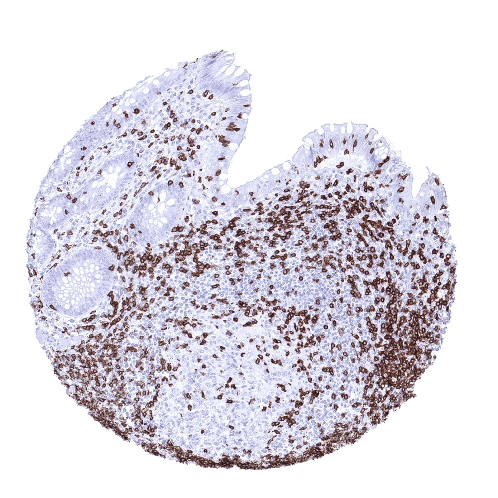
Immunohistochemistry-Paraffin:CD3 Antibody (MSVA-003R)NBP3-57213
Appendix mucosa In the appendix numerous CD3 positive T lymphocytes occur in the epithelium the lamina propria and in germinal centres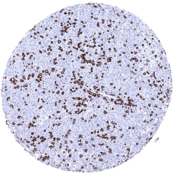
Immunohistochemistry-Paraffin:CD3 Antibody (MSVA-003R)NBP3-57213
Diffuse large B cell lymphoma infiltrated by numerous CD3 positive non neoplastic lymphocytes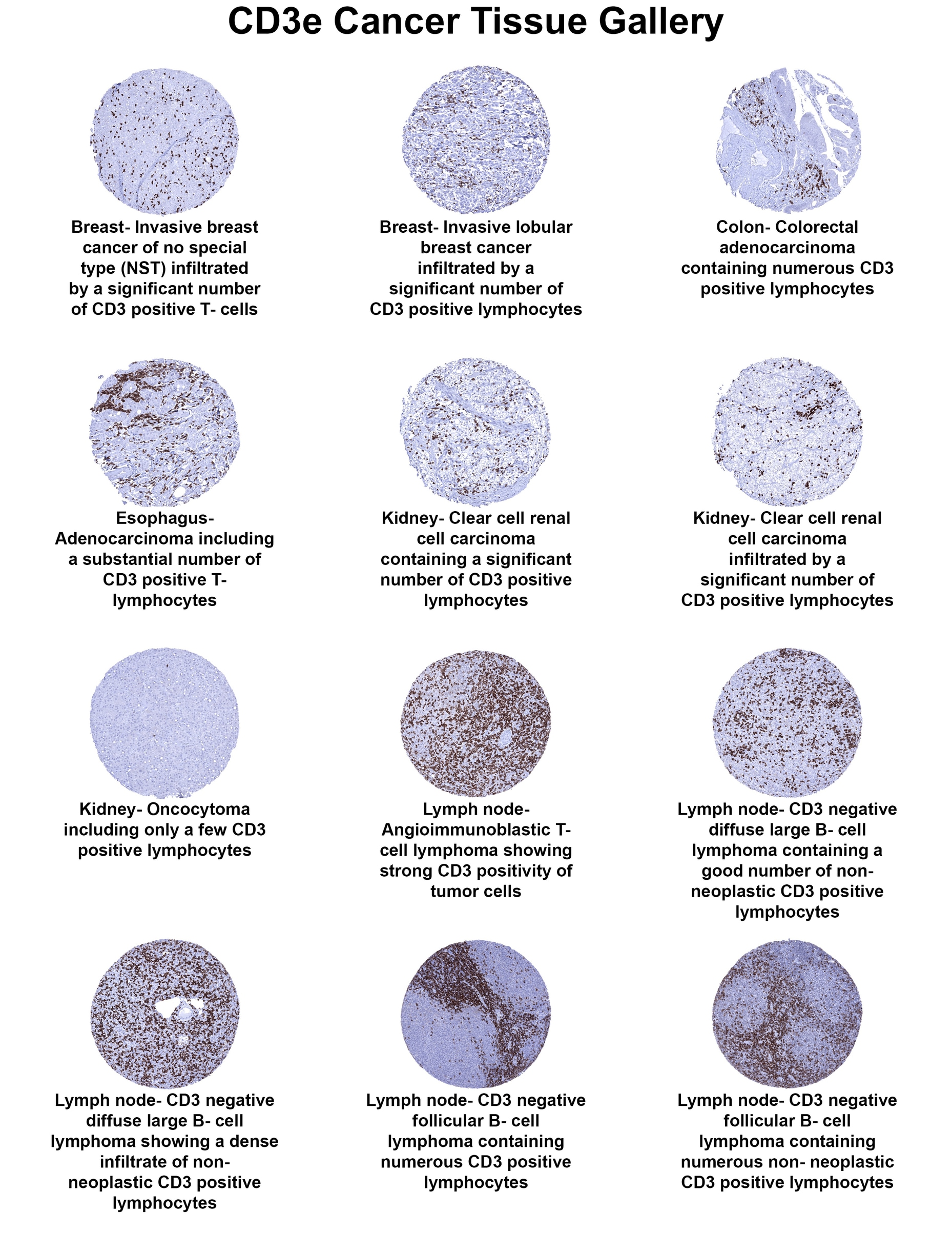
CD3 Cancer Tissue Gallery
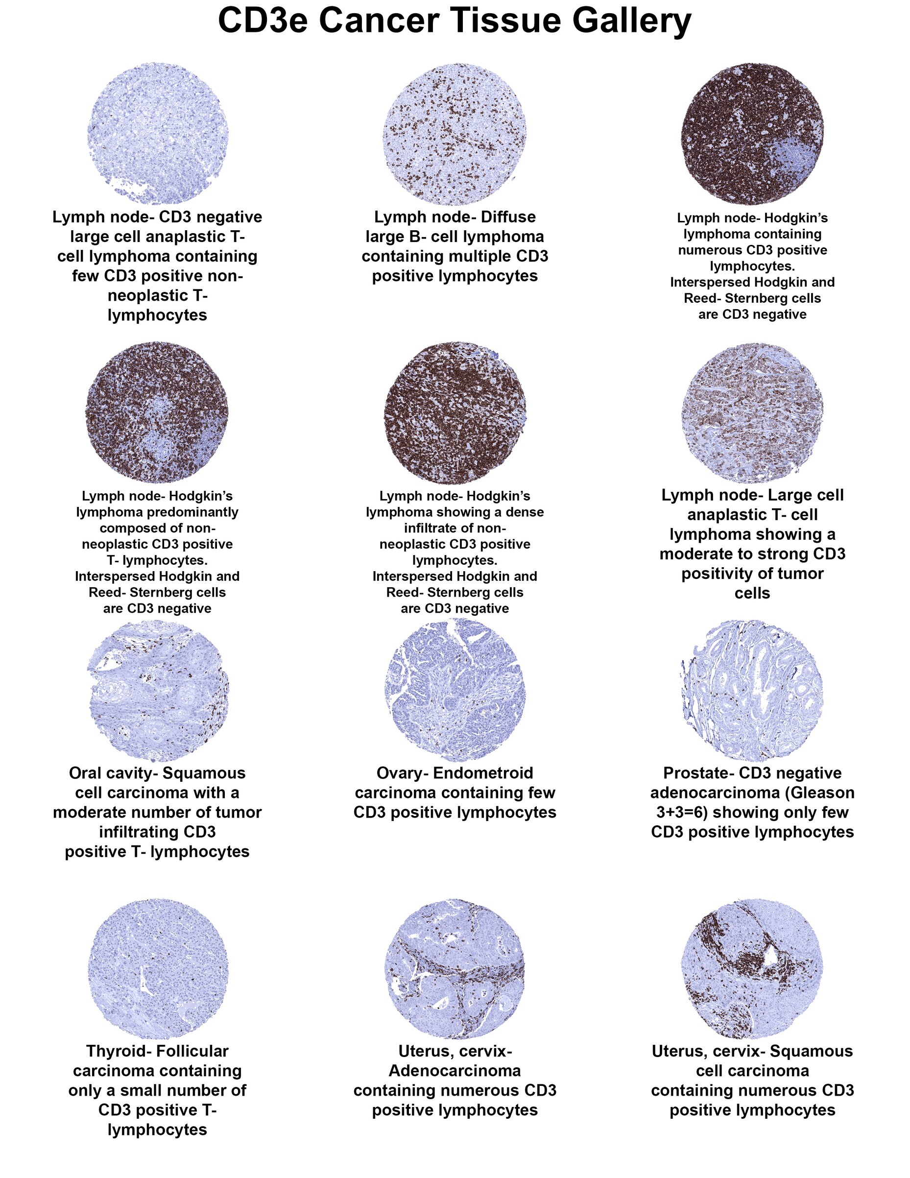
CD3 Cancer Tissue Gallery

CD3 Cancer Tissue Gallery
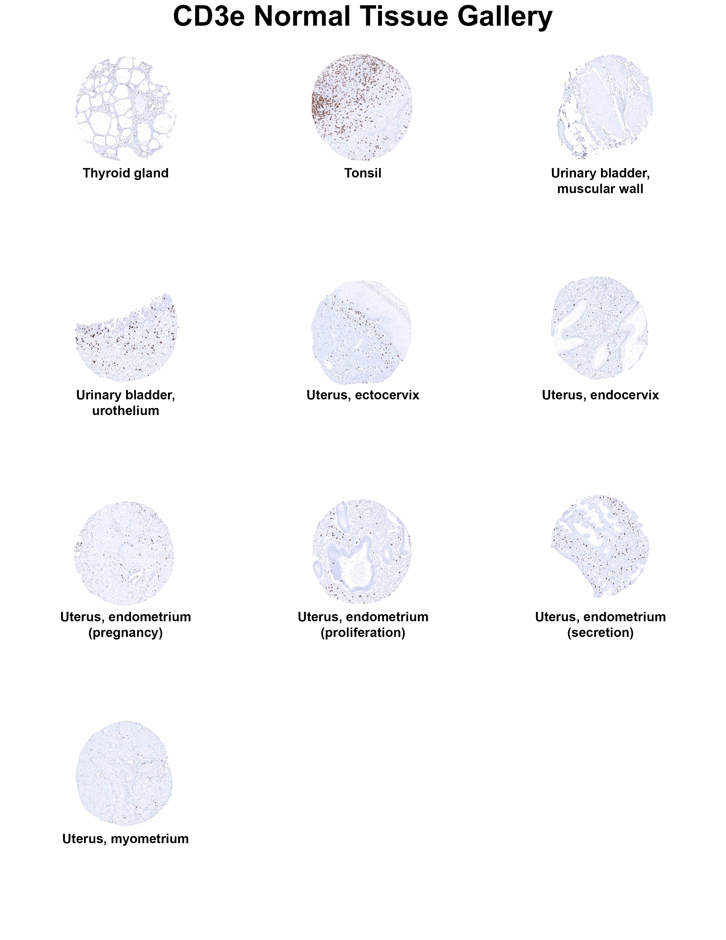
CD3 Normal Tissue Gallery
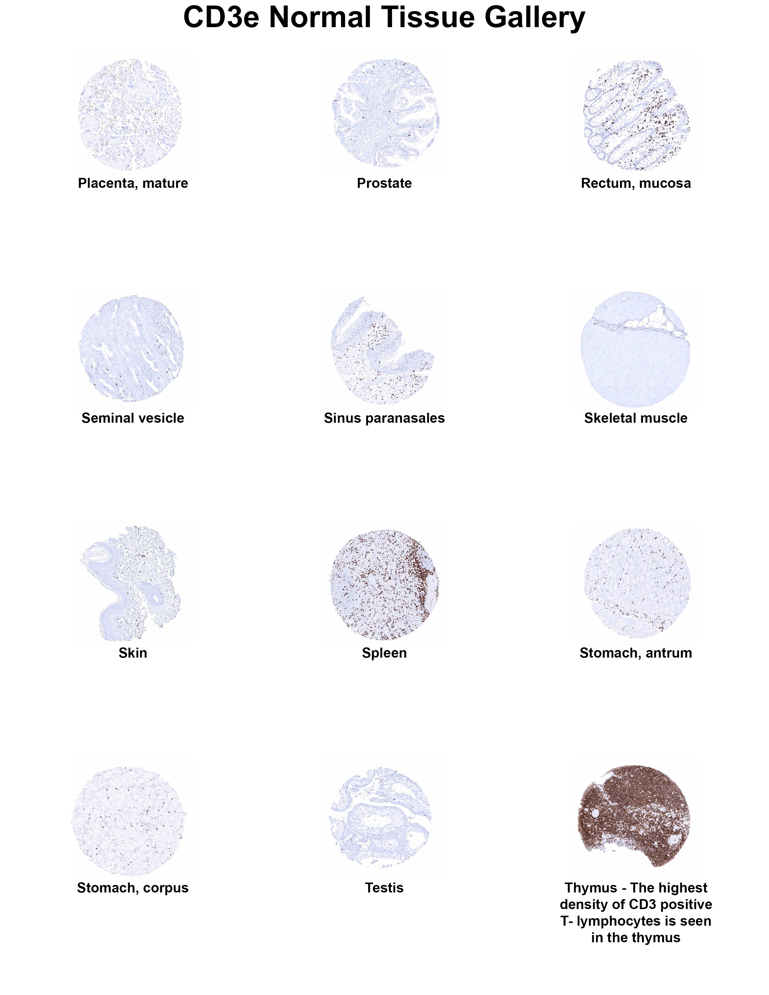
CD3 Normal Tissue Gallery
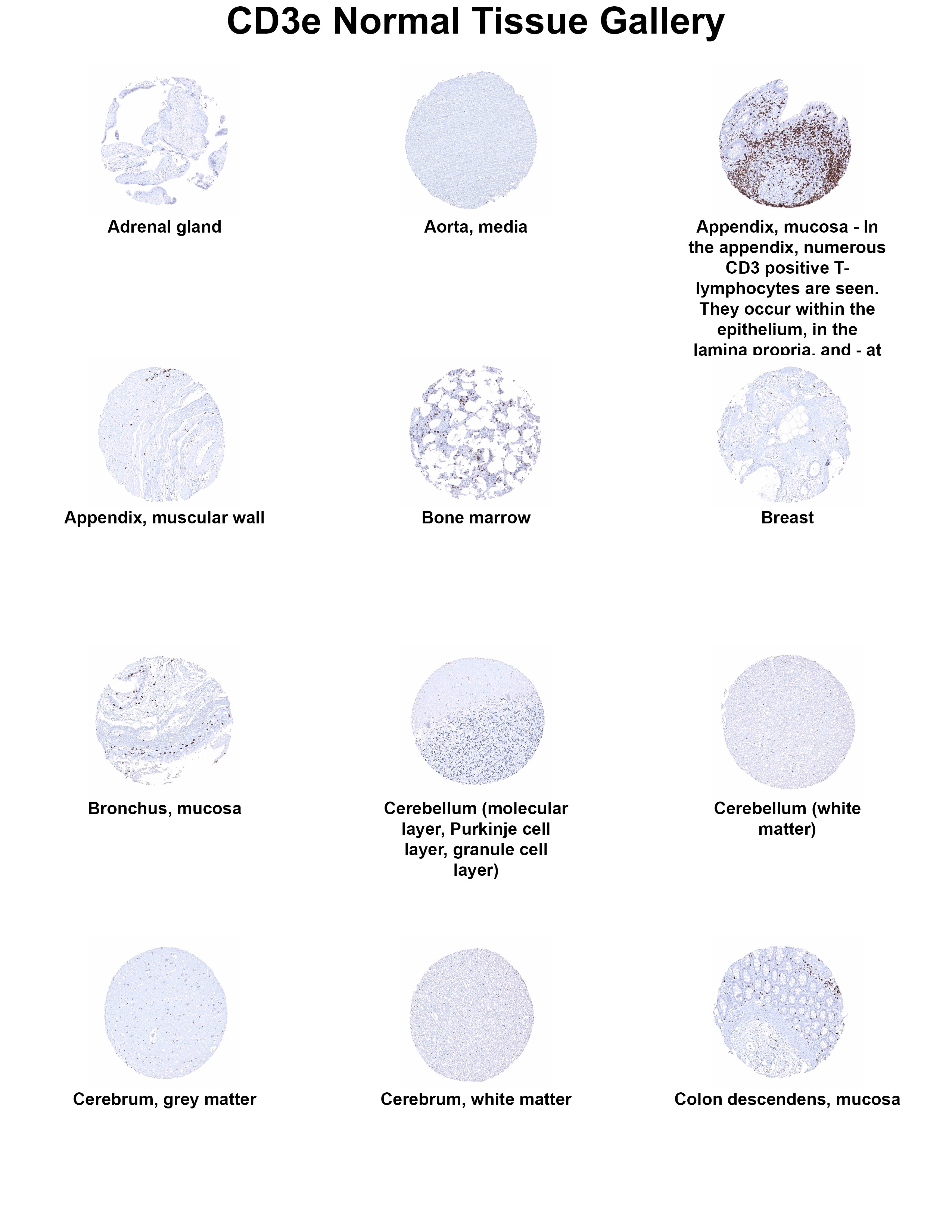
CD3 Normal Tissue Gallery
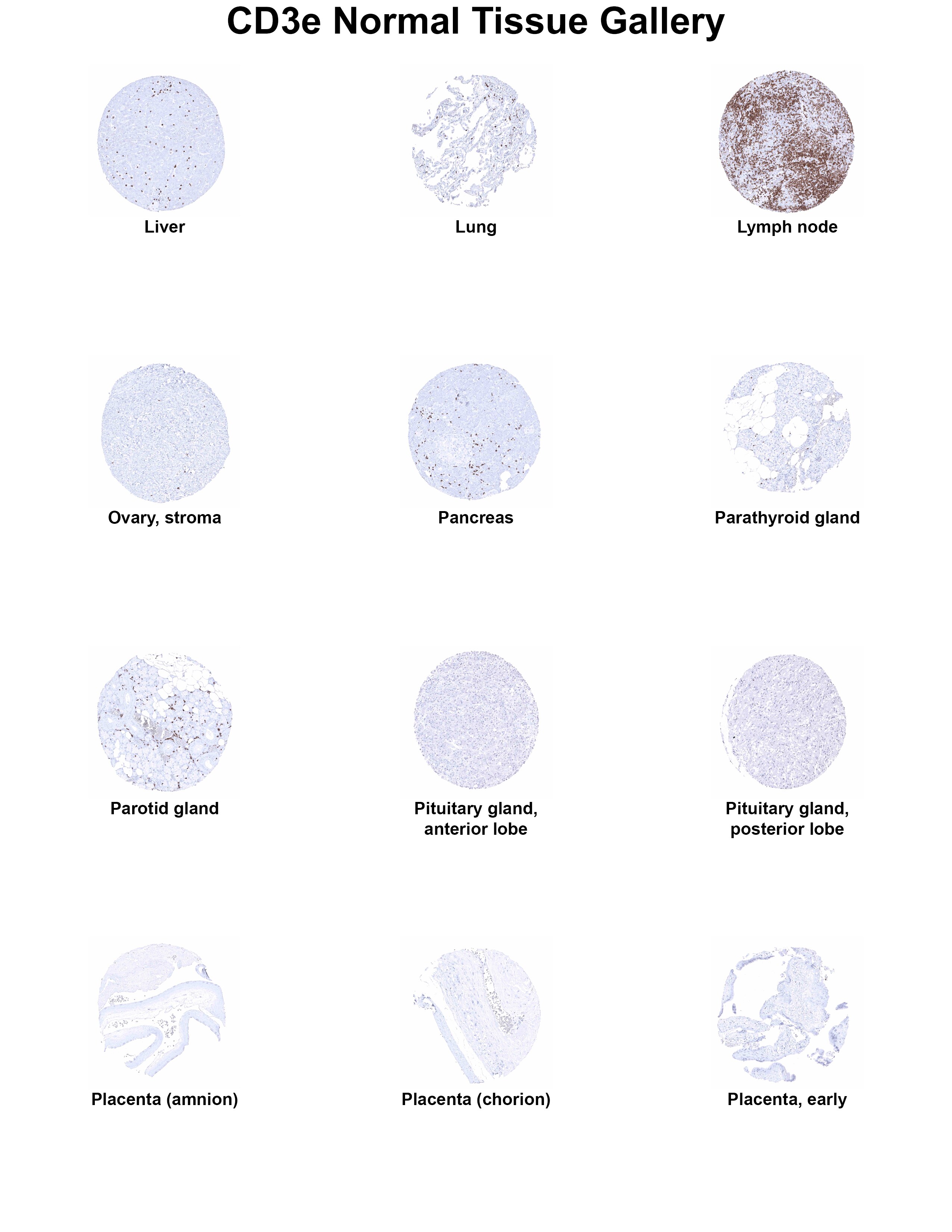
CD3 Normal Tissue Gallery
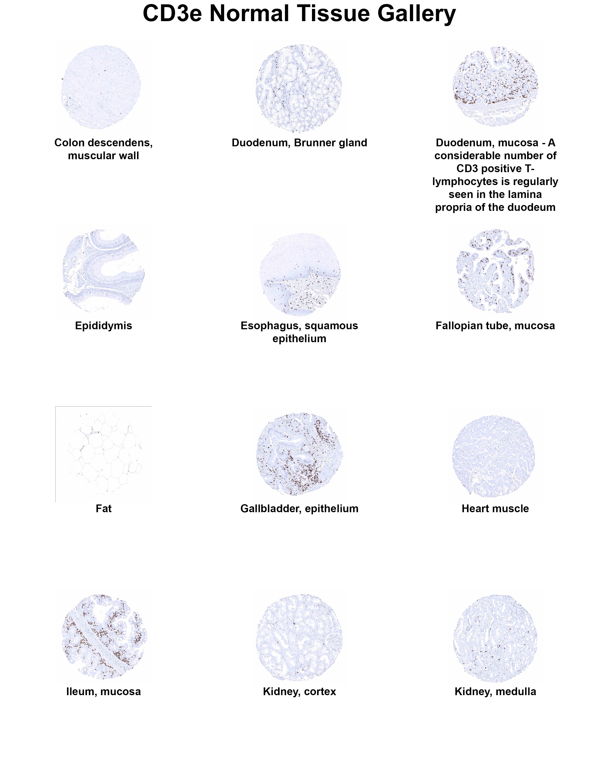
CD3 Normal Tissue Gallery
Applications for CD3 Antibody (MSVA-003R)
Application
Recommended Usage
Immunohistochemistry-Paraffin
1:100 1:200
Application Notes
Manual Protocol: Freshly cut sections should be used (less than 10 days between cutting and staining). Heat-induced antigen retrieval for 5 minutes in an autoclave at 121C in pH 7,8 Target Retrieval Solution buffer. Apply the antibody at a dilution of 1:150 at 37C for 60 minutes. Visualization of bound antibody by the EnVision Kit (Dako, Agilent) according to the manufacturer's directions.
Formulation, Preparation, and Storage
Purification
Protein A or G purified
Formulation
10mM PBS
Preservative
<0.1% Sodium Azide
Concentration
Please see the vial label for concentration. If unlisted please contact technical services.
Shipping
The product is shipped with polar packs. Upon receipt, store it immediately at the temperature recommended below.
Stability & Storage
Store at 4C.
Background: CD3
CD3 proteins are expressed on the surface of thymocytes during thymocyte development, proliferation, and maturation to T-cells (4, 6, 7). During T-cell development CD4-CD8- double negative (DN) cells differentiate to CD4+CD8+ double positive (DP) cells before progressing to single positive (SP) CD4+ helper T-cells or CD8+ cytotoxic T-cells (4, 6, 7). As CD3 plays an important role in thymocyte development, it is understandable that CD3 defects and mutations in CD3 protein chains cause severe combined immunodeficiencies (SCIDs) (8). Additionally, a subset of CD3+ T-cells that co-express CD20 are described in a variety of diseases including rheumatoid arthritis, multiple sclerosis, CD20+ T-cell leukemia/lymphoma, and HIV (9). Clinical trials and animal models have shown that anti-CD3 monoclonal antibodies are a promising treatment modality for inflammatory disorders and autoimmune diseases, such as type I diabetes (10).
References
1. Chetty, R., & Gatter, K. (1994). CD3: structure, function, and role of immunostaining in clinical practice. The Journal of pathology. https://doi.org/10.1002/path.1711730404
2. Mariuzza, R. A., Agnihotri, P., & Orban, J. (2020). The structural basis of T-cell receptor (TCR) activation: An enduring enigma. The Journal of biological chemistry. https://doi.org/10.1074/jbc.REV119.009411
3. Kuhns, M. S., Davis, M. M., & Garcia, K. C. (2006). Deconstructing the form and function of the TCR/CD3 complex. Immunity. https://doi.org/10.1016/j.immuni.2006.01.006
4. Clevers, H., Alarcon, B., Wileman, T., & Terhorst, C. (1988). The T cell receptor/CD3 complex: a dynamic protein ensemble. Annual review of immunology. https://doi.org/10.1146/annurev.iy.06.040188.003213
5. Uniprot: CD3-delta (P04234), CD3-epsilon (P07766), CD3-gamma (P09693), CD3-zeta (P20963)
6. D'Acquisto, F., & Crompton, T. (2011). CD3+CD4-CD8- (double negative) T cells: saviours or villains of the immune response?. Biochemical pharmacology. https://doi.org/10.1016/j.bcp.2011.05.019
7. Dave V. P. (2009). Hierarchical role of CD3 chains in thymocyte development. Immunological reviews. https://doi.org/10.1111/j.1600-065X.2009.00835.x
8. Fischer, A., de Saint Basile, G., & Le Deist, F. (2005). CD3 deficiencies. Current opinion in allergy and clinical immunology. https://doi.org/10.1097/01.all.0000191886.12645.79
9. Chen, Q., Yuan, S., Sun, H., & Peng, L. (2019). CD3+CD20+ T cells and their roles in human diseases. Human immunology. https://doi.org/10.1016/j.humimm.2019.01.001
10. Kuhn, C., & Weiner, H. L. (2016). Therapeutic anti-CD3 monoclonal antibodies: from bench to bedside. Immunotherapy. https://doi.org/10.2217/imt-2016-0049
Alternate Names
CD_antigen: CD3e, CD3 antigen, delta subunit, CD3d antigen, CD3d antigen, delta polypeptide (TiT3 complex), CD3d molecule, delta (CD3-TCR complex), CD3-DELTA, CD3e, CD3e antigen, CD3e antigen, epsilon polypeptide (TiT3 complex), CD3e molecule, epsilon (CD3-TCR complex), CD3-epsilon, CD3G, CD3g antigen, CD3g antigen, gamma polypeptide (TiT3 complex), CD3g molecule, epsilon (CD3-TCR complex), CD3g molecule, gamma (CD3-TCR complex), CD3-GAMMA, FLJ17620, FLJ17664, FLJ18683, FLJ79544, FLJ94613, IMD18, MGC138597, T3DOKT3, delta chain, T3E, T-cell antigen receptor complex, epsilon subunit of T3, T-cell receptor T3 delta chain, T-cell surface antigen T3/Leu-4 epsilon chain, T-cell surface glycoprotein CD3 delta chain, T-cell surface glycoprotein CD3 epsilon chain, TCRE
Gene Symbol
CD3E
Additional CD3 Products
Product Documents for CD3 Antibody (MSVA-003R)
Certificate of Analysis
To download a Certificate of Analysis, please enter a lot or batch number in the search box below.
Product Specific Notices for CD3 Antibody (MSVA-003R)
This product is for research use only and is not approved for use in humans or in clinical diagnosis. Primary Antibodies are guaranteed for 1 year from date of receipt.
Related Research Areas
Customer Reviews for CD3 Antibody (MSVA-003R)
There are currently no reviews for this product. Be the first to review CD3 Antibody (MSVA-003R) and earn rewards!
Have you used CD3 Antibody (MSVA-003R)?
Submit a review and receive an Amazon gift card!
$25/€18/£15/$25CAN/¥2500 for a review with an image
$10/€7/£6/$10CAN/¥1110 for a review without an image
Submit a review
Protocols
Find general support by application which include: protocols, troubleshooting, illustrated assays, videos and webinars.
- Antigen Retrieval Protocol (PIER)
- Antigen Retrieval for Frozen Sections Protocol
- Appropriate Fixation of IHC/ICC Samples
- Cellular Response to Hypoxia Protocols
- Chromogenic IHC Staining of Formalin-Fixed Paraffin-Embedded (FFPE) Tissue Protocol
- Chromogenic Immunohistochemistry Staining of Frozen Tissue
- ClariTSA™ Fluorophore Kits
- Detection & Visualization of Antibody Binding
- Fluorescent IHC Staining of Frozen Tissue Protocol
- Graphic Protocol for Heat-induced Epitope Retrieval
- Graphic Protocol for the Preparation and Fluorescent IHC Staining of Frozen Tissue Sections
- Graphic Protocol for the Preparation and Fluorescent IHC Staining of Paraffin-embedded Tissue Sections
- Graphic Protocol for the Preparation of Gelatin-coated Slides for Histological Tissue Sections
- IHC Sample Preparation (Frozen sections vs Paraffin)
- Immunofluorescent IHC Staining of Formalin-Fixed Paraffin-Embedded (FFPE) Tissue Protocol
- Immunohistochemistry (IHC) and Immunocytochemistry (ICC) Protocols
- Immunohistochemistry Frozen Troubleshooting
- Immunohistochemistry Paraffin Troubleshooting
- Preparing Samples for IHC/ICC Experiments
- Preventing Non-Specific Staining (Non-Specific Binding)
- Primary Antibody Selection & Optimization
- Protocol for Heat-Induced Epitope Retrieval (HIER)
- Protocol for Making a 4% Formaldehyde Solution in PBS
- Protocol for VisUCyte™ HRP Polymer Detection Reagent
- Protocol for the Preparation & Fixation of Cells on Coverslips
- Protocol for the Preparation and Chromogenic IHC Staining of Frozen Tissue Sections
- Protocol for the Preparation and Chromogenic IHC Staining of Frozen Tissue Sections - Graphic
- Protocol for the Preparation and Chromogenic IHC Staining of Paraffin-embedded Tissue Sections
- Protocol for the Preparation and Chromogenic IHC Staining of Paraffin-embedded Tissue Sections - Graphic
- Protocol for the Preparation and Fluorescent IHC Staining of Frozen Tissue Sections
- Protocol for the Preparation and Fluorescent IHC Staining of Paraffin-embedded Tissue Sections
- Protocol for the Preparation of Gelatin-coated Slides for Histological Tissue Sections
- TUNEL and Active Caspase-3 Detection by IHC/ICC Protocol
- The Importance of IHC/ICC Controls
- Troubleshooting Guide: Immunohistochemistry
- View all Protocols, Troubleshooting, Illustrated assays and Webinars