
CD3 epsilon Antibody (6G6K9)
Novus Biologicals | Catalog # NBP3-57370

Scientific images may be resized, cropped, or adjusted for brightness or contrast for presentation purposes. These updates are limited to presentation and do not affect the underlying scientific interpretation of the data. Where available, additional source data may be provided upon request.
Key Product Details
Species Reactivity
Applications
Label
Antibody Source
Product Specifications
Immunogen
Clonality
Host
Isotype
Description
This antibody is highly specific and optimized for use in Immunohistochemistry.
Normal Tissues Tested
Pancreas, Lung, Tonsil, Cervix, Placenta, Colon, Kidney, Spleen, Liver, esophagus
Cancer Tissues Tested
Breast cancer, Colon cancer, Thyroid cancer, Hepatocellular Carcinoma, Hepatocholangio carcinoma, Lung adenocarcinoma, Lung squamous carcinoma
Positive Controls
T cells, Tonsil (T-cells both in the interfollicular T-zones and in the germinal centers)
Negative Controls
Tonsil (epithelial cells, B cells, Fibroblasts)
Scientific Data Images for CD3 epsilon Antibody (6G6K9)
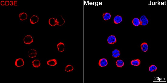
Immunocytochemistry/ Immunofluorescence: CD3 epsilon Antibody (6G6K9) [NBP3-57370]
Confocal imaging of Jurkat cells using NBP3-57370 (dilution 1:200) followed by a further incubation with Cy3 Goat Anti-Rabbit IgG (H+L) (dilution 1:500) (Red). DAPI was used for nuclear staining (Blue). Objective: 100x.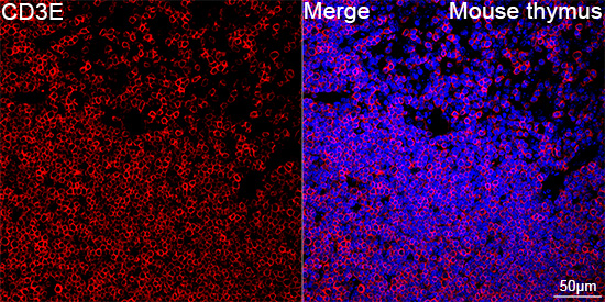
Immunohistochemistry-Paraffin: CD3 epsilon Antibody (6G6K9) [NBP3-57370]
Confocal imaging of paraffin-embedded Mouse thymus tissue using NBP3-57370 (dilution 1:200) followed by a further incubation with Cy3 Goat Anti-Rabbit IgG (H+L) (dilution 1:500) (Red). DAPI was used for nuclear staining (Blue). High pressure antigen retrieval performed with 0.01M Citrate Buffer (pH 6.0) prior to IF staining. Objective: 40x.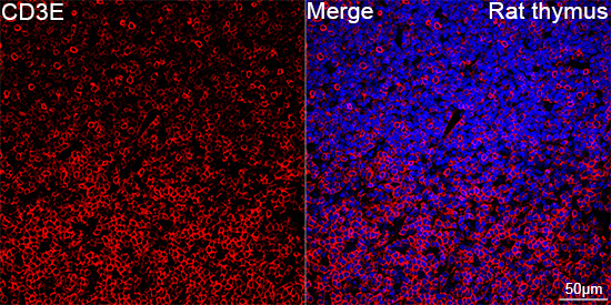
Immunohistochemistry-Paraffin: CD3 epsilon Antibody (6G6K9) [NBP3-57370]
Confocal imaging of paraffin-embedded Rat thymus tissue using NBP3-57370 (dilution 1:200) followed by a further incubation with Cy3 Goat Anti-Rabbit IgG (H+L) (dilution 1:500) (Red). DAPI was used for nuclear staining (Blue). High pressure antigen retrieval performed with 0.01M Citrate Buffer (pH 6.0) prior to IF staining. Objective: 40x.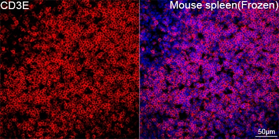
Immunohistochemistry-Frozen: CD3 epsilon Antibody (6G6K9) [NBP3-57370]
Confocal imaging of frozen sections of Mouse spleen tissue using NBP3-57370 (dilution 1:200) followed by a further incubation with Cy3-conjugated Goat anti-Rabbit IgG (H+L) (dilution 1:500) (Red). DAPI was used for nuclear staining (Blue). Microwave antigen retrieval performed with 0.01M Citrate Buffer (pH 6.0) prior to IF staining. Objective: 40x.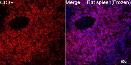
Immunohistochemistry-Frozen: CD3 epsilon Antibody (6G6K9) [NBP3-57370]
Confocal imaging of frozen sections of Rat spleen tissue using NBP3-57370 (dilution 1:200) followed by a further incubation with Cy3-conjugated Goat anti-Rabbit IgG (H+L) (dilution 1:500) (Red). DAPI was used for nuclear staining (Blue). Microwave antigen retrieval performed with 0.01M Citrate Buffer (pH 6.0) prior to IF staining. Objective: 40x.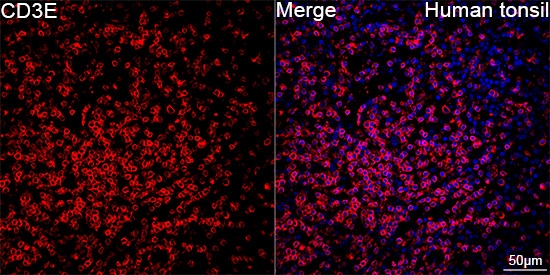
Immunohistochemistry-Paraffin: CD3 epsilon Antibody (6G6K9) [NBP3-57370]
Confocal imaging of paraffin-embedded Human tonsil using NBP3-57370 (dilution 1:200) followed by a further incubation with Cy3-conjugated Goat anti-Rabbit IgG (H+L) (dilution 1:500) (Red). DAPI was used for nuclear staining (Blue). High pressure antigen retrieval performed with 0.01M Citrate Buffer (pH 6.0) prior to IF staining. Objective: 40x.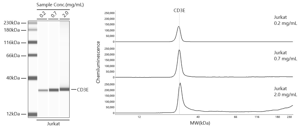
Simple Western: CD3 epsilon Antibody (6G6K9) [NBP3-57370]
Simple Western analysis of lysates from Jurkat cells using NBP3-57370 at 1:100 dilution. The virtual lane view (left) shows the target band (as indicated) with samples in concentrations of 0.2 mg/mL, 0.7 mg/mL and 2.0 mg/mL. The corresponding electropherogram view (right) plots chemiluminescence intensity against molecular weight along the capillary for sample concentrations of 0.2 mg/mL, 0.7 mg/mL and 2.0 mg/mL. This experiment was performed under reducing conditions on the Jess Simple Western instrument using the 12-230 kDa separation module.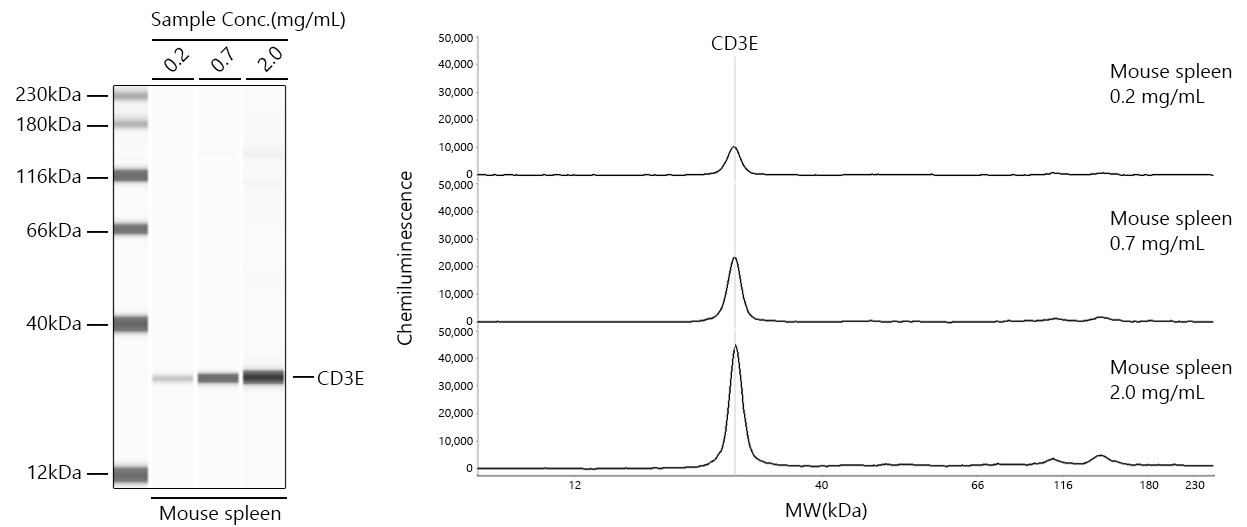
Simple Western: CD3 epsilon Antibody (6G6K9) [NBP3-57370]
Simple Western analysis of lysates from Mouse spleen using NBP3-57370 at 1:100 dilution. The virtual lane view (left) shows the target band (as indicated) with samples in concentrations of 0.2 mg/mL, 0.7 mg/mL and 2.0 mg/mL. The corresponding electropherogram view (right) plots chemiluminescence intensity against molecular weight along the capillary for sample concentrations of 0.2 mg/mL, 0.7 mg/mL and 2.0 mg/mL. This experiment was performed under reducing conditions on the Jess Simple Western instrument using the 12-230 kDa separation module.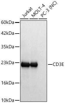
Western Blot: CD3 epsilon Antibody (6G6K9) [NBP3-57370]
Western blot analysis of various lysates using NBP3-57370 at 1:10000 dilution incubated overnight at 4C. Secondary antibody: HRP-conjugated Goat anti-Rabbit IgG (H+L) at 1:10000 dilution. Lysates/proteins: 25 ug per lane. Blocking buffer: 3% nonfat dry milk in TBST. Negative control (NC): PC-3 Exposure time: 5s.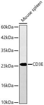
Western Blot: CD3 epsilon Antibody (6G6K9) [NBP3-57370]
Western blot analysis of lysates from Mouse spleen using NBP3-57370 at 1:10000 dilution incubated overnight at 4C. Secondary antibody: HRP-conjugated Goat anti-Rabbit IgG (H+L) at 1:10000 dilution. Lysates/proteins: 25 ug per lane. Blocking buffer: 3% nonfat dry milk in TBST. Exposure time: 10s.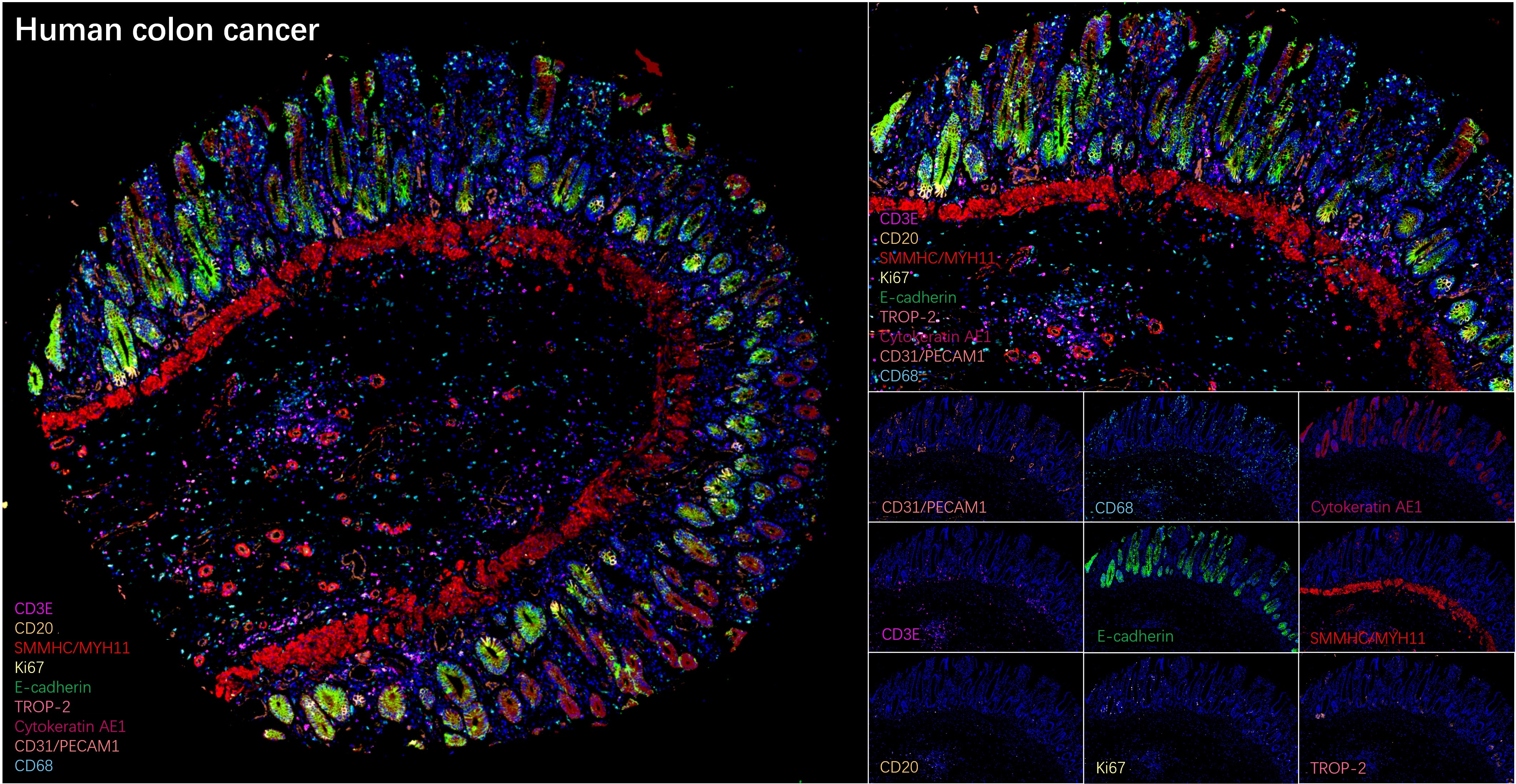
Multiplex Immunofluorescence: CD3 epsilon Antibody (6G6K9) [NBP3-57370]
The multiplex IHC analysis on paraffin-embedded Human colon cancer tissue using tyramide signal amplification (TSA) reagents with SMMHC/MYH11 Rabbit mAb (1:4000) (Red), and CD3 epsilon mAb (NBP3-57370, 1:40000) (Magenta), and CD20 Rabbit mAb (1:2000) (Pale Yellow), andE-Cadherin mAb (NBP3-57377, 1:1000) (Green), and CD31/PECAM-1 mAb (NBP3-57369, 1:200) (Light Coral), and CD68 Rabbit mAb (1:20000) (Cyan), and Ki67/MKI67 mAb (NBP3-57376, 1:500) (Light Yellow), and Cytokeratin, Acidic (Type I or LMW) mAb (NBP3-57402, 1:2000) (Magenta), and TROP-2 Rabbit mAb (1:10000) (Magenta). DAPI (Blue) was used for nuclear staining. Prior to multiplex IHC staining, high-pressure antigen retrieval was performed using 0.01M citrate buffer at pH 6.0. The analysis was completed using a 40x objective lens.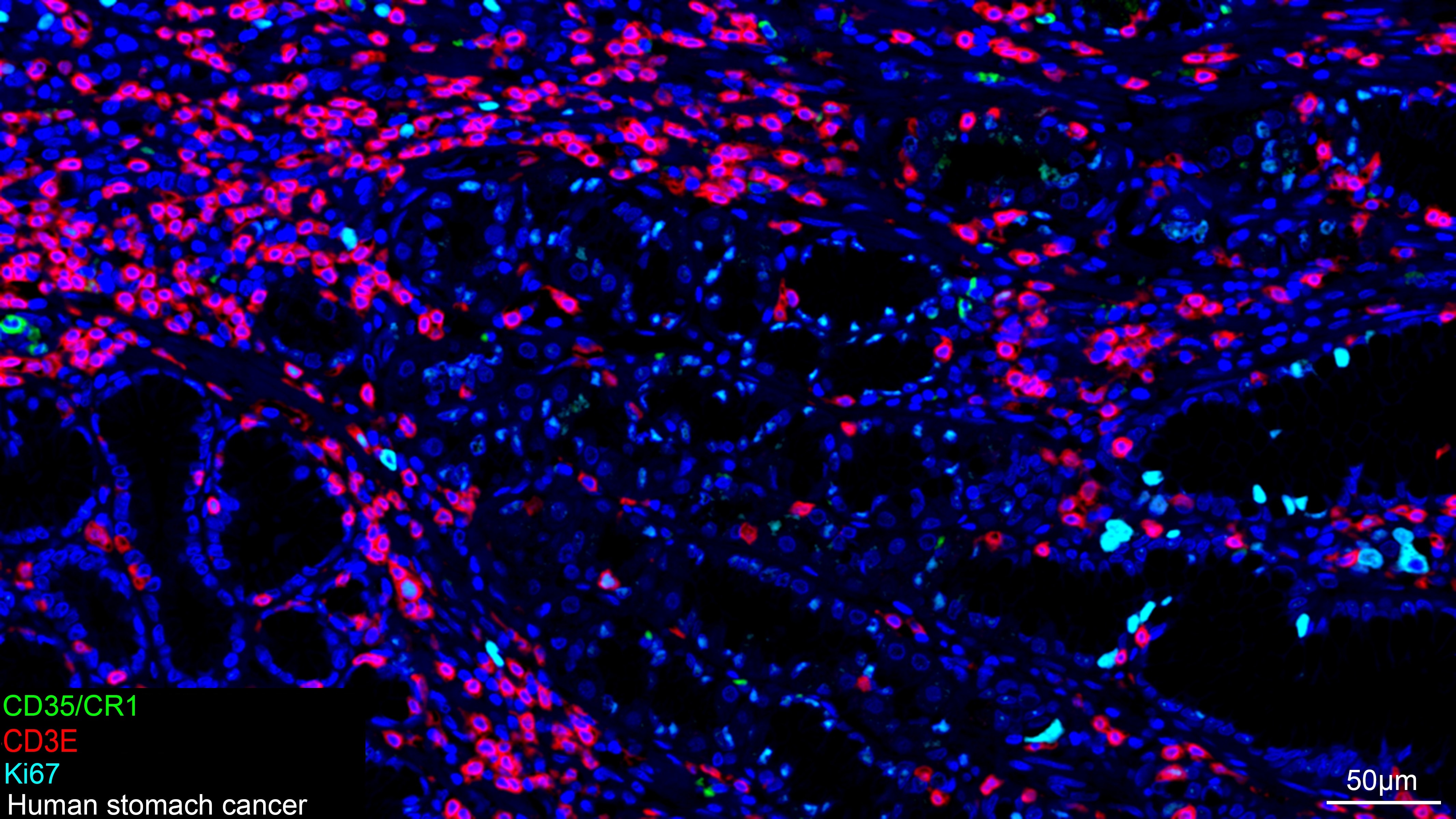
Multiplex Immunofluorescence: CD3 epsilon Antibody (6G6K9) [NBP3-57370]
The multiplex IHC analysis on paraffin-embedded Human stomach cancer tissue using tyramide signal amplification (TSA) reagents with CD35/CR1 Rabbit mAb (1:100) (Green), CD3 epsilon mAb (NBP3-57370, 1:2000) (Red), and Ki67/MKI67 mAb (NBP3-57376, 1:500) (cyan). DAPI (Blue) was used for nuclear staining. Prior to multiplex IHC staining, high-pressure antigen retrieval was performed using 0.01M citrate buffer at pH 6.0. The analysis was completed using a 20x objective lens.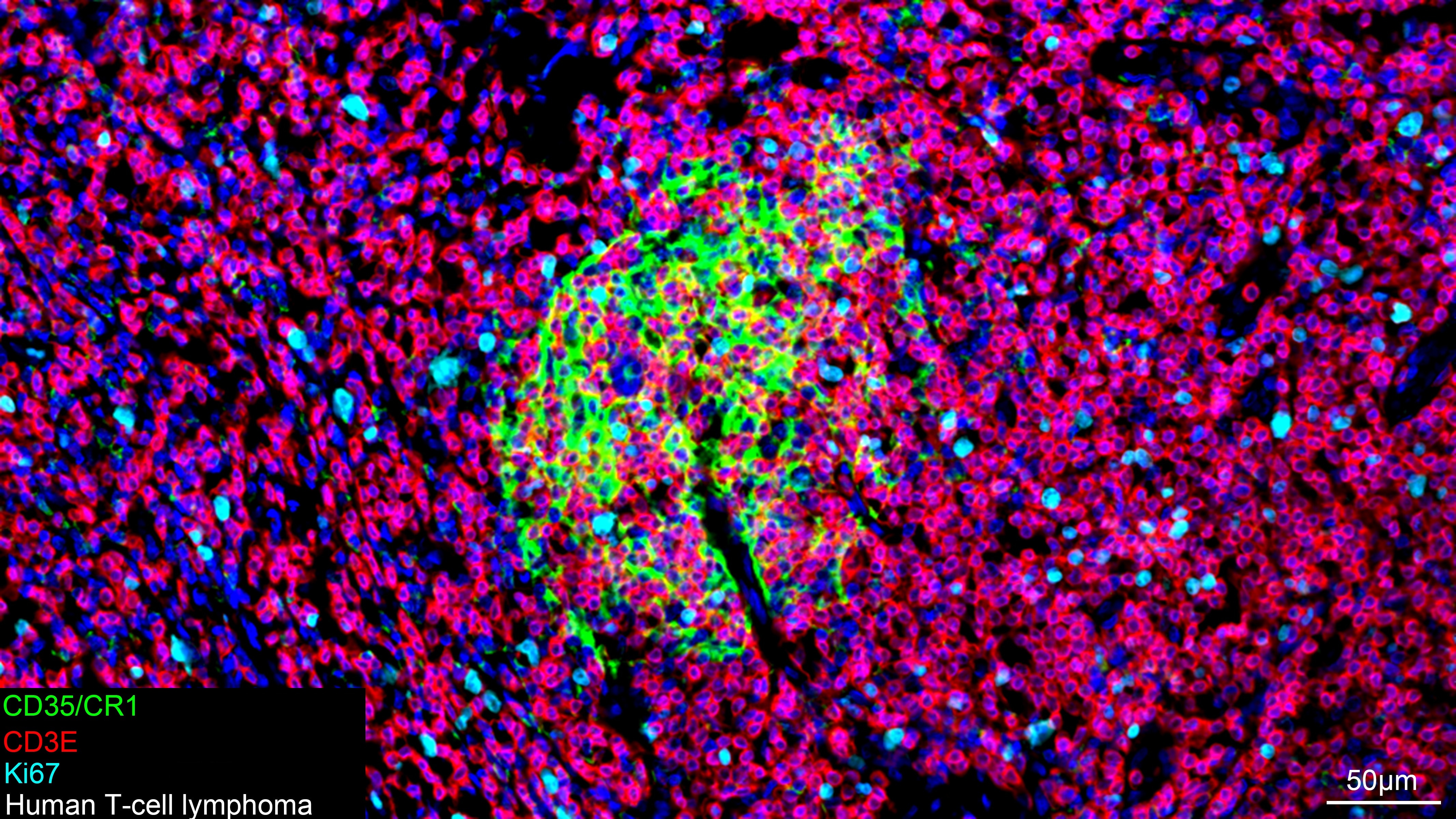
Multiplex Immunofluorescence: CD3 epsilon Antibody (6G6K9) [NBP3-57370]
The multiplex IHC analysis on paraffin-embedded Human T-cell lymphoma tissue using tyramide signal amplification (TSA) reagents with CD35/CR1 Rabbit mAb (1:100) (Green), CD3 epsilon mAb (NBP3-57370, 1:2000) (Red), and Ki67/MKI67 mAb (NBP3-57376, 1:500) (cyan). DAPI (Blue) was used for nuclear staining. Prior to multiplex IHC staining, high-pressure antigen retrieval was performed using 0.01M citrate buffer at pH 6.0. The analysis was completed using a 20x objective lens.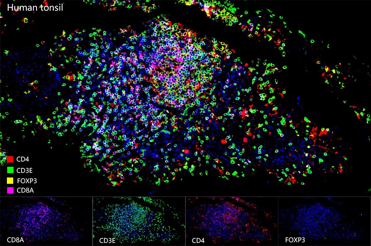
Multiplex Immunofluorescence: CD3 epsilon Antibody (6G6K9) [NBP3-57370]
The multiplex IHC analysis on paraffin-embedded Human tonsil tissue using tyramide signal amplification (TSA) reagents with FOXP3 Rabbit mAb (1:600) (Yellow), and CD8 mAb (NBP3-57757, 1:1000) (Magenta), and CD4 mAb (NBP3-57754, 1:400) (Red), and CD3 epsilon mAb (NBP3-57370, 1:4000) (Green). DAPI (Blue) was used for nuclear staining. Prior to multiplex IHC staining, high-pressure antigen retrieval was performed using 0.01M citrate buffer at pH 6.0. The analysis was completed using a 40x objective lens.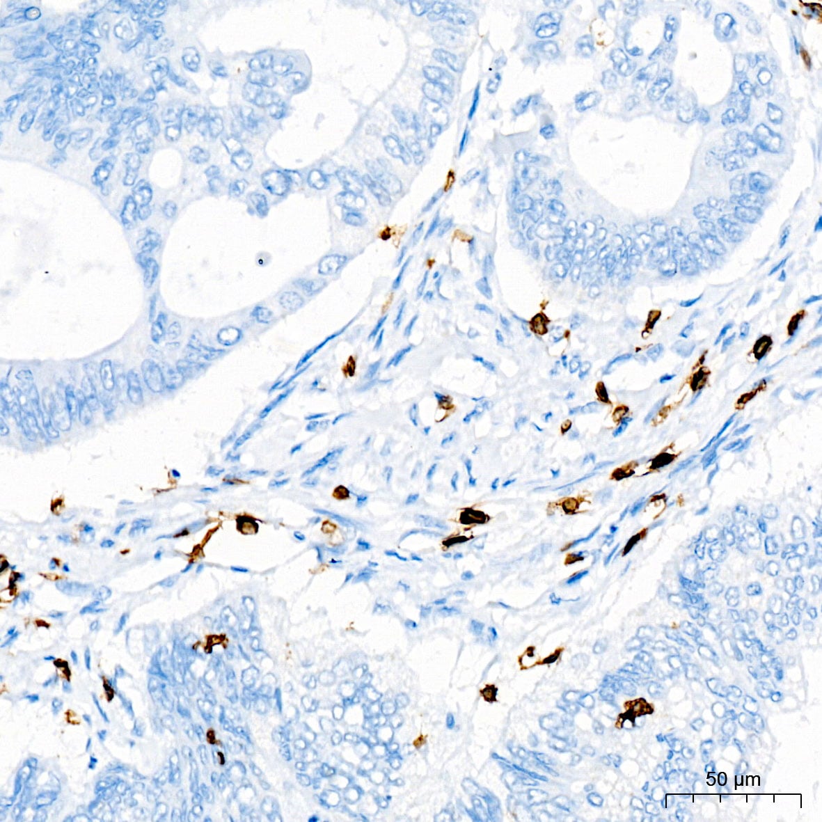
Immunohistochemistry-Paraffin: CD3 epsilon Antibody (6G6K9) [NBP3-57370]
Immunohistochemistry analysis of paraffin-embedded Human colon carcinoma tissue using NBP3-57370 at a dilution of 1:2000 (40x lens). High pressure antigen retrieval was performed with 0.01 M citrate buffer (pH 6.0) prior to IHC staining.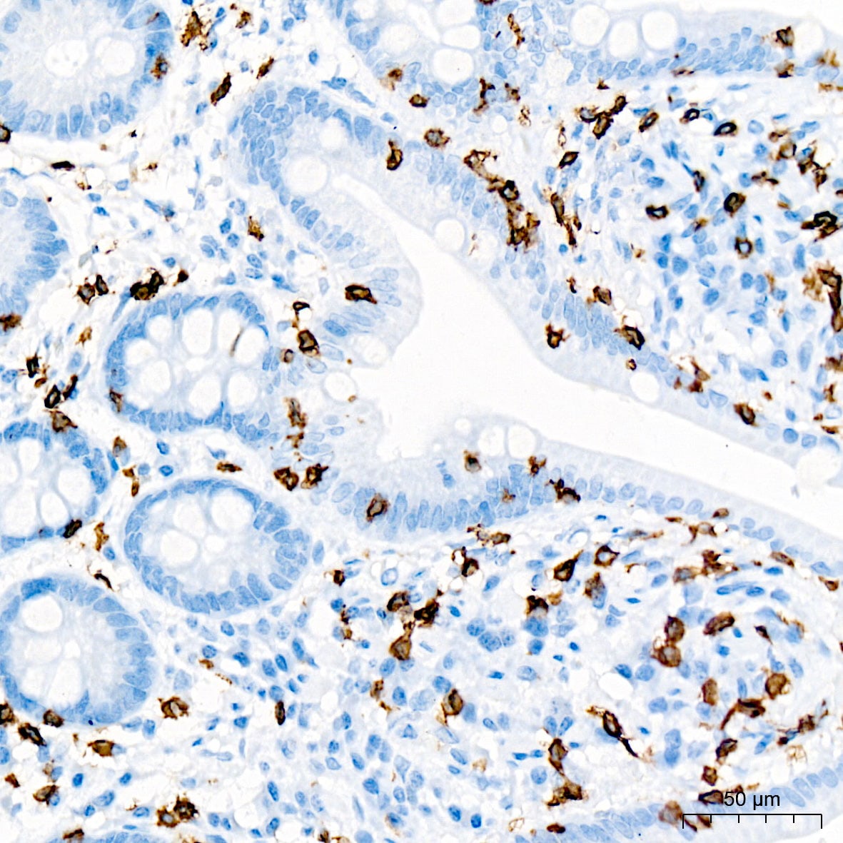
Immunohistochemistry-Paraffin: CD3 epsilon Antibody (6G6K9) [NBP3-57370]
Immunohistochemistry analysis of paraffin-embedded Human small intestine tissue using NBP3-57370 at a dilution of 1:2000 (40x lens). High pressure antigen retrieval was performed with 0.01 M citrate buffer (pH 6.0) prior to IHC staining.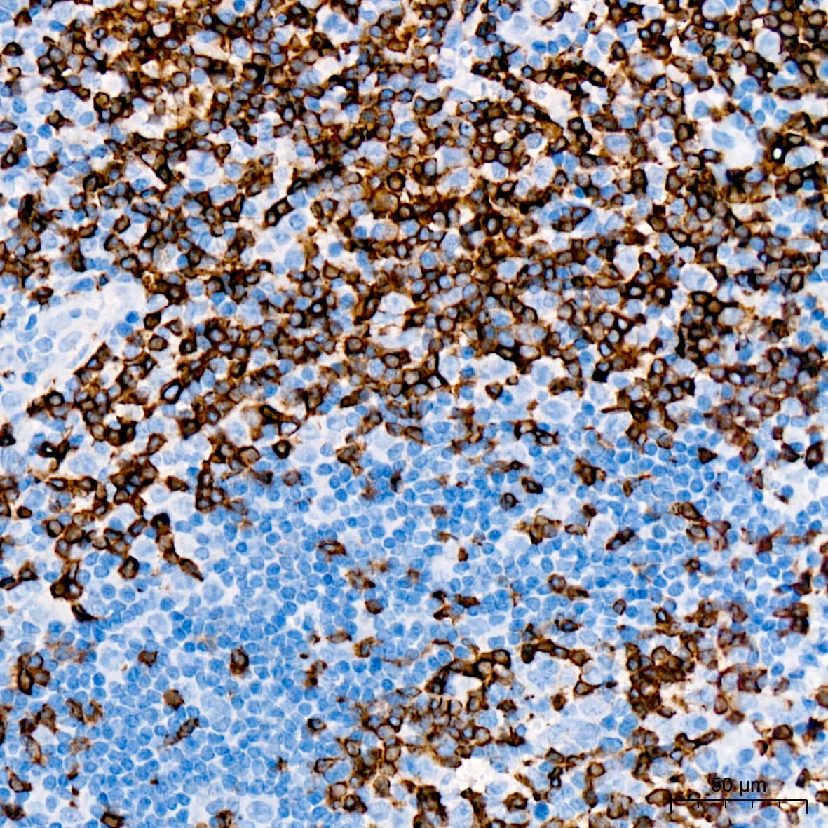
Immunohistochemistry-Paraffin: CD3 epsilon Antibody (6G6K9) [NBP3-57370]
Immunohistochemistry analysis of paraffin-embedded Human tonsil tissue using NBP3-57370 at a dilution of 1:2000 (40x lens). High pressure antigen retrieval was performed with 0.01 M citrate buffer (pH 6.0) prior to IHC staining.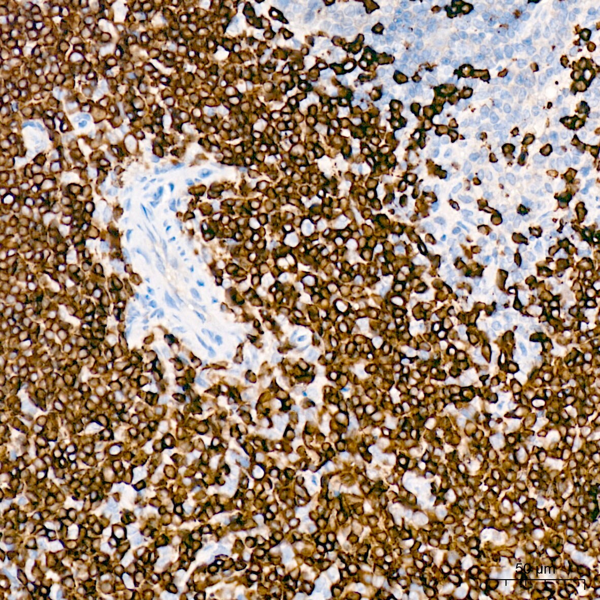
Immunohistochemistry-Paraffin: CD3 epsilon Antibody (6G6K9) [NBP3-57370]
Immunohistochemistry analysis of paraffin-embedded Mouse spleen tissue using NBP3-57370 at a dilution of 1:2000 (40x lens). High pressure antigen retrieval was performed with 0.01 M citrate buffer (pH 6.0) prior to IHC staining.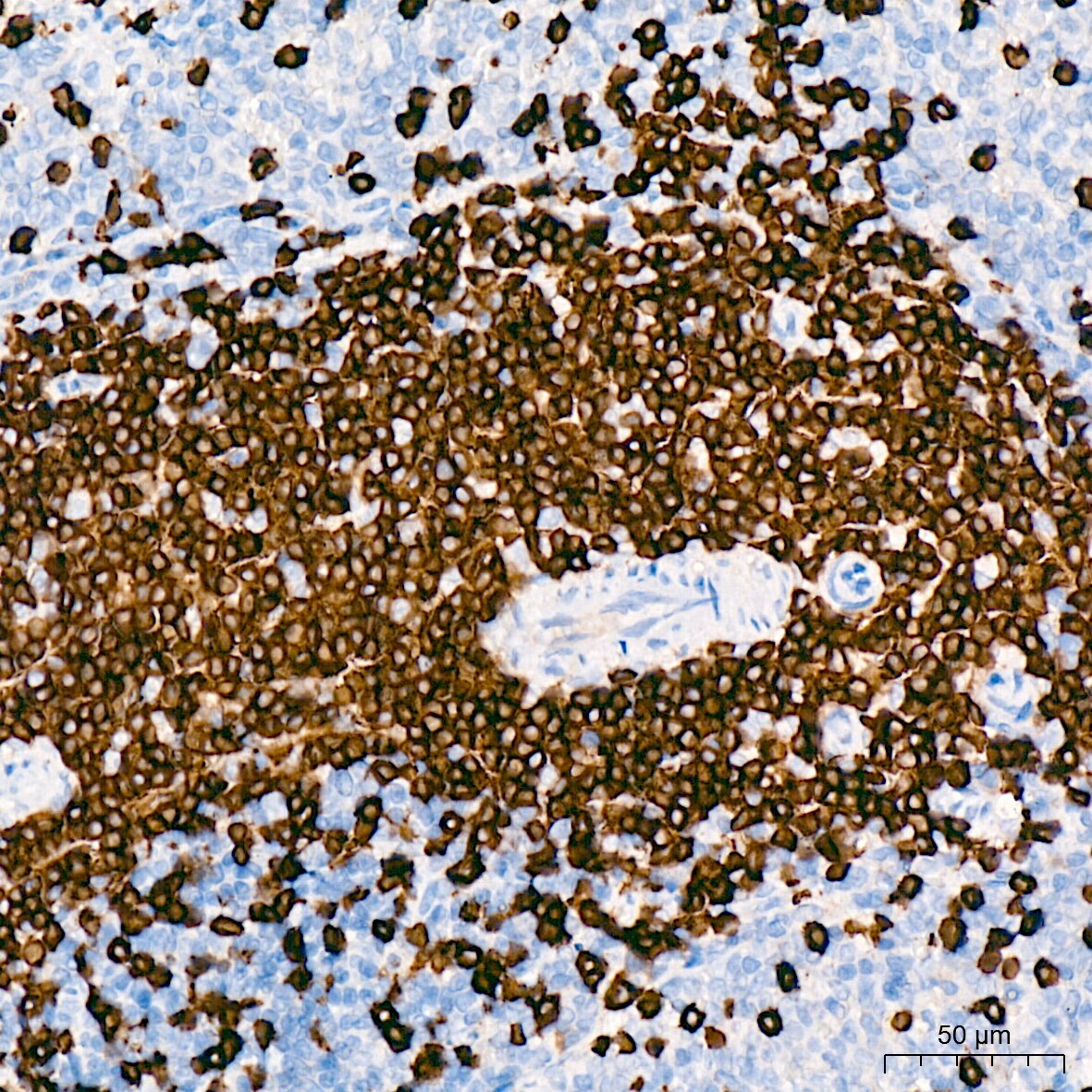
Immunohistochemistry-Paraffin: CD3 epsilon Antibody (6G6K9) [NBP3-57370]
Immunohistochemistry analysis of paraffin-embedded Rat spleen tissue using NBP3-57370 at a dilution of 1:2000 (40x lens). High pressure antigen retrieval was performed with 0.01 M citrate buffer (pH 6.0) prior to IHC staining.Applications for CD3 epsilon Antibody (6G6K9)
ELISA
Immunocytochemistry/ Immunofluorescence
Immunohistochemistry-Paraffin
Multiplex Immunoassay
Western Blot
Formulation, Preparation, and Storage
Purification
Formulation
Preservative
Concentration
Shipping
Stability & Storage
Background: CD3 epsilon
Alternate Names
Gene Symbol
Additional CD3 epsilon Products
Product Documents for CD3 epsilon Antibody (6G6K9)
Certificate of Analysis
To download a Certificate of Analysis, please enter a lot or batch number in the search box below.
Product Specific Notices for CD3 epsilon Antibody (6G6K9)
This product is for research use only and is not approved for use in humans or in clinical diagnosis. Primary Antibodies are guaranteed for 1 year from date of receipt.
Related Research Areas
Customer Reviews for CD3 epsilon Antibody (6G6K9)
There are currently no reviews for this product. Be the first to review CD3 epsilon Antibody (6G6K9) and earn rewards!
Have you used CD3 epsilon Antibody (6G6K9)?
Submit a review and receive an Amazon gift card!
$25/€18/£15/$25CAN/¥2500 for a review with an image
$10/€7/£6/$10CAN/¥1110 for a review without an image
Submit a review
Protocols
Find general support by application which include: protocols, troubleshooting, illustrated assays, videos and webinars.
- Antigen Retrieval Protocol (PIER)
- Antigen Retrieval for Frozen Sections Protocol
- Appropriate Fixation of IHC/ICC Samples
- Cellular Response to Hypoxia Protocols
- Chromogenic IHC Staining of Formalin-Fixed Paraffin-Embedded (FFPE) Tissue Protocol
- Chromogenic Immunohistochemistry Staining of Frozen Tissue
- ClariTSA™ Fluorophore Kits
- Detection & Visualization of Antibody Binding
- ELISA Sample Preparation & Collection Guide
- ELISA Troubleshooting Guide
- Fluorescent IHC Staining of Frozen Tissue Protocol
- Graphic Protocol for Heat-induced Epitope Retrieval
- Graphic Protocol for the Preparation and Fluorescent IHC Staining of Frozen Tissue Sections
- Graphic Protocol for the Preparation and Fluorescent IHC Staining of Paraffin-embedded Tissue Sections
- Graphic Protocol for the Preparation of Gelatin-coated Slides for Histological Tissue Sections
- How to Run an R&D Systems DuoSet ELISA
- How to Run an R&D Systems Quantikine ELISA
- How to Run an R&D Systems Quantikine™ QuicKit™ ELISA
- ICC Cell Smear Protocol for Suspension Cells
- ICC Immunocytochemistry Protocol Videos
- ICC for Adherent Cells
- IHC Sample Preparation (Frozen sections vs Paraffin)
- Immunocytochemistry (ICC) Protocol
- Immunocytochemistry Troubleshooting
- Immunofluorescence of Organoids Embedded in Cultrex Basement Membrane Extract
- Immunofluorescent IHC Staining of Formalin-Fixed Paraffin-Embedded (FFPE) Tissue Protocol
- Immunohistochemistry (IHC) and Immunocytochemistry (ICC) Protocols
- Immunohistochemistry Frozen Troubleshooting
- Immunohistochemistry Paraffin Troubleshooting
- Preparing Samples for IHC/ICC Experiments
- Preventing Non-Specific Staining (Non-Specific Binding)
- Primary Antibody Selection & Optimization
- Protocol for Heat-Induced Epitope Retrieval (HIER)
- Protocol for Making a 4% Formaldehyde Solution in PBS
- Protocol for VisUCyte™ HRP Polymer Detection Reagent
- Protocol for the Fluorescent ICC Staining of Cell Smears - Graphic
- Protocol for the Fluorescent ICC Staining of Cultured Cells on Coverslips - Graphic
- Protocol for the Preparation & Fixation of Cells on Coverslips
- Protocol for the Preparation and Chromogenic IHC Staining of Frozen Tissue Sections
- Protocol for the Preparation and Chromogenic IHC Staining of Frozen Tissue Sections - Graphic
- Protocol for the Preparation and Chromogenic IHC Staining of Paraffin-embedded Tissue Sections
- Protocol for the Preparation and Chromogenic IHC Staining of Paraffin-embedded Tissue Sections - Graphic
- Protocol for the Preparation and Fluorescent ICC Staining of Cells on Coverslips
- Protocol for the Preparation and Fluorescent ICC Staining of Non-adherent Cells
- Protocol for the Preparation and Fluorescent ICC Staining of Stem Cells on Coverslips
- Protocol for the Preparation and Fluorescent IHC Staining of Frozen Tissue Sections
- Protocol for the Preparation and Fluorescent IHC Staining of Paraffin-embedded Tissue Sections
- Protocol for the Preparation of Gelatin-coated Slides for Histological Tissue Sections
- Protocol for the Preparation of a Cell Smear for Non-adherent Cell ICC - Graphic
- Quantikine HS ELISA Kit Assay Principle, Alkaline Phosphatase
- Quantikine HS ELISA Kit Principle, Streptavidin-HRP Polymer
- R&D Systems Quality Control Western Blot Protocol
- Sandwich ELISA (Colorimetric) – Biotin/Streptavidin Detection Protocol
- Sandwich ELISA (Colorimetric) – Direct Detection Protocol
- TUNEL and Active Caspase-3 Detection by IHC/ICC Protocol
- The Importance of IHC/ICC Controls
- Troubleshooting Guide: ELISA
- Troubleshooting Guide: Immunohistochemistry
- Troubleshooting Guide: Western Blot Figures
- Western Blot Conditions
- Western Blot Protocol
- Western Blot Protocol for Cell Lysates
- Western Blot Troubleshooting
- Western Blot Troubleshooting Guide
- View all Protocols, Troubleshooting, Illustrated assays and Webinars