
CD68/SR-D1 Antibody (001) [DyLight 680]
Novus Biologicals | Catalog # NBP2-90914FR
Recombinant Monoclonal Antibody

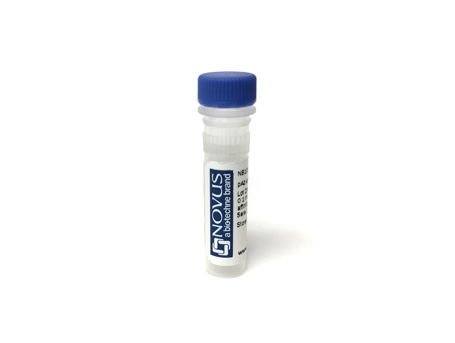
Key Product Details
Species Reactivity
Rat
Applications
Immunohistochemistry, Immunohistochemistry-Paraffin
Label
DyLight 680 (Excitation = 692 nm, Emission = 712 nm)
Antibody Source
Recombinant Monoclonal Rabbit IgG Clone # 001 expressed in HEK293
Loading...
Product Specifications
Immunogen
This antibody was obtained from a rabbit immunized with purified, recombinant Rat CD68/SR-D1 (Uniprot#: Q4FZY1; Met1-Ser295).
Clonality
Monoclonal
Host
Rabbit
Isotype
IgG
Description
This conjugate is made on demand and is not held in inventory. Each order represents a unique lot. All conjugates are generated using validated conjugation protocols and meet strict degree of labeling (F/P) specifications; additional information is available upon request. The antibody concentration is provided on the individual product label.
For specialized conjugation needs, like large quantities, specific concentrations, and defined F/P ratios, bulk and custom antibody conjugation services are available.
For specialized conjugation needs, like large quantities, specific concentrations, and defined F/P ratios, bulk and custom antibody conjugation services are available.
Applications for CD68/SR-D1 Antibody (001) [DyLight 680]
Application
Recommended Usage
Immunohistochemistry
Optimal dilutions of this antibody should be experimentally determined.
Immunohistochemistry-Paraffin
Optimal dilutions of this antibody should be experimentally determined.
Application Notes
Recommended applications are based on validated applications from the unconjugated base product NBP2-90914. This conjugated antibody is not kept in inventory and is made to order using validated conjugation protocols.
Formulation, Preparation, and Storage
Purification
Protein A purified
Formulation
50mM Sodium Borate
Preservative
0.05% Sodium Azide
Concentration
Please see the vial label for concentration. If unlisted please contact technical services.
Shipping
The product is shipped with polar packs. Upon receipt, store it immediately at the temperature recommended below.
Stability & Storage
Store at 4C in the dark.
Background: CD68/SR-D1
CD68 is highly expressed in cells of the mononuclear phagocyte system such as macrophages, microglia, osteoclasts, and myeloid dendritic cells (DCs); and is expressed to a lesser extent in lymphoid cells (CD19+ B lymphocytes and CD4+ T lymphocytes), human umbilical cord mesenchymal stem cells (MSCs), fibroblasts, endothelial cells, multiple non-hematopoietic cancer cell lines, and human arterial intimal smooth muscle cells (SMCs). Expression has been also observed in diseased states for granulocytes and neutrophils, in particular basophils from myeloproliferative disorders and intestinal neutrophils from inflammatory bowel disease (IBD), respectively (1).
Although the function of CD68 has yet to be established, it has often been used as an immunohistochemistry (IHC) marker of inflammation and for granular cell tumors (GCTs). CD68+ tumor associated macrophages (TAMs) has been suggested to be a predictive marker for poor cancer prognosis, but a meta-analysis showed the presence of CD68 is not correlated with survival (2). In addition, a role in hepatic malaria infection has been reported based on the finding that peptide P39 binds CD68, considered a receptor for malaria sporozoite, and inhibits parasite entry into Kupffer cells. CD68 was deemed a member of the Scavenger receptor family due to its upregulation in macrophages following inflammatory stimuli, ability to bind modified LDL, phosphatidylserine, and apoptotic cells, as well as shuttling between the plasma membrane and endosomes. CD68 has been linked to atherogenesis based on binding and internalization of its ligand, oxLDL (1).
References
1. Chistiakov, DA, Killingsworth, MC, Myasoedova, VA. Orekhov AN, Bobryshev YV. (2017) CD68/macrosialin: not just a histochemical marker. Lab Invest. 97:4-13. PMID: 27869795
2. Troiano G, Caponio VCA, Adipietro I, Tepedino M, Santoro R, Laino L, Lo Russo L, Cirillo N, Lo Muzio L. (2019) Prognostic significance of CD68+ and CD163+ tumor associated macrophages in head and neck squamous cell carcinoma: A systematic review and meta-analysis. Oral Oncol. 93:66-75. PMID: 31109698.
Alternate Names
CD68, gp110, Macrosialin, SCARD1, SR-D1, SRD1
Gene Symbol
CD68
Additional CD68/SR-D1 Products
Product Documents for CD68/SR-D1 Antibody (001) [DyLight 680]
Certificate of Analysis
To download a Certificate of Analysis, please enter a lot or batch number in the search box below.
Product Specific Notices for CD68/SR-D1 Antibody (001) [DyLight 680]
DyLight (R) is a trademark of Thermo Fisher Scientific Inc. and its subsidiaries.
This product is for research use only and is not approved for use in humans or in clinical diagnosis. Primary Antibodies are guaranteed for 1 year from date of receipt.
Customer Reviews for CD68/SR-D1 Antibody (001) [DyLight 680]
There are currently no reviews for this product. Be the first to review CD68/SR-D1 Antibody (001) [DyLight 680] and earn rewards!
Have you used CD68/SR-D1 Antibody (001) [DyLight 680]?
Submit a review and receive an Amazon gift card!
$25/€18/£15/$25CAN/¥2500 for a review with an image
$10/€7/£6/$10CAN/¥1110 for a review without an image
Submit a review
Protocols
Find general support by application which include: protocols, troubleshooting, illustrated assays, videos and webinars.
- Antigen Retrieval Protocol (PIER)
- Antigen Retrieval for Frozen Sections Protocol
- Appropriate Fixation of IHC/ICC Samples
- Cellular Response to Hypoxia Protocols
- Chromogenic IHC Staining of Formalin-Fixed Paraffin-Embedded (FFPE) Tissue Protocol
- Chromogenic Immunohistochemistry Staining of Frozen Tissue
- ClariTSA™ Fluorophore Kits
- Detection & Visualization of Antibody Binding
- Fluorescent IHC Staining of Frozen Tissue Protocol
- Graphic Protocol for Heat-induced Epitope Retrieval
- Graphic Protocol for the Preparation and Fluorescent IHC Staining of Frozen Tissue Sections
- Graphic Protocol for the Preparation and Fluorescent IHC Staining of Paraffin-embedded Tissue Sections
- Graphic Protocol for the Preparation of Gelatin-coated Slides for Histological Tissue Sections
- IHC Sample Preparation (Frozen sections vs Paraffin)
- Immunofluorescent IHC Staining of Formalin-Fixed Paraffin-Embedded (FFPE) Tissue Protocol
- Immunohistochemistry (IHC) and Immunocytochemistry (ICC) Protocols
- Immunohistochemistry Frozen Troubleshooting
- Immunohistochemistry Paraffin Troubleshooting
- Preparing Samples for IHC/ICC Experiments
- Preventing Non-Specific Staining (Non-Specific Binding)
- Primary Antibody Selection & Optimization
- Protocol for Heat-Induced Epitope Retrieval (HIER)
- Protocol for Making a 4% Formaldehyde Solution in PBS
- Protocol for VisUCyte™ HRP Polymer Detection Reagent
- Protocol for the Preparation & Fixation of Cells on Coverslips
- Protocol for the Preparation and Chromogenic IHC Staining of Frozen Tissue Sections
- Protocol for the Preparation and Chromogenic IHC Staining of Frozen Tissue Sections - Graphic
- Protocol for the Preparation and Chromogenic IHC Staining of Paraffin-embedded Tissue Sections
- Protocol for the Preparation and Chromogenic IHC Staining of Paraffin-embedded Tissue Sections - Graphic
- Protocol for the Preparation and Fluorescent IHC Staining of Frozen Tissue Sections
- Protocol for the Preparation and Fluorescent IHC Staining of Paraffin-embedded Tissue Sections
- Protocol for the Preparation of Gelatin-coated Slides for Histological Tissue Sections
- TUNEL and Active Caspase-3 Detection by IHC/ICC Protocol
- The Importance of IHC/ICC Controls
- Troubleshooting Guide: Immunohistochemistry
- View all Protocols, Troubleshooting, Illustrated assays and Webinars
Loading...
Associated Pathways
