CD8 Antibody (12.C7) - BSA Free
Novus Biologicals | Catalog # NB100-64021
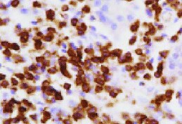
![Immunohistochemistry: CD8 Antibody (12.C7) - BSA Free [NB100-64021]](https://resources.rndsystems.com/images/products/CD8-Antibody-12-C7-Immunohistochemistry-NB100-64021-img0002.jpg "Immunohistochemistry: CD8 Antibody (12.C7) - BSA Free [NB100-64021]")
Key Product Details
Validated by
Biological Validation
Species Reactivity
Validated:
Rabbit
Cited:
Rabbit
Applications
Validated:
Immunohistochemistry, Immunohistochemistry-Paraffin, Flow Cytometry, Immunocytochemistry/ Immunofluorescence
Cited:
Immunohistochemistry-Paraffin, Flow Cytometry, Immunocytochemistry/ Immunofluorescence, IF/IHC
Label
Unconjugated
Antibody Source
Monoclonal Mouse IgG1 Clone # 12.C7
Format
BSA Free
Loading...
Product Specifications
Immunogen
CD8 Antibody (12.C7) was developed against Rabbit CD8
Specificity
CD8 Antibody (12.C7) recognizes the rabbit CD8 cell surface antigen, expressed by a subset of T lymphocytes with cytotoxic/suppressor activity.
Clonality
Monoclonal
Host
Mouse
Isotype
IgG1
Theoretical MW
26 kDa.
Disclaimer note: The observed molecular weight of the protein may vary from the listed predicted molecular weight due to post translational modifications, post translation cleavages, relative charges, and other experimental factors.
Disclaimer note: The observed molecular weight of the protein may vary from the listed predicted molecular weight due to post translational modifications, post translation cleavages, relative charges, and other experimental factors.
Scientific Data Images for CD8 Antibody (12.C7) - BSA Free
![Immunohistochemistry-Paraffin: CD8 Antibody (12.C7) - BSA Free [NB100-64021]](https://resources.rndsystems.com/images/products/CD8-Antibody-12-C7-Immunohistochemistry-Paraffin-NB100-64021-img0003.jpg "Immunohistochemistry-Paraffin: CD8 Antibody (12.C7) - BSA Free [NB100-64021]")
Immunohistochemistry-Paraffin: CD8 Antibody (12.C7) - BSA Free [NB100-64021]
Immunohistochemistry-Paraffin: CD8 Antibody (12.C7) [NB100-64021] - Human bile duct cancer tissue. IHC-P image submitted by a verified customer review.![Flow Cytometry: CD8 Antibody (12.C7) - BSA Free [NB100-64021]](https://resources.rndsystems.com/images/products/CD8-alpha-Antibody-12-C7-Flow-Cytometry-NB100-64021-img0001.jpg "Flow Cytometry: CD8 Antibody (12.C7) - BSA Free [NB100-64021]")
Flow Cytometry: CD8 Antibody (12.C7) - BSA Free [NB100-64021]
Flow Cytometry: CD8 alpha Antibody (12.C7) [NB100-64021] - Rabbit peripheral blood lymphocytes stained with Mouse anti Rabbit CD8 Alpha followed by Goat anti Mouse IgG (H/L):FITC - BSA Free [NB100-64021] -")
Immunohistochemistry: CD8 Antibody (12.C7) - BSA Free [NB100-64021] -
scAAV8G9-optHLA-G Combo Prevents Cornea Burn-induced Vascularization and Cytotoxic T-cell Infiltration. Rabbit cornea sections were acquired 60 days following the injection of indicated vectors into burn corneas and stained for an endothelial cell marker (CD31), T cell markers, transgene abundance, and alpha SMA in the indicated treatment groups. Scale bars = (A) 10 µm, (B) 5 µm, (C) 20 µm, (D) 200 µm. - BSA Free [NB100-64021] -")
Immunocytochemistry/ Immunofluorescence: CD8 Antibody (12.C7) - BSA Free [NB100-64021] -
scAAV8G9-optHLA-G Combo Prevents Cornea Burn-induced Vascularization and Cytotoxic T-cell Infiltration. Rabbit cornea sections were acquired 60 days following the injection of indicated vectors into burn corneas and stained for an endothelial cell marker (CD31), T cell markers, transgene abundance, and alpha SMA in the indicated treatment groups. Scale bars = (A) 10 um, (B) 5 um, (C) 20 um, (D) 200 um. Image collected and cropped by CiteAb from the following open publication (https://pubmed.ncbi.nlm.nih.gov/29259248), licensed under a CC-BY license. Not internally tested by Novus Biologicals.Applications for CD8 Antibody (12.C7) - BSA Free
Application
Recommended Usage
Flow Cytometry
1:100-1:200
Immunocytochemistry/ Immunofluorescence
1:10-1:500
Immunohistochemistry
1:10-1:500
Immunohistochemistry-Paraffin
1:10-1:500
Application Notes
Succerssful use in ICC/IF, IHC reported in scientific literature (PMID: 22796166).
Reviewed Applications
Read 2 reviews rated 5 using NB100-64021 in the following applications:
Flow Cytometry Panel Builder
Bio-Techne Knows Flow Cytometry
Save time and reduce costly mistakes by quickly finding compatible reagents using the Panel Builder Tool.
Advanced Features
- Spectra Viewer - Custom analysis of spectra from multiple fluorochromes
- Spillover Popups - Visualize the spectra of individual fluorochromes
- Antigen Density Selector - Match fluorochrome brightness with antigen density

Formulation, Preparation, and Storage
Purification
Protein A purified
Formulation
PBS
Format
BSA Free
Preservative
0.09% Sodium Azide
Concentration
1.0 mg/ml
Shipping
The product is shipped with polar packs. Upon receipt, store it immediately at the temperature recommended below.
Stability & Storage
Store at 4C short term. Aliquot and store at -20C long term. Avoid freeze-thaw cycles.
Background: CD8
Given its role in the immune system, CD8-deficiency in T-cells is a hallmark of many diseases and pathologies (8-10). Specifically, CD8+ T-cell deficiency is prevalent in chronic autoimmune diseases including multiple sclerosis, rheumatoid arthritis, ulcerative colitis, Crohn's disease, type 1 diabetes mellitus, and Graves' disease (8). Furthermore, cancers or chronic infection can lead to CD8 T-cell exhaustion as the continual antigen presentation and inflammatory signals eventually cause the CD8+ T-cells to lose functionality (9, 10). However, animal models and clinical studies have suggested that T-cells are capable of being reinvigorated using inhibitory receptor blockade resulting in better disease outcomes and these exhausted T-cells may be a potential therapeutic target (9, 10).
Alternative names for CD8 includes CD antigen: CD8a, CD8 antigen, alpha polypeptide (p32), CD8a molecule, CD8A, Leu2 T-lymphocyte antigen, LEU2, MAL, OKT8 T-cell antigen, p32, T cell co-receptor, T8 T-cell antigen, T-cell antigen Leu2, T-cell surface glycoprotein CD8 alpha chain, and T-lymphocyte differentiation antigen T8/Leu-2.
References
1. Littman D. R. (1987). The structure of the CD4 and CD8 genes. Annual review of immunology. https://doi.org/10.1146/annurev.iy.05.040187.003021
2. Naeim F. (2008). Chapter 2- Principles of Immunophenotyping. Hematopathology. https://doi.org/10.1016/B978-0-12-370607-2.00002-8.
3. Gao, G. F., & Jakobsen, B. K. (2000). Molecular interactions of coreceptor CD8 and MHC class I: the molecular basis for functional coordination with the T-cell receptor. Immunology today. https://doi.org/10.1016/s0167-5699(00)01750-3
4. UniProt (P01732)
5. UniProt (P01731)
6. Kappes D. J. (2007). CD4 and CD8: hogging all the Lck. Immunity. https://doi.org/10.1016/j.immuni.2007.11.002
7. Gangadharan, D., & Cheroutre, H. (2004). The CD8 isoform CD8alphaalpha is not a functional homologue of the TCR co-receptor CD8alphabeta. Current opinion in immunology. https://doi.org/10.1016/j.coi.2004.03.015
8. Pender M. P. (2012). CD8+ T-Cell Deficiency, Epstein-Barr Virus Infection, Vitamin D Deficiency, and Steps to Autoimmunity: A Unifying Hypothesis. Autoimmune diseases. https://doi.org/10.1155/2012/189096
9. Kurachi M. (2019). CD8+ T cell exhaustion. Seminars in immunopathology. https://doi.org/10.1007/s00281-019-00744-5
10. Hashimoto, M., Kamphorst, A. O., Im, S. J., Kissick, H. T., Pillai, R. N., Ramalingam, S. S., Araki, K., & Ahmed, R. (2018). CD8 T Cell Exhaustion in Chronic Infection and Cancer: Opportunities for Interventions. Annual review of medicine. https://doi.org/10.1146/annurev-med-012017-043208
Alternate Names
CD8, CD8A
Gene Symbol
CD8A
Additional CD8 Products
Product Documents for CD8 Antibody (12.C7) - BSA Free
Certificate of Analysis
To download a Certificate of Analysis, please enter a lot or batch number in the search box below.
Product Specific Notices for CD8 Antibody (12.C7) - BSA Free
This product is for research use only and is not approved for use in humans or in clinical diagnosis. Primary Antibodies are guaranteed for 1 year from date of receipt.
Citations for CD8 Antibody (12.C7) - BSA Free
 Powered by Bioz
Customer Reviews for CD8 Antibody (12.C7) - BSA Free (2)
5 out of 5
2 Customer Ratings
Have you used CD8 Antibody (12.C7) - BSA Free?
Submit a review and receive an Amazon gift card!
$25/€18/£15/$25CAN/¥2500 Yen for a review with an image
$10/€7/£6/$10CAN/¥1110 Yen for a review without an image
Submit a review
Customer Images
Showing
1
-
2 的
2 reviews
Showing All
Filter By:
-
Application: Immunohistochemistry-ParaffinSample Tested: Bile duct cancerSpecies: HumanVerified Customer | Posted 11/30/2021Bile duct cancer tissue

-
Application: Immunohistochemistry-ParaffinSample Tested: Rabbit tissueSpecies: OtherVerified Customer | Posted 12/27/2011
There are no reviews that match your criteria.
Protocols
Find general support by application which include: protocols, troubleshooting, illustrated assays, videos and webinars.
- 7-Amino Actinomycin D (7-AAD) Cell Viability Flow Cytometry Protocol
- Antigen Retrieval Protocol (PIER)
- Antigen Retrieval for Frozen Sections Protocol
- Appropriate Fixation of IHC/ICC Samples
- Cellular Response to Hypoxia Protocols
- Chromogenic IHC Staining of Formalin-Fixed Paraffin-Embedded (FFPE) Tissue Protocol
- Chromogenic Immunohistochemistry Staining of Frozen Tissue
- ClariTSA™ Fluorophore Kits
- Detection & Visualization of Antibody Binding
- Extracellular Membrane Flow Cytometry Protocol
- Flow Cytometry Protocol for Cell Surface Markers
- Flow Cytometry Protocol for Staining Membrane Associated Proteins
- Flow Cytometry Staining Protocols
- Flow Cytometry Troubleshooting Guide
- Fluorescent IHC Staining of Frozen Tissue Protocol
- Graphic Protocol for Heat-induced Epitope Retrieval
- Graphic Protocol for the Preparation and Fluorescent IHC Staining of Frozen Tissue Sections
- Graphic Protocol for the Preparation and Fluorescent IHC Staining of Paraffin-embedded Tissue Sections
- Graphic Protocol for the Preparation of Gelatin-coated Slides for Histological Tissue Sections
- ICC Cell Smear Protocol for Suspension Cells
- ICC Immunocytochemistry Protocol Videos
- ICC for Adherent Cells
- IHC Sample Preparation (Frozen sections vs Paraffin)
- Immunocytochemistry (ICC) Protocol
- Immunocytochemistry Troubleshooting
- Immunofluorescence of Organoids Embedded in Cultrex Basement Membrane Extract
- Immunofluorescent IHC Staining of Formalin-Fixed Paraffin-Embedded (FFPE) Tissue Protocol
- Immunohistochemistry (IHC) and Immunocytochemistry (ICC) Protocols
- Immunohistochemistry Frozen Troubleshooting
- Immunohistochemistry Paraffin Troubleshooting
- Intracellular Flow Cytometry Protocol Using Alcohol (Methanol)
- Intracellular Flow Cytometry Protocol Using Detergents
- Intracellular Nuclear Staining Flow Cytometry Protocol Using Detergents
- Intracellular Staining Flow Cytometry Protocol Using Alcohol Permeabilization
- Intracellular Staining Flow Cytometry Protocol Using Detergents to Permeabilize Cells
- Preparing Samples for IHC/ICC Experiments
- Preventing Non-Specific Staining (Non-Specific Binding)
- Primary Antibody Selection & Optimization
- Propidium Iodide Cell Viability Flow Cytometry Protocol
- Protocol for Heat-Induced Epitope Retrieval (HIER)
- Protocol for Liperfluo
- Protocol for Making a 4% Formaldehyde Solution in PBS
- Protocol for VisUCyte™ HRP Polymer Detection Reagent
- Protocol for the Characterization of Human Th22 Cells
- Protocol for the Characterization of Human Th9 Cells
- Protocol for the Fluorescent ICC Staining of Cell Smears - Graphic
- Protocol for the Fluorescent ICC Staining of Cultured Cells on Coverslips - Graphic
- Protocol for the Preparation & Fixation of Cells on Coverslips
- Protocol for the Preparation and Chromogenic IHC Staining of Frozen Tissue Sections
- Protocol for the Preparation and Chromogenic IHC Staining of Frozen Tissue Sections - Graphic
- Protocol for the Preparation and Chromogenic IHC Staining of Paraffin-embedded Tissue Sections
- Protocol for the Preparation and Chromogenic IHC Staining of Paraffin-embedded Tissue Sections - Graphic
- Protocol for the Preparation and Fluorescent ICC Staining of Cells on Coverslips
- Protocol for the Preparation and Fluorescent ICC Staining of Non-adherent Cells
- Protocol for the Preparation and Fluorescent ICC Staining of Stem Cells on Coverslips
- Protocol for the Preparation and Fluorescent IHC Staining of Frozen Tissue Sections
- Protocol for the Preparation and Fluorescent IHC Staining of Paraffin-embedded Tissue Sections
- Protocol for the Preparation of Gelatin-coated Slides for Histological Tissue Sections
- Protocol for the Preparation of a Cell Smear for Non-adherent Cell ICC - Graphic
- Protocol: Annexin V and PI Staining by Flow Cytometry
- Protocol: Annexin V and PI Staining for Apoptosis by Flow Cytometry
- TUNEL and Active Caspase-3 Detection by IHC/ICC Protocol
- The Importance of IHC/ICC Controls
- Troubleshooting Guide: Fluorokine Flow Cytometry Kits
- Troubleshooting Guide: Immunohistochemistry
- View all Protocols, Troubleshooting, Illustrated assays and Webinars


