CHCHD3 Antibody - BSA Free
Novus Biologicals | Catalog # NBP1-83656
![Immunocytochemistry/ Immunofluorescence: CHCHD3 Antibody [NBP1-83656]](https://resources.rndsystems.com/images/products/CHCHD3-Antibody-Immunocytochemistry-Immunofluorescence-NBP1-83656-img0011.jpg "Immunocytochemistry/ Immunofluorescence: CHCHD3 Antibody [NBP1-83656]")
Loading...
Key Product Details
Validated by
Knockout/Knockdown
Species Reactivity
Validated:
Human, Mouse, Rat
Cited:
Human
Applications
Validated:
Immunohistochemistry, Immunohistochemistry-Paraffin, Western Blot, Immunocytochemistry/ Immunofluorescence, Knockdown Validated
Cited:
Western Blot
Label
Unconjugated
Antibody Source
Polyclonal Rabbit IgG
Format
BSA Free
Loading...
Product Specifications
Immunogen
This antibody was developed against Recombinant Protein corresponding to amino acids: DENENITVVKGIRLSENVIDRMKESSPSGSKSQRYSGAYGASVSDEELKRRVAEELALEQAKKESEDQKRLKQAK
Clonality
Polyclonal
Host
Rabbit
Isotype
IgG
Scientific Data Images for CHCHD3 Antibody - BSA Free
Immunocytochemistry/ Immunofluorescence: CHCHD3 Antibody [NBP1-83656]
Immunocytochemistry/Immunofluorescence: CHCHD3 Antibody [NBP1-83656] - Staining of human cell line U-2 OS shows localization to mitochondria. Antibody staining is shown in green.![Immunohistochemistry-Paraffin: CHCHD3 Antibody [NBP1-83656]](https://resources.rndsystems.com/images/products/CHCHD3-Antibody-Immunohistochemistry-Paraffin-NBP1-83656-img0015.jpg "Immunohistochemistry-Paraffin: CHCHD3 Antibody [NBP1-83656]")
Immunohistochemistry-Paraffin: CHCHD3 Antibody [NBP1-83656]
Immunohistochemistry-Paraffin: CHCHD3 Antibody [NBP1-83656] - Staining of human skeletal muscle shows strong granular cytoplasmic positivity in myocytes.![Western Blot: CHCHD3 Antibody [NBP1-83656]](https://resources.rndsystems.com/images/products/CHCHD3-Antibody-Western-Blot-NBP1-83656-img0010.jpg "Western Blot: CHCHD3 Antibody [NBP1-83656]")
Western Blot: CHCHD3 Antibody [NBP1-83656]
Western Blot: CHCHD3 Antibody [NBP1-83656] - Analysis in mouse cell line NIH-3T3 and rat cell line NBT-II.![Immunohistochemistry-Paraffin: CHCHD3 Antibody [NBP1-83656]](https://resources.rndsystems.com/images/products/CHCHD3-Antibody-Immunohistochemistry-Paraffin-NBP1-83656-img0012.jpg "Immunohistochemistry-Paraffin: CHCHD3 Antibody [NBP1-83656]")
Immunohistochemistry-Paraffin: CHCHD3 Antibody [NBP1-83656]
Immunohistochemistry-Paraffin: CHCHD3 Antibody [NBP1-83656] - Staining of human duodenum shows strong granular cytoplasmic positivity in glandular cells.![Immunohistochemistry-Paraffin: CHCHD3 Antibody [NBP1-83656]](https://resources.rndsystems.com/images/products/CHCHD3-Antibody-Immunohistochemistry-Paraffin-NBP1-83656-img0013.jpg "Immunohistochemistry-Paraffin: CHCHD3 Antibody [NBP1-83656]")
Immunohistochemistry-Paraffin: CHCHD3 Antibody [NBP1-83656]
Immunohistochemistry-Paraffin: CHCHD3 Antibody [NBP1-83656] - Staining of human kidney shows strong granular cytoplasmic positivity in cells in tubules.![Immunohistochemistry-Paraffin: CHCHD3 Antibody [NBP1-83656]](https://resources.rndsystems.com/images/products/CHCHD3-Antibody-Immunohistochemistry-Paraffin-NBP1-83656-img0014.jpg "Immunohistochemistry-Paraffin: CHCHD3 Antibody [NBP1-83656]")
Immunohistochemistry-Paraffin: CHCHD3 Antibody [NBP1-83656]
Immunohistochemistry-Paraffin: CHCHD3 Antibody [NBP1-83656] - Staining of human liver shows moderate granular cytoplasmic positivity in hepatocytes.![CHCHD3 Antibody - BSA Free Western Blot: CHCHD3 Antibody - BSA Free [NBP1-83656]](https://resources.rndsystems.com/images/products/nbp1-83656_rabbit-polyclonal-chchd3-antibody-84202517254737.jpg "Western Blot: CHCHD3 Antibody - BSA Free [NBP1-83656]")
![CHCHD3 Antibody - BSA Free Western Blot: CHCHD3 Antibody - BSA Free [NBP1-83656]](https://resources.rndsystems.com/images/products/nbp1-83656_rabbit-polyclonal-chchd3-antibody-84202517195657.jpg "Western Blot: CHCHD3 Antibody - BSA Free [NBP1-83656]")
Western Blot: CHCHD3 Antibody - BSA Free [NBP1-83656]
Analysis in human cell line HEK 293.![CHCHD3 Antibody - BSA Free Western Blot: CHCHD3 Antibody - BSA Free [NBP1-83656]](https://resources.rndsystems.com/images/products/nbp1-83656_rabbit-polyclonal-chchd3-antibody-8420251720240.jpg "Western Blot: CHCHD3 Antibody - BSA Free [NBP1-83656]")
Western Blot: CHCHD3 Antibody - BSA Free [NBP1-83656]
Analysis in mouse cell line NIH-3T3 and rat cell line NBT-II.
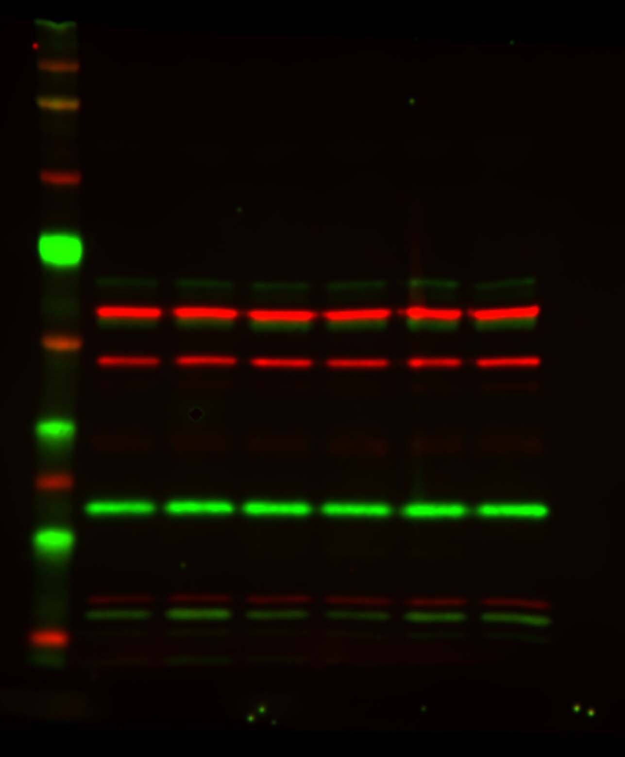
Western Blot: CHCHD3 Antibody - BSA Free [NBP1-83656] -
Mitochondrial targeting of mutant renin isoforms. (A) Western blot analysis showing renin expression in the lysates and conditioned media of transiently transfected Calu-6 cells. GAPDH is shown as a loading control. The presence of a non-glycosylated renin isoform (red asterisk) can be seen for all mutants. (B) Cell lysates were deglycosylated with PNGase F. The lower band indicated in panel A with the asterisk is not sensitive to PNGase F treatment, indicating the absence of N-glycans. (C) Immunofluorescence analysis in Calu-6 cells showing merged pictures of renin (red) and TIM44 (marker of mitochondria, green) with nuclei (DAPI, blue). Scale bar: 20 μm. Colocalisation of renin and mitochondria can be assessed by the presence of merged yellow signal. The graph below shows Pearson's correlation coefficient between signals of TIM44 and renin. *P<0.05; **P<0.01; ***P<0.001; ****P<0.0001; two-way ANOVA with Bonferroni post hoc test versus WT. Data are shown as mean+/-s.e.m. (n=3 independent experiments). (D) Protease protection assay performed on isolated mitochondria from HEK293 cells expressing p.L16R or p.W17R mutant isoforms. Western blot experiments showed digestion of renin isoforms by trypsin only when the inner membrane was permeabilised. This result suggests that mutant isoforms are imported within the mitochondrial matrix. Images are representative of three independent experiments. Image collected and cropped by CiteAb from the following open publication (https://pubmed.ncbi.nlm.nih.gov/37283036), licensed under a CC-BY license. Not internally tested by Novus Biologicals.Applications for CHCHD3 Antibody - BSA Free
Application
Recommended Usage
Immunocytochemistry/ Immunofluorescence
0.25-2 ug/ml
Immunohistochemistry
1:500 - 1:1000
Immunohistochemistry-Paraffin
1:500 - 1:1000
Western Blot
0.04-0.4 ug/ml
Application Notes
For IHC-Paraffin, HIER pH 6 retrieval is recommended. ICC/IF Fixation Permeabilization, Use PFA/Triton X-100.
Reviewed Applications
Read 1 review rated 4 using NBP1-83656 in the following applications:
Formulation, Preparation, and Storage
Purification
Affinity purified
Formulation
PBS (pH 7.2) and 40% Glycerol
Format
BSA Free
Preservative
0.02% Sodium Azide
Concentration
Concentrations vary lot to lot. See vial label for concentration. If unlisted please contact technical services.
Shipping
The product is shipped with polar packs. Upon receipt, store it immediately at the temperature recommended below.
Stability & Storage
Store at 4C short term. Aliquot and store at -20C long term. Avoid freeze-thaw cycles.
Background: CHCHD3
Alternate Names
coiled-coil-helix-coiled-coil-helix domain containing 3, FLJ20420, mitochondrial
Gene Symbol
CHCHD3
Additional CHCHD3 Products
Product Documents for CHCHD3 Antibody - BSA Free
Certificate of Analysis
To download a Certificate of Analysis, please enter a lot or batch number in the search box below.
Product Specific Notices for CHCHD3 Antibody - BSA Free
This product is for research use only and is not approved for use in humans or in clinical diagnosis. Primary Antibodies are guaranteed for 1 year from date of receipt.
Citations for CHCHD3 Antibody - BSA Free
 Powered by Bioz
Customer Reviews for CHCHD3 Antibody - BSA Free (1)
4 out of 5
1 Customer Rating
Have you used CHCHD3 Antibody - BSA Free?
Submit a review and receive an Amazon gift card!
$25/€18/£15/$25CAN/¥2500 Yen for a review with an image
$10/€7/£6/$10CAN/¥1110 Yen for a review without an image
Submit a review
Customer Images
Showing
1
-
1 的
1 review
Showing All
Filter By:
-
Application: Western BlotSample Tested: Protein lysate from mitochondria isolated from SH-SY5Y cellsSpecies: HumanVerified Customer | Posted 06/15/2016

There are no reviews that match your criteria.
Protocols
Find general support by application which include: protocols, troubleshooting, illustrated assays, videos and webinars.
- Antigen Retrieval Protocol (PIER)
- Antigen Retrieval for Frozen Sections Protocol
- Appropriate Fixation of IHC/ICC Samples
- Cellular Response to Hypoxia Protocols
- Chromogenic IHC Staining of Formalin-Fixed Paraffin-Embedded (FFPE) Tissue Protocol
- Chromogenic Immunohistochemistry Staining of Frozen Tissue
- ClariTSA™ Fluorophore Kits
- Detection & Visualization of Antibody Binding
- Fluorescent IHC Staining of Frozen Tissue Protocol
- Graphic Protocol for Heat-induced Epitope Retrieval
- Graphic Protocol for the Preparation and Fluorescent IHC Staining of Frozen Tissue Sections
- Graphic Protocol for the Preparation and Fluorescent IHC Staining of Paraffin-embedded Tissue Sections
- Graphic Protocol for the Preparation of Gelatin-coated Slides for Histological Tissue Sections
- ICC Cell Smear Protocol for Suspension Cells
- ICC Immunocytochemistry Protocol Videos
- ICC for Adherent Cells
- IHC Sample Preparation (Frozen sections vs Paraffin)
- Immunocytochemistry (ICC) Protocol
- Immunocytochemistry Troubleshooting
- Immunofluorescence of Organoids Embedded in Cultrex Basement Membrane Extract
- Immunofluorescent IHC Staining of Formalin-Fixed Paraffin-Embedded (FFPE) Tissue Protocol
- Immunohistochemistry (IHC) and Immunocytochemistry (ICC) Protocols
- Immunohistochemistry Frozen Troubleshooting
- Immunohistochemistry Paraffin Troubleshooting
- Preparing Samples for IHC/ICC Experiments
- Preventing Non-Specific Staining (Non-Specific Binding)
- Primary Antibody Selection & Optimization
- Protocol for Heat-Induced Epitope Retrieval (HIER)
- Protocol for Making a 4% Formaldehyde Solution in PBS
- Protocol for VisUCyte™ HRP Polymer Detection Reagent
- Protocol for the Fluorescent ICC Staining of Cell Smears - Graphic
- Protocol for the Fluorescent ICC Staining of Cultured Cells on Coverslips - Graphic
- Protocol for the Preparation & Fixation of Cells on Coverslips
- Protocol for the Preparation and Chromogenic IHC Staining of Frozen Tissue Sections
- Protocol for the Preparation and Chromogenic IHC Staining of Frozen Tissue Sections - Graphic
- Protocol for the Preparation and Chromogenic IHC Staining of Paraffin-embedded Tissue Sections
- Protocol for the Preparation and Chromogenic IHC Staining of Paraffin-embedded Tissue Sections - Graphic
- Protocol for the Preparation and Fluorescent ICC Staining of Cells on Coverslips
- Protocol for the Preparation and Fluorescent ICC Staining of Non-adherent Cells
- Protocol for the Preparation and Fluorescent ICC Staining of Stem Cells on Coverslips
- Protocol for the Preparation and Fluorescent IHC Staining of Frozen Tissue Sections
- Protocol for the Preparation and Fluorescent IHC Staining of Paraffin-embedded Tissue Sections
- Protocol for the Preparation of Gelatin-coated Slides for Histological Tissue Sections
- Protocol for the Preparation of a Cell Smear for Non-adherent Cell ICC - Graphic
- R&D Systems Quality Control Western Blot Protocol
- TUNEL and Active Caspase-3 Detection by IHC/ICC Protocol
- The Importance of IHC/ICC Controls
- Troubleshooting Guide: Immunohistochemistry
- Troubleshooting Guide: Western Blot Figures
- Western Blot Conditions
- Western Blot Protocol
- Western Blot Protocol for Cell Lysates
- Western Blot Troubleshooting
- Western Blot Troubleshooting Guide
- View all Protocols, Troubleshooting, Illustrated assays and Webinars
Loading...