
Glutathione S Transferase kappa 1 Antibody (16A11.E4.E3.E7) - BSA Free
Novus Biologicals | Catalog # NBP2-44172

![SDS-PAGE: Glutathione S Transferase kappa 1 Antibody (16A11.E4.E3.E7) [NBP2-44172]](https://resources.rndsystems.com/images/products/Glutathione-S-Transferase-kappa-1-Antibody-16A11-E4-E3-E7-SDS-Page-NBP2-44172-img0001.jpg "SDS-PAGE: Glutathione S Transferase kappa 1 Antibody (16A11.E4.E3.E7) [NBP2-44172]")
Loading...
Key Product Details
Species Reactivity
Human, Mouse
Applications
Immunohistochemistry, Western Blot, ELISA, Immunoprecipitation, SDS-Page
Label
Unconjugated
Antibody Source
Monoclonal Mouse IgG2a Lambda Clone # 16A11.E4.E3.E7
Format
BSA Free
Loading...
Product Specifications
Immunogen
This Protein A purified Glutathione S Transferase kappa 1 Antibody (16A11.E4.E3.E7) was prepared by repeated immunizations in mice with Glutathione S Transferase kappa 1 Protein. (Uniprot: Q9Y2Q3)
Reactivity Notes
Cross reactivity from other sources has not been determined
Specificity
Reacts specifically with Glutathione S Transferase kappa 1 protein.
Clonality
Monoclonal
Host
Mouse
Isotype
IgG2a Lambda
Description
This product is purified from roller bottle culture by Protein A chromatography followed by extensive dialysis against the buffer stated above
Store vial at -20C prior to opening. Aliquot contents and freeze at -20C or below for extended storage. Avoid cycles of freezing and thawing. Centrifuge product if not completely clear after standing at room temperature. This product is stable for several weeks at 4C as an undiluted liquid. Dilute only prior to immediate use.
Store vial at -20C prior to opening. Aliquot contents and freeze at -20C or below for extended storage. Avoid cycles of freezing and thawing. Centrifuge product if not completely clear after standing at room temperature. This product is stable for several weeks at 4C as an undiluted liquid. Dilute only prior to immediate use.
Scientific Data Images
SDS-PAGE: Glutathione S Transferase kappa 1 Antibody (16A11.E4.E3.E7) [NBP2-44172]
SDS-Page: Glutathione S Transferase kappa 1 Antibody (16A11.E4.E3.E7) [NBP2-44172] - SDS-PAGE of Mouse anti-GSTK1 Monoclonal Antibody. Lane 1: Reduced Mouse anti-GSTK1 Monoclonal Antibody. Lane 2: 3 UL OPAL Pre-stained Marker Lane 3: Non-reduced Mouse anti-GSTK1 Monoclonal Antibody. Load: 1 Ug per lane. Predicted/Observed size: Non-reduced at 160 kDa/observed at 180-200 kDa; Reduced at 55, 25 kDa. Non-reduced migrates at slightly higher molecular weight.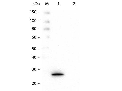
Glutathione S Transferase kappa 1 Antibody (16A11.E4.E3.E7)
Western Blot of Mouse anti-GSTK1 Monoclonal Antibody. Lane 1: Recombinant GSTK1 protein. Lane 2: GST. Load: 50 ng per lane. Primary antibody: Mouse anti-Glutathione S Transferase kappa 1 Antibody (16A11.E4.E3.E7)at 1:1,000 overnight at 4C. Secondary antibody: HRP Mouse Secondary Antibody at 1:40,000 for 30 min at RT. Block for 30 min at RT. Predicted/Observed size: 27 kDa, 27 kDa for GSTK1.Applications
Application
Recommended Usage
ELISA
1:50000-1:200000
Immunohistochemistry
1:10-1:500
Immunoprecipitation
1:10-1:500
SDS-Page
1:100-1:2000
Western Blot
1:500-1:1000
Application Notes
This product is tested by in ELISA, SDS-Page, and Western Blot. Suitable for most immunological techniques requiring high titer binding and lot-to-lot consistency. Specific conditions for reactivity should be optimized by the end user.
Formulation, Preparation, and Storage
Purification
Protein A purified
Formulation
0.02 M Potassium Phosphate, 0.15 M Sodium Chloride, pH 7.2
Format
BSA Free
Preservative
0.01% Sodium Azide
Concentration
Please see the vial label for concentration. If unlisted please contact technical services.
Shipping
The product is shipped with polar packs. Upon receipt, store it immediately at the temperature recommended below.
Stability & Storage
Store at -20C short term. Aliquot and store at -80C long term. Avoid freeze-thaw cycles.
Background: Glutathione S Transferase kappa 1
Alternate Names
EC 2.5.1.18, glutathione S-transferase k1, glutathione S-transferase kappa 1, Glutathione S-transferase subunit 13, glutathione S-transferase subunit 13 homolog, GST, GST 13-13, GST class-kappa, GST13, GST13-13, GSTK1-1, hGSTK1
Gene Symbol
GSTK1
UniProt
Additional Glutathione S Transferase kappa 1 Products
Product Documents
Certificate of Analysis
To download a Certificate of Analysis, please enter a lot or batch number in the search box below.
Product Specific Notices
This product is for research use only and is not approved for use in humans or in clinical diagnosis. Primary Antibodies are guaranteed for 1 year from date of receipt.
Customer Reviews
There are currently no reviews for this product. Be the first to review Glutathione S Transferase kappa 1 Antibody (16A11.E4.E3.E7) - BSA Free and earn rewards!
Have you used Glutathione S Transferase kappa 1 Antibody (16A11.E4.E3.E7) - BSA Free?
Submit a review and receive an Amazon gift card!
$25/€18/£15/$25CAN/¥2500 Yen for a review with an image
$10/€7/£6/$10CAN/¥1110 Yen for a review without an image
Submit a review
Protocols
Find general support by application which include: protocols, troubleshooting, illustrated assays, videos and webinars.
- Antigen Retrieval Protocol (PIER)
- Antigen Retrieval for Frozen Sections Protocol
- Appropriate Fixation of IHC/ICC Samples
- Cellular Response to Hypoxia Protocols
- Chromogenic IHC Staining of Formalin-Fixed Paraffin-Embedded (FFPE) Tissue Protocol
- Chromogenic Immunohistochemistry Staining of Frozen Tissue
- ClariTSA™ Fluorophore Kits
- Detection & Visualization of Antibody Binding
- ELISA Sample Preparation & Collection Guide
- ELISA Troubleshooting Guide
- Fluorescent IHC Staining of Frozen Tissue Protocol
- Graphic Protocol for Heat-induced Epitope Retrieval
- Graphic Protocol for the Preparation and Fluorescent IHC Staining of Frozen Tissue Sections
- Graphic Protocol for the Preparation and Fluorescent IHC Staining of Paraffin-embedded Tissue Sections
- Graphic Protocol for the Preparation of Gelatin-coated Slides for Histological Tissue Sections
- How to Run an R&D Systems DuoSet ELISA
- How to Run an R&D Systems Quantikine ELISA
- How to Run an R&D Systems Quantikine™ QuicKit™ ELISA
- IHC Sample Preparation (Frozen sections vs Paraffin)
- Immunofluorescent IHC Staining of Formalin-Fixed Paraffin-Embedded (FFPE) Tissue Protocol
- Immunohistochemistry (IHC) and Immunocytochemistry (ICC) Protocols
- Immunohistochemistry Frozen Troubleshooting
- Immunohistochemistry Paraffin Troubleshooting
- Immunoprecipitation Protocol
- Preparing Samples for IHC/ICC Experiments
- Preventing Non-Specific Staining (Non-Specific Binding)
- Primary Antibody Selection & Optimization
- Protocol for Heat-Induced Epitope Retrieval (HIER)
- Protocol for Making a 4% Formaldehyde Solution in PBS
- Protocol for VisUCyte™ HRP Polymer Detection Reagent
- Protocol for the Preparation & Fixation of Cells on Coverslips
- Protocol for the Preparation and Chromogenic IHC Staining of Frozen Tissue Sections
- Protocol for the Preparation and Chromogenic IHC Staining of Frozen Tissue Sections - Graphic
- Protocol for the Preparation and Chromogenic IHC Staining of Paraffin-embedded Tissue Sections
- Protocol for the Preparation and Chromogenic IHC Staining of Paraffin-embedded Tissue Sections - Graphic
- Protocol for the Preparation and Fluorescent IHC Staining of Frozen Tissue Sections
- Protocol for the Preparation and Fluorescent IHC Staining of Paraffin-embedded Tissue Sections
- Protocol for the Preparation of Gelatin-coated Slides for Histological Tissue Sections
- Quantikine HS ELISA Kit Assay Principle, Alkaline Phosphatase
- Quantikine HS ELISA Kit Principle, Streptavidin-HRP Polymer
- R&D Systems Quality Control Western Blot Protocol
- Sandwich ELISA (Colorimetric) – Biotin/Streptavidin Detection Protocol
- Sandwich ELISA (Colorimetric) – Direct Detection Protocol
- TUNEL and Active Caspase-3 Detection by IHC/ICC Protocol
- The Importance of IHC/ICC Controls
- Troubleshooting Guide: ELISA
- Troubleshooting Guide: Immunohistochemistry
- Troubleshooting Guide: Western Blot Figures
- Western Blot Conditions
- Western Blot Protocol
- Western Blot Protocol for Cell Lysates
- Western Blot Troubleshooting
- Western Blot Troubleshooting Guide
- View all Protocols, Troubleshooting, Illustrated assays and Webinars
Loading...