
Aggrecan, also known as aggrecan 1, chondroitin sulfate proteoglycan, and large aggregating proteoglycan, is encoded by the AGC1 gene with gene aliases of SEDK; CSPG1; MSK16; CSPGCP (1). As the key component of the cartilage extracellular matrix, aggrecan hydrates the collagen network and provides cartilage with its properties of compressibility and elasticity. Maintenance of aggrecan content is therefore critical to the function of the tissue and aggrecan degradation is an important factor in the erosion of articular cartilage in arthritic diseases (2). The deduced amino acid sequence of human aggrecan core protein consists of 2415 residues and predicts a signal peptide and domains of G1, IGD, G2, KS, CS-1, CS-2 and G3 (3). Two globular domains, G1 and G2, comprise the N-terminus of the proteoglycan and also contain link domains. The third globular domain, G3, corresponds to the C-terminus. The keratan sulfate (KS) and the chondroitin sulfate (CS) attachment domains are between G2 and G3. With KS and CS attached to the 250 kDa core protein, aggrecan monomers exist as a 1,000 to 2,000 kDa molecule. In addition, aggrecan monomers interact with hyaluronan through their G1 domain, resulting in larger aggregates containing 10 to 100 aggrecan monomers on a hyaluronan backbone (2). Aggrecan can be cleaved by MMPs and ADAMTSs at the Asn360-Phe361 and Glu392-Ala393 bond in the IGD (residues are numbered based on Accession # NP_037359), respectively (2). Inhibition of ADATMS4 and ADAMTS5 cleavage prevents aggrecan degradation in osteoarthritic cartilage, while mice with aggrecan resistant to MMP cleavage do not accumulate aggrean and develop normally (2, 4).
Human Aggrecan G1-IGD-G2 Domains Antibody (179509)
R&D Systems | Catalog # MAB1220

Scientific images may be resized, cropped, or adjusted for brightness or contrast for presentation purposes. These updates are limited to presentation and do not affect the underlying scientific interpretation of the data. Where available, additional source data may be provided upon request.
Key Product Details
Species Reactivity
Validated:
Cited:
Applications
Validated:
Cited:
Label
Antibody Source
Product Specifications
Immunogen
Val20-Gly675
Accession # NP_037359
Specificity
Clonality
Host
Isotype
Scientific Data Images for Human Aggrecan G1-IGD-G2 Domains Antibody (179509)
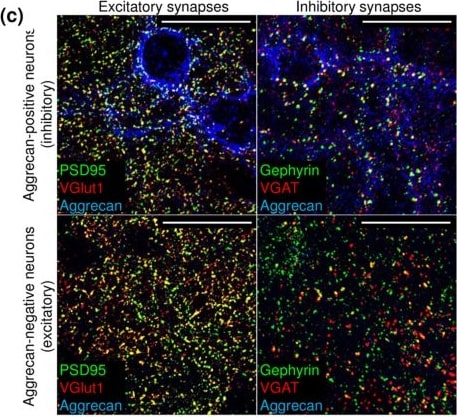
Detection of Mouse Human Aggrecan G1-IGD-G2 Domains Antibody by Immunocytochemistry/ Immunofluorescence
Olanzapine (Oz) and haloperidol (Hp) modify the density of excitatory and inhibitory synapses to excitatory and inhibitory neurons. (a) The principle of the synapse quantification method is based on the detection of presynaptic and postsynaptic markers. (b) The overlap between presynaptic and postsynaptic fluorescence allows to identify the structurally complete synapses. Neurotransmitter transporter proteins are labeled in red at the presynapse, scaffolding protein is marked green at the postsynapse. (c) Glutamatergic and GABAergic synapses are detected with reference to aggrecan expression in 21 days in vitro (21 DIV) neuronal cultures. The representative 66.5 × 66.5 µm single plane confocal micrographs exemplify the staining patterns of excitatory (colocalization of PSD95 and VGlut1) and inhibitory (colocalization of gephyrin and VGAT) synapses in proximity to inhibitory (aggrecan-positive) and excitatory (aggrecan-negative) neuron somata (See explanation in the text). High-resolution scans were obtained from the regions proximal to neuronal somata. Scale bar, 30 µm. (d) The percent of increase/decrease in synapse density upon treatment with antipsychotics is shown as median (square center), and the inter quartile range (25–75% IQR whiskers). Each data point reflects the quantification of minimum 35 images (66.5 × 66.5 µm area, containing a single cell body of a 21 DIV neuron, exemplified in (c), N = 5. The asterisks indicate significant differences with the corresponding control, based on the Kruskal-Wallis ANOVA test (*p < 0.05; ***p < 0.001). Image collected and cropped by CiteAb from the following publication (https://pubmed.ncbi.nlm.nih.gov/28912551), licensed under a CC-BY license. Not internally tested by R&D Systems.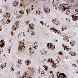
Aggrecan in Human Cartilage.
Aggrecan was detected in immersion fixed paraffin-embedded sections of human cartilage using Mouse Anti-Human Aggrecan G1-IGD-G2 Domains Monoclonal Antibody (Catalog # MAB1220) at 5 µg/mL for 1 hour at room temperature followed by incubation with the Anti-Mouse IgG VisUCyte™ HRP Polymer Antibody (VC001). Before incubation with the primary antibody, tissue was subjected to heat-induced epitope retrieval using Antigen Retrieval Reagent-Basic (CTS013). Tissue was stained using DAB (brown) and counterstained with hematoxylin (blue). Specific staining was localized to chondrocytes. Staining was performed using our protocol for IHC Staining with VisUCyte HRP Polymer Detection Reagents.Applications for Human Aggrecan G1-IGD-G2 Domains Antibody (179509)
Immunohistochemistry
Sample: Immersion fixed paraffin-embedded sections of human cartilage
Immunoprecipitation
Sample: Conditioned cell culture medium spiked with Recombinant Human Aggrecan G1-IGD-G2 Domains (Catalog # 1220-PG), see our available Western blot detection antibodies
Western Blot
Sample: Recombinant Human Aggrecan G1-IGD-G2 Domains (Catalog # 1220-PG)
under non-reducing conditions only
Formulation, Preparation, and Storage
Purification
Reconstitution
Reconstitute at 0.5 mg/mL in sterile PBS. For liquid material, refer to CoA for concentration.
Formulation
Shipping
Stability & Storage
- 12 months from date of receipt, -20 to -70 °C as supplied.
- 1 month, 2 to 8 °C under sterile conditions after reconstitution.
- 6 months, -20 to -70 °C under sterile conditions after reconstitution.
Calculators
Background: Aggrecan
References
- Doege. K.J. et al. (1991) J. Biol. Chem. 266:894.
- Malfait, A.-M. et al. (2002) J. Biol. Chem. 277:22201.
- Caterson, B. et al. (2000) Matrix Biol. 19:333.
- Little, C.B. et al. (2005) Mol. Cel. Biol. 25:3388.
Alternate Names
Entrez Gene IDs
Gene Symbol
UniProt
Additional Aggrecan Products
Product Documents for Human Aggrecan G1-IGD-G2 Domains Antibody (179509)
Certificate of Analysis
To download a Certificate of Analysis, please enter a lot or batch number in the search box below.
Note: Certificate of Analysis not available for kit components.
Product Specific Notices for Human Aggrecan G1-IGD-G2 Domains Antibody (179509)
For research use only
Citations for Human Aggrecan G1-IGD-G2 Domains Antibody (179509)
 Powered by Bioz
Customer Reviews for Human Aggrecan G1-IGD-G2 Domains Antibody (179509)
There are currently no reviews for this product. Be the first to review Human Aggrecan G1-IGD-G2 Domains Antibody (179509) and earn rewards!
Have you used Human Aggrecan G1-IGD-G2 Domains Antibody (179509)?
Submit a review and receive an Amazon gift card!
$25/€18/£15/$25CAN/¥2500 for a review with an image
$10/€7/£6/$10CAN/¥1110 for a review without an image
Submit a review
Protocols
Find general support by application which include: protocols, troubleshooting, illustrated assays, videos and webinars.
- Antigen Retrieval Protocol (PIER)
- Antigen Retrieval for Frozen Sections Protocol
- Appropriate Fixation of IHC/ICC Samples
- Cellular Response to Hypoxia Protocols
- Chromogenic IHC Staining of Formalin-Fixed Paraffin-Embedded (FFPE) Tissue Protocol
- Chromogenic Immunohistochemistry Staining of Frozen Tissue
- ClariTSA™ Fluorophore Kits
- Detection & Visualization of Antibody Binding
- Fluorescent IHC Staining of Frozen Tissue Protocol
- Graphic Protocol for Heat-induced Epitope Retrieval
- Graphic Protocol for the Preparation and Fluorescent IHC Staining of Frozen Tissue Sections
- Graphic Protocol for the Preparation and Fluorescent IHC Staining of Paraffin-embedded Tissue Sections
- Graphic Protocol for the Preparation of Gelatin-coated Slides for Histological Tissue Sections
- IHC Sample Preparation (Frozen sections vs Paraffin)
- Immunofluorescent IHC Staining of Formalin-Fixed Paraffin-Embedded (FFPE) Tissue Protocol
- Immunohistochemistry (IHC) and Immunocytochemistry (ICC) Protocols
- Immunohistochemistry Frozen Troubleshooting
- Immunohistochemistry Paraffin Troubleshooting
- Immunoprecipitation Protocol
- Preparing Samples for IHC/ICC Experiments
- Preventing Non-Specific Staining (Non-Specific Binding)
- Primary Antibody Selection & Optimization
- Protocol for Heat-Induced Epitope Retrieval (HIER)
- Protocol for Making a 4% Formaldehyde Solution in PBS
- Protocol for VisUCyte™ HRP Polymer Detection Reagent
- Protocol for the Preparation & Fixation of Cells on Coverslips
- Protocol for the Preparation and Chromogenic IHC Staining of Frozen Tissue Sections
- Protocol for the Preparation and Chromogenic IHC Staining of Frozen Tissue Sections - Graphic
- Protocol for the Preparation and Chromogenic IHC Staining of Paraffin-embedded Tissue Sections
- Protocol for the Preparation and Chromogenic IHC Staining of Paraffin-embedded Tissue Sections - Graphic
- Protocol for the Preparation and Fluorescent IHC Staining of Frozen Tissue Sections
- Protocol for the Preparation and Fluorescent IHC Staining of Paraffin-embedded Tissue Sections
- Protocol for the Preparation of Gelatin-coated Slides for Histological Tissue Sections
- R&D Systems Quality Control Western Blot Protocol
- TUNEL and Active Caspase-3 Detection by IHC/ICC Protocol
- The Importance of IHC/ICC Controls
- Troubleshooting Guide: Immunohistochemistry
- Troubleshooting Guide: Western Blot Figures
- Western Blot Conditions
- Western Blot Protocol
- Western Blot Protocol for Cell Lysates
- Western Blot Troubleshooting
- Western Blot Troubleshooting Guide
- View all Protocols, Troubleshooting, Illustrated assays and Webinars
Associated Pathways

