Arginase 1 (ARG1) is a 35‑40 kDa member of the arginase family of enzymes. It is expressed in multiple cell types, including erythrocytes, hepatocytes, neutrophils, smooth muscle and macrophages. ARG1 demonstrates two distinct functions: in the hepatocyte cytoplasm, it catalyzes the conversion of arginine to ornithine and urea, while in multiple cells, it degrades arginine, thus indirectly downregulating NO synthase (NOS) activity by depriving this enzyme of its substrate. Human AGR1 is 322 amino acids (aa) in length. Its enzyme region comprises aa 9‑309 and contains two Mn atoms. ARG1 is moderately active as a monomer, but highly active as a 105 kDa homotrimer. Trimerization is promoted by nitrosylation of Cys303, creating a regulatory feedback loop with NOS. There are two isoform variants, one that shows an eight aa insertion after Gln43, and another that shows a deletion of aa 204‑289. Full-length human ARG1 shares 87% aa identity with mouse and rat ARG1.

Key Product Details
Species Reactivity
Human
Applications
Multiplex Immunofluorescence, Immunohistochemistry, Western Blot, Simple Western, Immunoprecipitation, COMET
Label
Unconjugated
Antibody Source
Monoclonal Mouse IgG2B Clone # 658934
Loading...
Product Specifications
Immunogen
E. coli-derived recombinant human Arginase 1/ARG1
Met1-Lys322
Accession # P05089
Met1-Lys322
Accession # P05089
Specificity
Detects human Arginase 1/ARG1 in direct ELISAs and Western blots.
Clonality
Monoclonal
Host
Mouse
Isotype
IgG2B
Scientific Data Images for Human Arginase 1/ARG1 Antibody
Detection of Arginase1 in Human Liver Tumor via seqIF™ staining on COMET™
Arginase 1 was detected in immersion fixed paraffin-embedded sections of human Liver Tumor using Mouse Anti-Human Arginase 1, Monoclonal Antibody (Catalog # MAB5868) at 3ug/mL at 37° Celsius for 4 minutes. Before incubation with the primary antibody, tissue underwent an all-in-one dewaxing and antigen retrieval preprocessing using PreTreatment Module (PT Module) and Dewax and HIER Buffer H (pH 9; Epredia Catalog # TA-999-DHBH). Tissue was stained using the Alexa Fluor™ 647 Goat anti-Mouse IgG Secondary Antibody at 1:200 at 37 ° Celsius for 2 minutes. (Yellow; Lunaphore Catalog # DR647MS) and counterstained with DAPI (blue; Lunaphore Catalog # DR100). Specific staining was localized to the cytoplasm and nucleus. Protocol available in COMET™ Panel Builder.
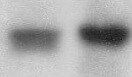
Detection of Human Arginase 1/ARG1 by Western Blot.
Western blot shows lysates of human liver tissue. PVDF Membrane was probed with 1 µg/mL of Human Arginase 1/ARG1 Monoclonal Antibody (Catalog # MAB5868) followed by HRP-conjugated Anti-Mouse IgG Secondary Antibody (Catalog # HAF007). A specific band was detected for Arginase 1/ARG1 at approximately 40 kDa (as indicated). This experiment was conducted under reducing conditions and using Immunoblot Buffer Group 1.
Detection of Human Arginase 1/ARG1 by Simple WesternTM.
Simple Western lane view shows lysates of human liver tissue, loaded at 0.2 mg/mL. A specific band was detected for Arginase 1/ARG1 at approximately 42 kDa (as indicated) using 20 µg/mL of Mouse Anti-Human Arginase 1/ARG1 Monoclonal Antibody (Catalog # MAB5868). This experiment was conducted under reducing conditions and using the 12-230 kDa separation system.*Non-specific interaction with the 230kDa Simple Western standard may be seen with this antibody.
Detection of Arginase 1/ARG1 in Human Liver Cancer.
Arginase 1/ARG1 was detected in immersion fixed paraffin-embedded sections of human liver cancer using Mouse Anti-Human Arginase 1/ARG1 Monoclonal Antibody (Catalog # MAB5868) at 1 µg/ml overnight at 4 °C. Before incubation with the primary antibody, tissue was subjected to heat-induced epitope retrieval using VisUCyte Antigen Retrieval Reagent-Basic (Catalog # VCTS021). Tissue was stained using the HRP-conjugated Anti-Mouse IgG Secondary Antibody (Catalog # HAF007) and counterstained with hematoxylin (blue). Specific staining was localized to the nucleus and cytoplasm. View our protocol for Chromogenic IHC Staining of Paraffin-embedded Tissue Sections.
Detection of Arginase 1/ARG1 by Western Blot
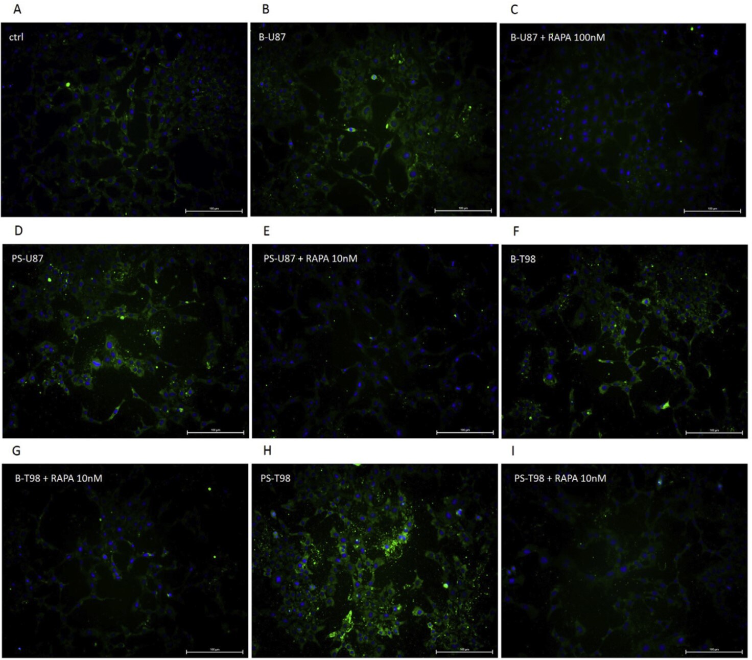
Hypoxic glioma cell‐derived EVs promote M2 macrophage polarization. (A) Images of EVs observed under a TEM (Scale bar = 100 nm). (B) Diameter of EVs detected by dynamic light scattering. (C) The expression of CD9, CD63, TSG101, and calnexin on the surface of EVs determined by Immunoblotting. (D) Detection of the uptake of EVs from normoxic and hypoxic glioma cells by macrophages U937 through immunofluorescence (400×). (E) The mRNA expression of iNOS, TNF‐ alpha, Arg‐1, and IL‐10 assessed by RT‐qPCR after EV treatment. (F) Immunoblotting of protein expression of iNOS and Arg‐1 in macrophages U937 after EV treatment. (G) The expression of TNF‐ alpha and IL‐10 in supernatant of macrophages U937 evaluated by ELISA assay. (H) The proportion of CD11b+CD163+ cells in U937 cells examined by flow cytometry. *p < 0.05 versus macrophages treated with PBS. #p < 0.05 versus macrophages treated with normal oxygen‐induced glioma cell‐derived EVs. The experiment was repeated three times Image collected and cropped by CiteAb from the following open publication (https://pubmed.ncbi.nlm.nih.gov/36052751), licensed under a CC-BY license. Not internally tested by R&D Systems.
Detection of Arginase 1/ARG1 by Western Blot
Hypoxic glioma cell‐derived EVs promote M2 macrophage polarization. (A) Images of EVs observed under a TEM (Scale bar = 100 nm). (B) Diameter of EVs detected by dynamic light scattering. (C) The expression of CD9, CD63, TSG101, and calnexin on the surface of EVs determined by Immunoblotting. (D) Detection of the uptake of EVs from normoxic and hypoxic glioma cells by macrophages U937 through immunofluorescence (400×). (E) The mRNA expression of iNOS, TNF‐ alpha, Arg‐1, and IL‐10 assessed by RT‐qPCR after EV treatment. (F) Immunoblotting of protein expression of iNOS and Arg‐1 in macrophages U937 after EV treatment. (G) The expression of TNF‐ alpha and IL‐10 in supernatant of macrophages U937 evaluated by ELISA assay. (H) The proportion of CD11b+CD163+ cells in U937 cells examined by flow cytometry. *p < 0.05 versus macrophages treated with PBS. #p < 0.05 versus macrophages treated with normal oxygen‐induced glioma cell‐derived EVs. The experiment was repeated three times Image collected and cropped by CiteAb from the following open publication (https://pubmed.ncbi.nlm.nih.gov/36052751), licensed under a CC-BY license. Not internally tested by R&D Systems.Applications for Human Arginase 1/ARG1 Antibody
Application
Recommended Usage
COMET
Optimal dilutions of this antibody should be experimentally determined.
Immunohistochemistry
1-15 µg/mL
Sample: Immersion fixed paraffin-embedded sections of human liver cancer
Sample: Immersion fixed paraffin-embedded sections of human liver cancer
Immunoprecipitation
25 µg/mL
Sample: Cell lysates spiked with Recombinant Human Arginase 1/ARG1 (Catalog # 5868-AR), see our available Western blot detection antibodies
Sample: Cell lysates spiked with Recombinant Human Arginase 1/ARG1 (Catalog # 5868-AR), see our available Western blot detection antibodies
Multiplex Immunofluorescence
3 µg/mL
Sample: Immersion fixed paraffin-embedded sections of human liver tumor
Sample: Immersion fixed paraffin-embedded sections of human liver tumor
Simple Western
20 µg/mL
Sample: Human liver tissue
Sample: Human liver tissue
Western Blot
1 µg/mL
Sample: Human liver tissue
Sample: Human liver tissue
Reviewed Applications
Read 2 reviews rated 4.5 using MAB5868 in the following applications:
Formulation, Preparation, and Storage
Purification
Protein A or G purified from hybridoma culture supernatant
Reconstitution
Reconstitute at 0.5 mg/mL in sterile PBS. For liquid material, refer to CoA for concentration.
Loading...
Formulation
Lyophilized from a 0.2 μm filtered solution in PBS with Trehalose. See Certificate of Analysis for details.
*Small pack size (-SP) is supplied either lyophilized or as a 0.2 µm filtered solution in PBS.
*Small pack size (-SP) is supplied either lyophilized or as a 0.2 µm filtered solution in PBS.
Shipping
Lyophilized product is shipped at ambient temperature. Liquid small pack size (-SP) is shipped with polar packs. Upon receipt, store immediately at the temperature recommended below.
Stability & Storage
Use a manual defrost freezer and avoid repeated freeze-thaw cycles.
- 12 months from date of receipt, -20 to -70 °C as supplied.
- 1 month, 2 to 8 °C under sterile conditions after reconstitution.
- 6 months, -20 to -70 °C under sterile conditions after reconstitution.
Calculators
Background: Arginase 1/ARG1
Long Name
Liver-Type Arginase
Alternate Names
AI, ARG1, Arginase-1, Liver Arginase, PGIF, Type I Arginase
Gene Symbol
ARG1
UniProt
Additional Arginase 1/ARG1 Products
Product Documents for Human Arginase 1/ARG1 Antibody
Certificate of Analysis
To download a Certificate of Analysis, please enter a lot or batch number in the search box below.
Note: Certificate of Analysis not available for kit components.
Product Specific Notices for Human Arginase 1/ARG1 Antibody
For research use only
Related Research Areas
Citations for Human Arginase 1/ARG1 Antibody
 Powered by Bioz
Customer Reviews for Human Arginase 1/ARG1 Antibody (2)
4.5 out of 5
2 Customer Ratings
Have you used Human Arginase 1/ARG1 Antibody?
Submit a review and receive an Amazon gift card!
$25/€18/£15/$25CAN/¥2500 Yen for a review with an image
$10/€7/£6/$10CAN/¥1110 Yen for a review without an image
Submit a review
Customer Images
Showing
1
-
2 的
2 reviews
Showing All
Filter By:
-
Application: Western BlotSample Tested: Liver tissueSpecies: HumanVerified Customer | Posted 01/21/2022

-
Application: Immunocytochemistry/ImmunofluorescenceSample Tested: MICROGLIASpecies: HumanVerified Customer | Posted 05/19/2021
There are no reviews that match your criteria.
Protocols
Find general support by application which include: protocols, troubleshooting, illustrated assays, videos and webinars.
- Antigen Retrieval Protocol (PIER)
- Antigen Retrieval for Frozen Sections Protocol
- Appropriate Fixation of IHC/ICC Samples
- Cellular Response to Hypoxia Protocols
- Chromogenic IHC Staining of Formalin-Fixed Paraffin-Embedded (FFPE) Tissue Protocol
- Chromogenic Immunohistochemistry Staining of Frozen Tissue
- ClariTSA™ Fluorophore Kits
- Detection & Visualization of Antibody Binding
- Fluorescent IHC Staining of Frozen Tissue Protocol
- Graphic Protocol for Heat-induced Epitope Retrieval
- Graphic Protocol for the Preparation and Fluorescent IHC Staining of Frozen Tissue Sections
- Graphic Protocol for the Preparation and Fluorescent IHC Staining of Paraffin-embedded Tissue Sections
- Graphic Protocol for the Preparation of Gelatin-coated Slides for Histological Tissue Sections
- IHC Sample Preparation (Frozen sections vs Paraffin)
- Immunofluorescent IHC Staining of Formalin-Fixed Paraffin-Embedded (FFPE) Tissue Protocol
- Immunohistochemistry (IHC) and Immunocytochemistry (ICC) Protocols
- Immunohistochemistry Frozen Troubleshooting
- Immunohistochemistry Paraffin Troubleshooting
- Immunoprecipitation Protocol
- Preparing Samples for IHC/ICC Experiments
- Preventing Non-Specific Staining (Non-Specific Binding)
- Primary Antibody Selection & Optimization
- Protocol for Heat-Induced Epitope Retrieval (HIER)
- Protocol for Making a 4% Formaldehyde Solution in PBS
- Protocol for VisUCyte™ HRP Polymer Detection Reagent
- Protocol for the Preparation & Fixation of Cells on Coverslips
- Protocol for the Preparation and Chromogenic IHC Staining of Frozen Tissue Sections
- Protocol for the Preparation and Chromogenic IHC Staining of Frozen Tissue Sections - Graphic
- Protocol for the Preparation and Chromogenic IHC Staining of Paraffin-embedded Tissue Sections
- Protocol for the Preparation and Chromogenic IHC Staining of Paraffin-embedded Tissue Sections - Graphic
- Protocol for the Preparation and Fluorescent IHC Staining of Frozen Tissue Sections
- Protocol for the Preparation and Fluorescent IHC Staining of Paraffin-embedded Tissue Sections
- Protocol for the Preparation of Gelatin-coated Slides for Histological Tissue Sections
- R&D Systems Quality Control Western Blot Protocol
- TUNEL and Active Caspase-3 Detection by IHC/ICC Protocol
- The Importance of IHC/ICC Controls
- Troubleshooting Guide: Immunohistochemistry
- Troubleshooting Guide: Western Blot Figures
- Western Blot Conditions
- Western Blot Protocol
- Western Blot Protocol for Cell Lysates
- Western Blot Troubleshooting
- Western Blot Troubleshooting Guide
- View all Protocols, Troubleshooting, Illustrated assays and Webinars
Loading...