Human BMP-2 is one of at least 15 structurally and functionally related BMPs, which are members of the transforming growth factor beta (TGF-beta ) superfamily. BMPs were originally identified as protein regulators of cartilage and bone formation. However, they have since been shown to be involved in embryogenesis and morphogenesis of various tissues and organs. BMPs have also been shown to regulate the growth, differentiation, chemotaxis and apoptosis of various cell types, including mesenchymal cells, epithelial cells, hematopoietic cells and neuronal cells. Similarly to other TGF-beta family proteins, BMPs are highly conserved across animal species. At the amino acid sequence level, mature human, mouse and rat BMP-2 are 100% identical. BMP-2 is synthesized as a large precursor protein that is cleaved at the dibasic cleavage site (RXXR) to release the carboxy-terminal domain. Biologically active BMP-2 is a disulfide-linked homodimer of the carboxy-terminal 114 amino acid residues that contains the characteristic seven conserved cysteine residues involved in the formation of the cysteine knot and the single interchain disulfide bond. Cellular responses to BMP-2 have been shown to be mediated by the formation of hetero-oligomeric complexes of type I and type II serine/threonine kinase receptors. One BMP type II and two BMP type I receptors have been identified. In contrast to the TGF-beta type I receptor, which does not bind the ligand in the absence of the TGF-beta receptor type II, both BMP receptor type Is can bind BMP-2 with high-affinity in the absence of BMP receptor type II.

Key Product Details
Species Reactivity
Validated:
Human
Cited:
Human, Mouse, Canine, Hamster
Applications
Validated:
Immunohistochemistry, Western Blot
Cited:
Immunohistochemistry, Immunohistochemistry-Paraffin, Western Blot, Immunocytochemistry
Label
Unconjugated
Antibody Source
Polyclonal Goat IgG
Loading...
Product Specifications
Immunogen
Chinese hamster ovary cell line CHO-derived recombinant human BMP-2
Gln283-Arg396
Accession # P12643
Gln283-Arg396
Accession # P12643
Specificity
Detects BMP-2 and BMP-4 in direct ELISAs and Western blots.
Clonality
Polyclonal
Host
Goat
Isotype
IgG
Scientific Data Images for Human BMP-2/BMP-4 Antibody
Detection of BMP‑2/BMP‑4 in Human Lung Cancer.
BMP‑2/BMP‑4 was detected in immersion fixed paraffin-embedded sections of human lung cancer using Goat Anti-Human BMP‑2/BMP‑4 Antigen Affinity-purified Polyclonal Antibody (Catalog # AF355) at 0.5 µg/ml for 1 hour at room temperature followed by incubation with the Anti-Goat IgG VisUCyte™ HRP Polymer Antibody (Catalog # VC004). Before incubation with the primary antibody, tissue was subjected to heat-induced epitope retrieval using VisUCyte Antigen Retrieval Reagent-Basic (Catalog # VCTS021). Tissue was stained using DAB (brown) and counterstained with hematoxylin (blue). Specific staining was localized to the cytoplasm. View our protocol for IHC Staining with VisUCyte HRP Polymer Detection Reagents.Applications for Human BMP-2/BMP-4 Antibody
Application
Recommended Usage
Immunohistochemistry
5-15 µg/mL
Sample: Immersion fixed human BMP-2 transfected cells
Sample: Immersion fixed human BMP-2 transfected cells
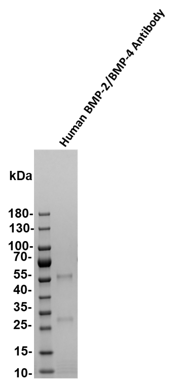
Western Blot
0.1 µg/mL
Sample: Recombinant Human BMP‑2 (Catalog # 355-BEC)
Recombinant Human BMP‑4 (Catalog # 314-BP)
Sample: Recombinant Human BMP‑2 (Catalog # 355-BEC)
Recombinant Human BMP‑4 (Catalog # 314-BP)
Reviewed Applications
Read 1 review rated 5 using AF355 in the following applications:
Formulation, Preparation, and Storage
Purification
Antigen Affinity-purified
Reconstitution
Reconstitute at 0.2 mg/mL in sterile PBS. For liquid material, refer to CoA for concentration.
Loading...
Formulation
Lyophilized from a 0.2 μm filtered solution in PBS with Trehalose. See Certificate of Analysis for details.
*Small pack size (-SP) is supplied either lyophilized or as a 0.2 µm filtered solution in PBS.
*Small pack size (-SP) is supplied either lyophilized or as a 0.2 µm filtered solution in PBS.
Shipping
Lyophilized product is shipped at ambient temperature. Liquid small pack size (-SP) is shipped with polar packs. Upon receipt, store immediately at the temperature recommended below.
Stability & Storage
Use a manual defrost freezer and avoid repeated freeze-thaw cycles.
- 12 months from date of receipt, -20 to -70 °C as supplied.
- 1 month, 2 to 8 °C under sterile conditions after reconstitution.
- 6 months, -20 to -70 °C under sterile conditions after reconstitution.
Calculators
Background: BMP-2/BMP-4
References
- Kawabata, M. et al. (1998) Cytokine and Growth Factor Reviews 9:49
- Ebendal, T. et al. (1998) J. Neurosci. Res. 51:139.
- Reddi, A.H. (1998) Nature Biotechnology 16: 247.
Long Name
Bone Morphogenetic Protein 2/4
Alternate Names
BDA2, BMP-2, BMP2A, BMP-2A, BMP2ABone morphogenetic protein 2A, bone morphogenetic protein 2
Gene Symbol
BMP2
UniProt
Additional BMP-2/BMP-4 Products
Product Documents for Human BMP-2/BMP-4 Antibody
Certificate of Analysis
To download a Certificate of Analysis, please enter a lot or batch number in the search box below.
Note: Certificate of Analysis not available for kit components.
Product Specific Notices for Human BMP-2/BMP-4 Antibody
For research use only
Related Research Areas
Citations for Human BMP-2/BMP-4 Antibody
 Powered by Bioz
Customer Reviews for Human BMP-2/BMP-4 Antibody (1)
5 out of 5
1 Customer Rating
Have you used Human BMP-2/BMP-4 Antibody?
Submit a review and receive an Amazon gift card!
$25/€18/£15/$25CAN/¥2500 Yen for a review with an image
$10/€7/£6/$10CAN/¥1110 Yen for a review without an image
Submit a review
Customer Images
Showing
1
-
1 的
1 review
Showing All
Filter By:
-
Application: Functional AssaySample Tested: PlasmaSpecies: HumanVerified Customer | Posted 11/29/2020

There are no reviews that match your criteria.
Protocols
Find general support by application which include: protocols, troubleshooting, illustrated assays, videos and webinars.
- Antigen Retrieval Protocol (PIER)
- Antigen Retrieval for Frozen Sections Protocol
- Appropriate Fixation of IHC/ICC Samples
- Cellular Response to Hypoxia Protocols
- Chromogenic IHC Staining of Formalin-Fixed Paraffin-Embedded (FFPE) Tissue Protocol
- Chromogenic Immunohistochemistry Staining of Frozen Tissue
- ClariTSA™ Fluorophore Kits
- Detection & Visualization of Antibody Binding
- Fluorescent IHC Staining of Frozen Tissue Protocol
- Graphic Protocol for Heat-induced Epitope Retrieval
- Graphic Protocol for the Preparation and Fluorescent IHC Staining of Frozen Tissue Sections
- Graphic Protocol for the Preparation and Fluorescent IHC Staining of Paraffin-embedded Tissue Sections
- Graphic Protocol for the Preparation of Gelatin-coated Slides for Histological Tissue Sections
- IHC Sample Preparation (Frozen sections vs Paraffin)
- Immunofluorescent IHC Staining of Formalin-Fixed Paraffin-Embedded (FFPE) Tissue Protocol
- Immunohistochemistry (IHC) and Immunocytochemistry (ICC) Protocols
- Immunohistochemistry Frozen Troubleshooting
- Immunohistochemistry Paraffin Troubleshooting
- Preparing Samples for IHC/ICC Experiments
- Preventing Non-Specific Staining (Non-Specific Binding)
- Primary Antibody Selection & Optimization
- Protocol for Heat-Induced Epitope Retrieval (HIER)
- Protocol for Making a 4% Formaldehyde Solution in PBS
- Protocol for VisUCyte™ HRP Polymer Detection Reagent
- Protocol for the Preparation & Fixation of Cells on Coverslips
- Protocol for the Preparation and Chromogenic IHC Staining of Frozen Tissue Sections
- Protocol for the Preparation and Chromogenic IHC Staining of Frozen Tissue Sections - Graphic
- Protocol for the Preparation and Chromogenic IHC Staining of Paraffin-embedded Tissue Sections
- Protocol for the Preparation and Chromogenic IHC Staining of Paraffin-embedded Tissue Sections - Graphic
- Protocol for the Preparation and Fluorescent IHC Staining of Frozen Tissue Sections
- Protocol for the Preparation and Fluorescent IHC Staining of Paraffin-embedded Tissue Sections
- Protocol for the Preparation of Gelatin-coated Slides for Histological Tissue Sections
- R&D Systems Quality Control Western Blot Protocol
- TUNEL and Active Caspase-3 Detection by IHC/ICC Protocol
- The Importance of IHC/ICC Controls
- Troubleshooting Guide: Immunohistochemistry
- Troubleshooting Guide: Western Blot Figures
- Western Blot Conditions
- Western Blot Protocol
- Western Blot Protocol for Cell Lysates
- Western Blot Troubleshooting
- Western Blot Troubleshooting Guide
- View all Protocols, Troubleshooting, Illustrated assays and Webinars
Loading...