
Human CCL7/MCP-3/MARC Antibody (36320)

R&D Systems | Catalog # MAB282

R&D Systems

Key Product Details

Species Reactivity

Validated:

Human

Cited:

Human

Applications

Validated:

Western Blot, ELISA Capture (Matched Antibody Pair), Neutralization, Intracellular Staining by Flow Cytometry, CyTOF-ready

Cited:

Immunohistochemistry-Paraffin, Immunohistochemistry-Frozen, Neutralization, Immunocytochemistry

Label

Unconjugated

Antibody Source

Monoclonal Mouse IgG1 Clone # 36320
Loading...

Product Specifications

Immunogen

E. coli-derived recombinant human CCL7/MCP-3/MARC
Gln34-109
Accession # Q7Z7Q8

Specificity

Detects human CCL7/MCP-3/MARC in ELISAs and Western blots. In ELISAs, no cross-reactivity with recombinant human (rh) CCL2/MCP-1, rhCCL8/MCP‑2, or rhCCL13/MCP‑4 is observed.

Clonality

Monoclonal

Host

Mouse

Isotype

IgG1

Endotoxin Level

<0.10 EU per 1 μg of the antibody by the LAL method.

Scientific Data Images for Human CCL7/MCP-3/MARC Antibody (36320)

Chemotaxis Induced by CCL7/MCP‑3 and Neutralization by Human CCL7/MCP‑3 Antibody.

Chemotaxis Induced by CCL7/MCP‑3 and Neutralization by Human CCL7/MCP‑3 Antibody.

Recombinant Human CCL7/MCP-3 (Catalog # 282-P3) chemoattracts the BaF3 mouse pro-B cell line transfected with human CCR2A in a dose-dependent manner (orange line). The amount of cells that migrated through to the lower chemotaxis chamber was measured by Resazurin (Catalog # AR002). Chemotaxis elicited by Recombinant Human CCL7/MCP-3 (1 µg/mL) is neutralized (green line) by increasing concentrations of Mouse Anti-Human CCL7/MCP-3 Monoclonal Antibody (Catalog # MAB282). The ND50 is typically 1.5-4.5 µg/mL.
Human CCL7 / MCP-3 / MARC ELISA Standard Curve

Human CCL7 / MCP-3 / MARC ELISA Standard Curve

Recombinant Human CCL7/MCP‑3/MARC (Catalog # 282-P3) was serially diluted and captured by Mouse Anti-Human CCL7/MCP‑3/MARC Monoclonal Antibody (Catalog # MAB282) coated on a Clear Polystyrene Microplate (Catalog # DY990). Goat Anti-Human CCL7/MCP‑3/MARC Antigen Affinity-purified Polyclonal Antibody (Catalog # AF-282-NA) was biotinylated and incubated with the protein captured on the plate. Detection of the standard curve was achieved by incubating Streptavidin-HRP (Catalog # DY998)
Detection of CCL7/MCP-3 by Immunohistochemistry

Detection of CCL7/MCP-3 by Immunohistochemistry

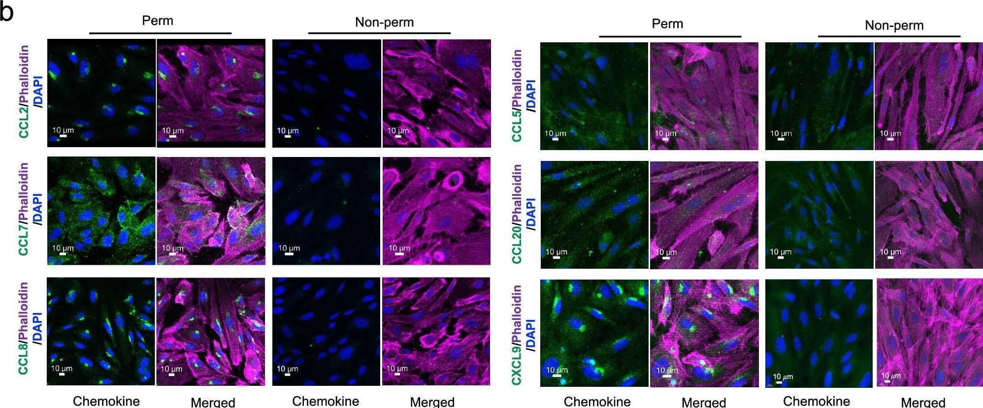
CCR2 ligands are secreted but do not bind to HUVECs.a Culture medium collected from the upper and lower wells of transwells with HUVEC monolayers either not stimulated (NS) or stimulated with TNF-alpha + IFN-gamma were assayed for CCL2, CCL7, and CCL8 by ELISA (n = 3 separate experiments). Each symbol shows data from one experiment, with assays done in duplicate, and bars indicate means ± SEM. b Confocal microscopy images at × 40 magnification of permeabilized and non-permeabilized TNF-alpha + IFN-gamma -stimulated HUVECs immunostained for CCL2, CCL7, CCL8, CCL5, CCL20, and CXCL9 (green) and stained using phalloidin for polymerized actin (magenta) and DAPI for nuclei (blue). The scale bars indicate 10 µm. Images are representative of three experiments. c Biotinylated CCL5 (0.1 μM) and CCL2 (1 μM) were incubated with CHO-K1 cells and the heparan-sulfate deficient cell line, D-677. Chemokine binding was detected using streptavidin-phycoerythrin (PE) and flow cytometry. Data shown are representative of three separate experiments. Image collected and cropped by CiteAb from the following open publication (https://www.nature.com/articles/s41467-025-57002-6), licensed under a CC-BY license. Not internally tested by R&D Systems.

Applications for Human CCL7/MCP-3/MARC Antibody (36320)

Application
Recommended Usage

CyTOF-ready

Ready to be labeled using established conjugation methods. No BSA or other carrier proteins that could interfere with conjugation.

Intracellular Staining by Flow Cytometry

2.5 µg/106 cells
Sample: THP‑1 human acute monocytic leukemia cell line fixed with paraformaldehyde and permeabilized with saponin

Western Blot

1 µg/mL
Sample: Recombinant Human CCL7/MCP‑3/MARC (Catalog # 282-P3)

Neutralization

Measured by its ability to neutralize CCL7/MCP‑3/MARC-induced chemotaxis in the BaF3 mouse pro‑B cell line transfected with human CCR2A. The Neutralization Dose (ND50) is typically 1.5-4.5 µg/mL in the presence of 1 µg/mL Recombinant Human CCL7/MCP‑3/MARC.

Human CCL7/MCP-3 Sandwich Immunoassay

ELISA Capture (Matched Antibody Pair)
Recommended Concentration: 2-8 µg/mL
Use in combination with these reagents:
  • Detection Reagent: Human CCL7/MCP‑3/MARC Biotinylated Antibody (Catalog # BAF282)
  • Standard: Recombinant Human CCL7/MCP-3 Protein (Catalog # 282-P3)
Please Note: Optimal dilutions of this antibody should be experimentally determined.

Flow Cytometry Panel Builder

Bio-Techne Knows Flow Cytometry

Save time and reduce costly mistakes by quickly finding compatible reagents using the Panel Builder Tool.

Advanced Features

  • Spectra Viewer - Custom analysis of spectra from multiple fluorochromes
  • Spillover Popups - Visualize the spectra of individual fluorochromes
  • Antigen Density Selector - Match fluorochrome brightness with antigen density
Build Your Panel Now

Formulation, Preparation, and Storage

Purification

Protein A or G purified from ascites

Reconstitution

Reconstitute at 0.5 mg/mL in sterile PBS. For liquid material, refer to CoA for concentration.


Loading...

Formulation

Lyophilized from a 0.2 μm filtered solution in PBS with Trehalose. *Small pack size (SP) is supplied either lyophilized or as a 0.2 µm filtered solution in PBS.

Shipping

Lyophilized product is shipped at ambient temperature. Liquid small pack size (-SP) is shipped with polar packs. Upon receipt, store immediately at the temperature recommended below.

Stability & Storage

Use a manual defrost freezer and avoid repeated freeze-thaw cycles.
  • 12 months from date of receipt, -20 to -70 °C as supplied.
  • 1 month, 2 to 8 °C under sterile conditions after reconstitution.
  • 6 months, -20 to -70 °C under sterile conditions after reconstitution.

Calculators

The reconstitution calculator allows you to quickly calculate the volume of a reagent to reconstitute your vial. Simply enter the mass of reagent and the target concentration and the calculator will determine the rest.

=
÷

Background: CCL7/MCP-3/MARC

MCP-2 and CCL7 are two monocyte chemotactic proteins produced by human MG-63 osteosarcoma cells. Both MCP-2 and CCL7 are members of the C-C family of chemokines and share 62% and 71% amino acid sequence identity, respectively, with MCP-1. CCL7 also shares 58% amino acid identity with MCP-2.

CCL7 cDNA encodes a 99 amino acid residue precursor protein from which the N-terminal 23 amino acid residues are cleaved to generate the 76 amino acid residue mature CCL7. Mature CCL7 contains a potential N-linked and several possible O-linked glycosylation sites.

Similarly to other C-C chemokines, all three MCP proteins are monocyte chemoattractants. In addition, the three MCPs can chemoattract activated NK cells as well as CD4+ and CD8+ T lymphocytes. All three cytokines have also been shown to attract eosinophils and induce histamine secretion from basophils.

Alternate Names

MARC, MCP-3

Entrez Gene IDs

6354 (Human); 20306 (Mouse)

Gene Symbol

CCL7

UniProt

Additional CCL7/MCP-3/MARC Products

Product Documents for Human CCL7/MCP-3/MARC Antibody (36320)

Certificate of Analysis

To download a Certificate of Analysis, please enter a lot or batch number in the search box below.

Note: Certificate of Analysis not available for kit components.

Product Specific Notices for Human CCL7/MCP-3/MARC Antibody (36320)

For research use only

Citations for Human CCL7/MCP-3/MARC Antibody (36320)

Customer Reviews for Human CCL7/MCP-3/MARC Antibody (36320)

There are currently no reviews for this product. Be the first to review Human CCL7/MCP-3/MARC Antibody (36320) and earn rewards!

Have you used Human CCL7/MCP-3/MARC Antibody (36320)?

Submit a review and receive an Amazon gift card!

$25/€18/£15/$25CAN/¥2500 for a review with an image

$10/€7/£6/$10CAN/¥1110 for a review without an image

Submit a review
Amazon Gift Card

FAQs

No product specific FAQs exist for this product.

View all FAQs for Antibodies
