
CXCL4, also known as Platelet Factor 4 (PF4), is an 8 kDa CXC chemokine that is stored in platelet alpha -granules as a
homotetramer and secreted abundantly during platelet activation. Human CXCL4 is a 101 amino acid (aa) protein with a 32 aa signal sequence and a 70 aa mature protein that includes granule targeting and
heparin-binding sequences. CXCL4 has homology with IL-8 and beta -thromboglobulin and can form heteromultimers with IL-8. Mature human and mouse CXCL4 share 76% aa identity. The active protein consists of a tetramer composed of individual CXCL4 subunits. Megakaryocytes synthesize CXCL4 and store it as tetramers in
alpha -granules. The CXCL4 tetramers are secreted by activated platelets and can be measured at micromolar levels in serum. In contrast to other CXC chemokines, CXCL4 lacks chemotactic activity for polymorphonuclear granulocytes. CXCL4 does not contain an ELR motif. However, many other functions have been observed for CXCL4. CXCL4 is involved in monocyte survivial and differentiation into macrophages, has anti-angiogenic activity and promotes granule Protein C activation. CXCL4 has been demonstrated to inhibit the binding of FGF-2 to high-affinity receptors and its subsequent internalization. Cell surface neutrophil chondroitin sulfate chains serve as CXCL4 binding sites; affinity is controlled by the degree of sulfation of these chains.
Human CXCL4/PF4 Antibody (170138)
R&D Systems | Catalog # MAB7952

Scientific images may be resized, cropped, or adjusted for brightness or contrast for presentation purposes. These updates are limited to presentation and do not affect the underlying scientific interpretation of the data. Where available, additional source data may be provided upon request.
Key Product Details
Species Reactivity
Validated:
Cited:
Applications
Validated:
Cited:
Label
Antibody Source
Product Specifications
Immunogen
Glu32-Ser101
Accession # P02776.2
Specificity
Clonality
Host
Isotype
Scientific Data Images for Human CXCL4/PF4 Antibody (170138)
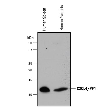
Detection of Human CXCL4/PF4 by Western Blot.
Western blot shows lysates of human spleen tissue and human platelets. PVDF membrane was probed with 0.5 µg/mL of Mouse Anti-Human CXCL4/PF4 Monoclonal Antibody (Catalog # MAB7952) followed by HRP-conjugated Anti-Mouse IgG Secondary Antibody (HAF018). A specific band was detected for CXCL4/PF4 at approximately 11 kDa (as indicated). This experiment was conducted under reducing conditions and using Immunoblot Buffer Group 1.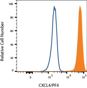
Detection of CXCL4/PF4 in PBMC monocytes by Flow Cytometry.
PBMC monocytes were stained with Mouse Anti-Human CXCL4/PF4 Monoclonal Antibody (Catalog # MAB7952, filled histogram) or isotype control antibody (Catalog # MAB0041, open histogram), followed by Allophycocyanin-conjugated Anti-Mouse IgG Secondary Antibody (Catalog # F0101B). To facilitate intracellular staining, cells were fixed with Flow Cytometry Fixation Buffer (Catalog # FC004) and permeabilized with Flow Cytometry Permeabilization/Wash Buffer I (Catalog # FC005). View our protocol for Staining Intracellular Molecules.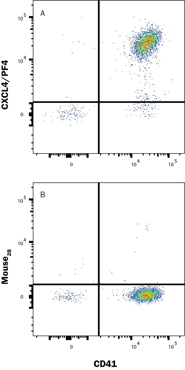
Detection of CXCL4/PF4 in Platelets by Flow Cytometry.
Platelets were stained with Mouse Anti-Human Integrin alpha 2b/CD41 APC‑conjugated Monoclonal Antibody (Catalog # FAB7616A) and either (A) Mouse Anti-Human CXCL4/PF4 Monoclonal Antibody (Catalog # MAB7952) or (B) Mouse IgG2B Flow Cytometry Isotype Control (Catalog # MAB0041) followed by Phycoerythrin-conjugated Anti-Mouse IgG Secondary Antibody (Catalog # F0102B). To facilitate intracellular staining, cells were fixed with Flow Cytometry Fixation Buffer (Catalog # FC004) and permeabilized with Flow Cytometry Permeabilization/Wash Buffer I (FC005). View our protocol for Staining Intracellular Molecules.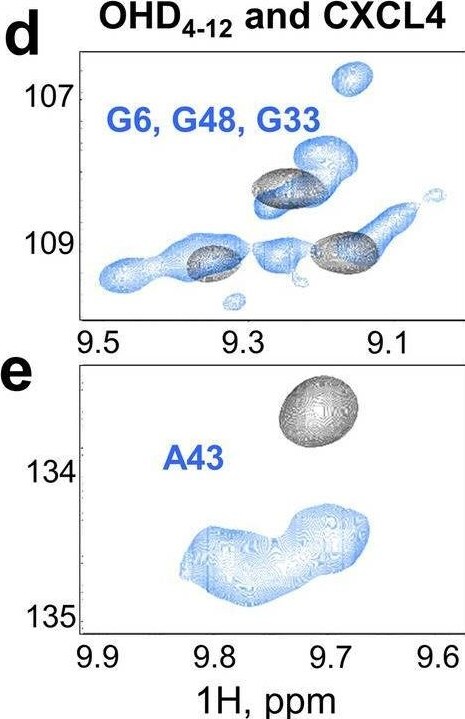
Detection of kfpxvaa9ffykrjoxefaf.jpg by Flow Cytometry
(a) Size-exclusion chromatograms. The elution profiles of lysozyme (14.4 kDa) and myoglobin (17 kDa) in blue and OHD4–12 (15.9 kDa) in orange are shown. (b) WB analysis in non-reduced (left bands, N.R.) and reduced (right bands, R.) conditions demonstrates the presence of OHD4–12 obtained following the mixing of CXCL4-S26C and CXCL12-L29C mutants in the presence of Cu2+ detected with anti-CXCL4 ( alpha CXCL4) or anti-CXCL12 ( alpha CXCL12) antibodies (see also Supplementary Fig. S1). (c) NMR spectroscopic analysis of OHD4–12 folding state. The 15N-HSQC NMR spectrum of the uniformly 15N-labeled 68 μM OHD4–12 in 90% H2O/10% D2O at pH 6.9 in the presence of 20 mM NaCl, collected at 40 °C. (d–f) Expansions from the OHD4–12 spectrum overlaid with CXCL4wt (blue) and CXCL12wt (red) 15N-HSQC NMR spectra. Known signal assignments for CXCL4 and CXCL12 are indicated. The 15N-HSQC NMR spectrum of the uniformly 15N-labeled 150 μM CXCL4 in 90% H2O/10% D2O at pH 5.0 in the presence of 20 mM NaCl was collected at 40 °C. The 15N-HSQC NMR spectrum of the uniformly 15N-labeled 51 μM CXCL12 in 20 MES buffer prepared with 90% H2O/10% D2O at pH 6.8 was collected at 25 °C. The difference in experimental conditions was due to the difference in solubility properties of CXCL4 and CXCL12. Image collected and cropped by CiteAb from the following open publication (https://pubmed.ncbi.nlm.nih.gov/36229490), licensed under a CC-BY license. Not internally tested by R&D Systems.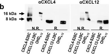
Detection of CXCL4/PF4 by Western Blot
(a) Size-exclusion chromatograms. The elution profiles of lysozyme (14.4 kDa) and myoglobin (17 kDa) in blue and OHD4–12 (15.9 kDa) in orange are shown. (b) WB analysis in non-reduced (left bands, N.R.) and reduced (right bands, R.) conditions demonstrates the presence of OHD4–12 obtained following the mixing of CXCL4-S26C and CXCL12-L29C mutants in the presence of Cu2+ detected with anti-CXCL4 ( alpha CXCL4) or anti-CXCL12 ( alpha CXCL12) antibodies (see also Supplementary Fig. S1). (c) NMR spectroscopic analysis of OHD4–12 folding state. The 15N-HSQC NMR spectrum of the uniformly 15N-labeled 68 μM OHD4–12 in 90% H2O/10% D2O at pH 6.9 in the presence of 20 mM NaCl, collected at 40 °C. (d–f) Expansions from the OHD4–12 spectrum overlaid with CXCL4wt (blue) and CXCL12wt (red) 15N-HSQC NMR spectra. Known signal assignments for CXCL4 and CXCL12 are indicated. The 15N-HSQC NMR spectrum of the uniformly 15N-labeled 150 μM CXCL4 in 90% H2O/10% D2O at pH 5.0 in the presence of 20 mM NaCl was collected at 40 °C. The 15N-HSQC NMR spectrum of the uniformly 15N-labeled 51 μM CXCL12 in 20 MES buffer prepared with 90% H2O/10% D2O at pH 6.8 was collected at 25 °C. The difference in experimental conditions was due to the difference in solubility properties of CXCL4 and CXCL12. Image collected and cropped by CiteAb from the following open publication (https://pubmed.ncbi.nlm.nih.gov/36229490), licensed under a CC-BY license. Not internally tested by R&D Systems.Applications for Human CXCL4/PF4 Antibody (170138)
CyTOF-ready
Intracellular Staining by Flow Cytometry
Sample: PBMC monocytes and Platelets fixed with Flow Cytometry Fixation Buffer (Catalog # FC004) and permeabilized with Flow Cytometry Permeabilization/Wash Buffer I (Catalog # FC005)
Western Blot
Sample: Human spleen tissue and human platelets
Flow Cytometry Panel Builder
Bio-Techne Knows Flow Cytometry
Save time and reduce costly mistakes by quickly finding compatible reagents using the Panel Builder Tool.
Advanced Features
- Spectra Viewer - Custom analysis of spectra from multiple fluorochromes
- Spillover Popups - Visualize the spectra of individual fluorochromes
- Antigen Density Selector - Match fluorochrome brightness with antigen density

Formulation, Preparation, and Storage
Purification
Reconstitution
Reconstitute at 0.5 mg/mL in sterile PBS. For liquid material, refer to CoA for concentration.
Formulation
Shipping
Stability & Storage
- 12 months from date of receipt, -20 to -70 °C as supplied.
- 1 month, 2 to 8 °C under sterile conditions after reconstitution.
- 6 months, -20 to -70 °C under sterile conditions after reconstitution.
Calculators
Background: CXCL4/PF4
References
- Poncz, M. et al. (1987) Blood 69:219.
- Scheuerer, B. et al. (2000) Blood 95:1158.
- Perollet, C. et al. (1998) Blood 91:3289.
- Petersen, F. et al. (1998) J. Immunol. 161:4347.
- Petersen, F. et al. (1999) J. Biol. Chem. 274:12376.
- Watanabe, O. et al. (1999) J. Hum. Genet. 44:173.
Alternate Names
Gene Symbol
UniProt
Additional CXCL4/PF4 Products
Product Documents for Human CXCL4/PF4 Antibody (170138)
Certificate of Analysis
To download a Certificate of Analysis, please enter a lot or batch number in the search box below.
Note: Certificate of Analysis not available for kit components.
Product Specific Notices for Human CXCL4/PF4 Antibody (170138)
For research use only
Related Research Areas
Citations for Human CXCL4/PF4 Antibody (170138)
 Powered by Bioz
Customer Reviews for Human CXCL4/PF4 Antibody (170138)
There are currently no reviews for this product. Be the first to review Human CXCL4/PF4 Antibody (170138) and earn rewards!
Have you used Human CXCL4/PF4 Antibody (170138)?
Submit a review and receive an Amazon gift card!
$25/€18/£15/$25CAN/¥2500 for a review with an image
$10/€7/£6/$10CAN/¥1110 for a review without an image
Submit a review
Protocols
Find general support by application which include: protocols, troubleshooting, illustrated assays, videos and webinars.
- 7-Amino Actinomycin D (7-AAD) Cell Viability Flow Cytometry Protocol
- Cellular Response to Hypoxia Protocols
- Extracellular Membrane Flow Cytometry Protocol
- Flow Cytometry Protocol for Cell Surface Markers
- Flow Cytometry Protocol for Staining Membrane Associated Proteins
- Flow Cytometry Staining Protocols
- Flow Cytometry Troubleshooting Guide
- Intracellular Flow Cytometry Protocol Using Alcohol (Methanol)
- Intracellular Flow Cytometry Protocol Using Detergents
- Intracellular Nuclear Staining Flow Cytometry Protocol Using Detergents
- Intracellular Staining Flow Cytometry Protocol Using Alcohol Permeabilization
- Intracellular Staining Flow Cytometry Protocol Using Detergents to Permeabilize Cells
- Propidium Iodide Cell Viability Flow Cytometry Protocol
- Protocol for Liperfluo
- Protocol for the Characterization of Human Th22 Cells
- Protocol for the Characterization of Human Th9 Cells
- Protocol: Annexin V and PI Staining by Flow Cytometry
- Protocol: Annexin V and PI Staining for Apoptosis by Flow Cytometry
- R&D Systems Quality Control Western Blot Protocol
- Troubleshooting Guide: Fluorokine Flow Cytometry Kits
- Troubleshooting Guide: Western Blot Figures
- Western Blot Conditions
- Western Blot Protocol
- Western Blot Protocol for Cell Lysates
- Western Blot Troubleshooting
- Western Blot Troubleshooting Guide
- View all Protocols, Troubleshooting, Illustrated assays and Webinars
Associated Pathways
