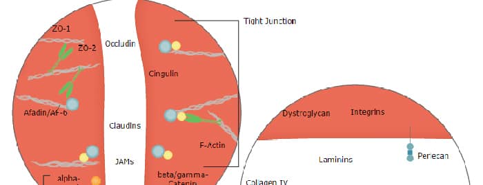
Dystroglycan, also DAG-1 (Dystrophin-associated glycoprotein 1) is a 180-200 kDa heterodimeric adhesion molecule that links the cell cytoskeleton to the extracellular matrix. It is found on skeletal muscle, cardiac muscle, fibroblasts, smooth muscle and keratinocytes. DAG-1 binds multiple matrix molecules, including laminin-1 and -2, agrin, and perlecan. Intracellularly, the cytoplasmic tail of DAG-1 contributes to a large 400 kDa complex that interacts with the cytoskeleton. The human DAG-1 preprocursor is a type I transmembrane protein 895 amino acids (aa) in length. It contains a 27 aa signal sequence plus an 868 aa proform that undergoes autocatalysis to generate a 626 aa alpha -chain (aa 28-653), and a 242 aa beta -chain. Mature DAG-1 is a heterodimer composed of noncovalently linked alpha ‑ and beta ‑chains. The alpha -chain possesses one potential Ig-like domain (aa 64-162), a mucin-like region (aa 316-485), and a peptidase S72 domain (aa 500-733). It is O‑glycosylated and runs from 100-160 kDa in SDS-PAGE. The beta -chain is N-glycosylated and runs at 42-44 kDa in SDS-Page. It possesses a short 95 aa extracellular region (aa 654-749) plus a 120 aa cytoplasmic domain (aa 776-895). Membrane cleavage of the beta -chain causes dissociation of the heterodimer and generates a 30 kDa truncated form. Over aa 28-749, human DAG-1 shares 93% aa identity with mouse DAG-1.
Human Dystroglycan Antibody
R&D Systems | Catalog # AF6868

Scientific images may be resized, cropped, or adjusted for brightness or contrast for presentation purposes. These updates are limited to presentation and do not affect the underlying scientific interpretation of the data. Where available, additional source data may be provided upon request.
Key Product Details
Validated by
Species Reactivity
Validated:
Cited:
Applications
Validated:
Cited:
Label
Antibody Source
Product Specifications
Immunogen
Gln28-Val749
Accession # Q14118
Specificity
Clonality
Host
Isotype
Scientific Data Images for Human Dystroglycan Antibody
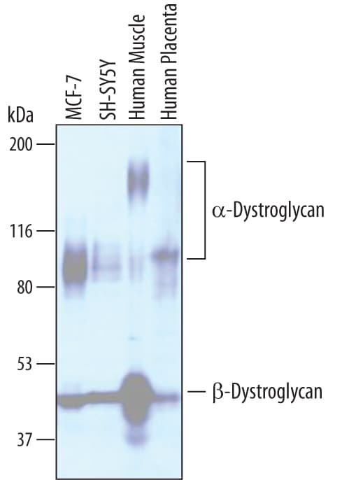
Detection of Human Dystroglycan by Western Blot.
Western blot shows lysates of MCF-7 human breast cancer cell line, SH-SY5Y human neuroblastoma cell line, human muscle tissue, and human placenta tissue. PVDF membrane was probed with 1 µg/mL of Sheep Anti-Human Dystroglycan Antigen Affinity-purified Polyclonal Antibody (Catalog # AF6868) followed by HRP-conjugated Anti-Sheep IgG Secondary Antibody (Catalog # HAF016). Specific bands were detected for a-Dystroglycan at approximately 100-160 kDa (as indicated) and beta -Dystroglycan at approximately 42-44 kDa (as indicated). This experiment was conducted under reducing conditions and using Immunoblot Buffer Group 1.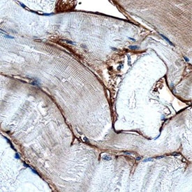
Dystroglycan in Human Skeletal Muscle.
Dystroglycan was detected in immersion fixed paraffin-embedded sections of human skeletal muscle using Sheep Anti-Human Dystroglycan Antigen Affinity-purified Polyclonal Antibody (Catalog # AF6868) at 3 µg/mL overnight at 4 °C. Before incubation with the primary antibody, tissue was subjected to heat-induced epitope retrieval using Antigen Retrieval Reagent-Basic (Catalog # CTS013). Tissue was stained using the Anti-Sheep HRP-DAB Cell & Tissue Staining Kit (brown; Catalog # CTS019) and counterstained with hematoxylin (blue). Specific staining was localized to basement membrane. View our protocol for Chromogenic IHC Staining of Paraffin-embedded Tissue Sections.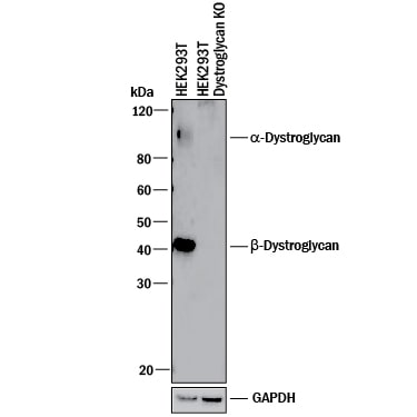
Western Blot Shows Human Dystroglycan Specificity by Using Knockout Cell Line.
Western blot shows lysates of HEK293T human embryonic kidney parental cell line and Dystroglycan knockout HEK293T cell line (KO). PVDF membrane was probed with 1 µg/mL of Sheep Anti-Human Dystroglycan Antigen Affinity-purified Polyclonal Antibody (Catalog # AF6868) followed by HRP-conjugated Anti-Sheep IgG Secondary Antibody (Catalog # HAF016). Specific bands were detected for a--Dystroglycan at approximately 110 kDa -and beta -Dystroglycan at approximately 42 kDa (as indicated) in the parental HEK293T cell line, but is not detectable in knockout HEK293T cell line. GAPDH (Catalog # AF5718) is shown as a loading control. This experiment was conducted under reducing conditions and using Immunoblot Buffer Group 1.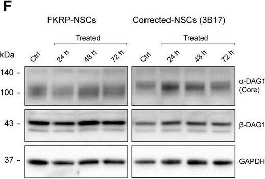
Detection of Human Dystroglycan by Western Blot
Hit compound 4BPPNit validation in the FKRP dystroglycanopathy human iPSC modelsFKRP‐NSCs treated with 4BPPNit showed increased IIH6 reactivity, compared with untreated control cells. Note that the augmented IIH6 reactivity by 4BPPNit is much weaker than that in CRISPR‐corrected‐NSCs. Under the same exposure time, the signal intensity of IIH6 reactivity is saturated in corrected‐NSCs.Representative immunoblots with the IIH6 antibody using FKRP‐ and corrected‐NSCs treated with 4BPPNit for 24, 48, and 72 h, and DMSO only controls.Quantification of alpha ‐dystroglycan glycosylation (IIH6), compared with DMSO only controls.Representative immunoblots of laminin‐binding analysis using FKRP‐ and corrected‐NSCs treated with 4BPPNit and DMSO only controls.Quantification of laminin‐binding activities, compared with DMSO only controls.Representative immunoblots with the AF6868 antibody detecting both alpha ‐dystroglycan core protein and beta ‐dystroglycan in FKRP‐ and corrected‐NSCs treated with 4BPPNit and DMSO only controls.Quantification of alpha ‐dystroglycan core protein and beta ‐dystroglycan expression, compared with DMSO only controls.Data information: Intensities of glycosylation, laminin‐binding activity, or protein expression are normalized to GAPDH protein expression. Note that the left and right panels of the immunoblots in (B) and (D) are not equivalent exposure time. See also Appendix Fig S5. Values indicate mean ± s.d. (n = 3 or 4 biological replicates; one‐way ANOVA; NS, not significant; *P < 0.05; **P < 0.01). Image collected and cropped by CiteAb from the following publication (https://pubmed.ncbi.nlm.nih.gov/31566294), licensed under a CC-BY license. Not internally tested by R&D Systems.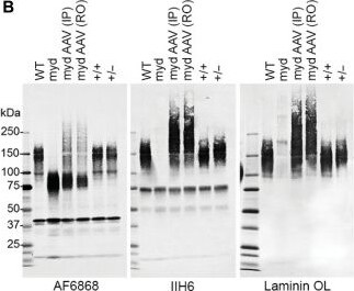
Detection of Dystroglycan by Western Blot
AAVLarge1 restores matriglycan on alpha -DG and laminin binding in skeletal muscles, which alleviates muscle pathology in myd mice.(A) Large1 expression. (B) Western blot of core alpha -DG and beta -DG (AF6868), matriglycan-positive alpha -DG (IIH6), or laminin (Laminin OL) in (A). (C) Solid-phase binding assay. (D) Representative cryosections stained with hematoxylin and eosin (H&E) or Sirius red and Fast Green, or used for immunofluorescence: beta -DG (AP83); matriglycan-positive alpha -DG (VIA41). Scale bar, 50 μm. (E to H) Quantitative analysis of sections in (D). (E) VIA41-positive fibers. (F) Connective tissue deposition. (G) Average Feret’s diameter of fibers. (H) Percentage of fibers of indicated diameter. Symbols, individual mice; bars, means ± SEM. WT, C57Bl/6J; +/+, Large+/+; +/−, Large+/−; myd, untreated myd; myd AAV, AAVLarge1-injected myd mice; IP, intraperitoneal; RO, retro-orbital. Image collected and cropped by CiteAb from the following open publication (https://pubmed.ncbi.nlm.nih.gov/35613260), licensed under a CC-BY license. Not internally tested by R&D Systems.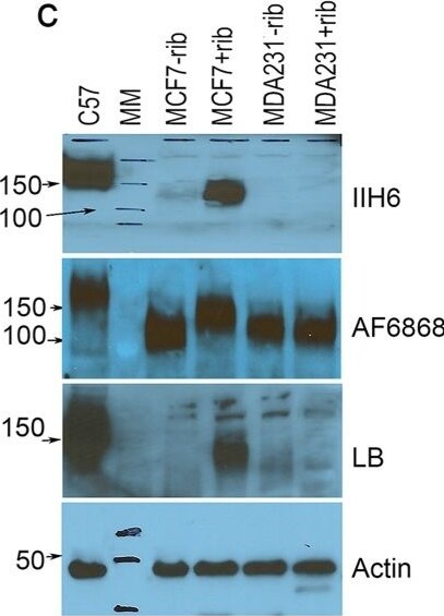
Detection of Dystroglycan by Western Blot
Analysis of matriglycan expression in MCF7 cells treated with 10 mM ribitol. Matriglycan is detected with IIH6 antibody by FACS (a) and immunocytochemistry (b). No Ab, untreated cells stained without primary antibody; + and − ribitol, cells treated with and without ribitol respectively and detected with IIH6. (c) Alpha DG is detected with antibody IIH6, AF6868 and laminin-overlay (LB). Antibody AF6868 detects core and matriglycan modified alpha -DG and shows increase in signal intensity of the higher molecular weight band and decrease in lower molecular weight band in the ribitol-treated MCF7 cells (+rib) when compared to the untreated cells (−rib). Stronger signal for laminin binding is detected in the ribitol-treated MCF7 cells over the untreated cells. Matriglycan is not detected in both treated and untreated MDA231 cells. C57, lysate from C57 skeletal muscle is used as positive control. MM, Lane of molecular weight marker. Scale bar, 50 μM. Image collected and cropped by CiteAb from the following open publication (https://pubmed.ncbi.nlm.nih.gov/32188898), licensed under a CC-BY license. Not internally tested by R&D Systems.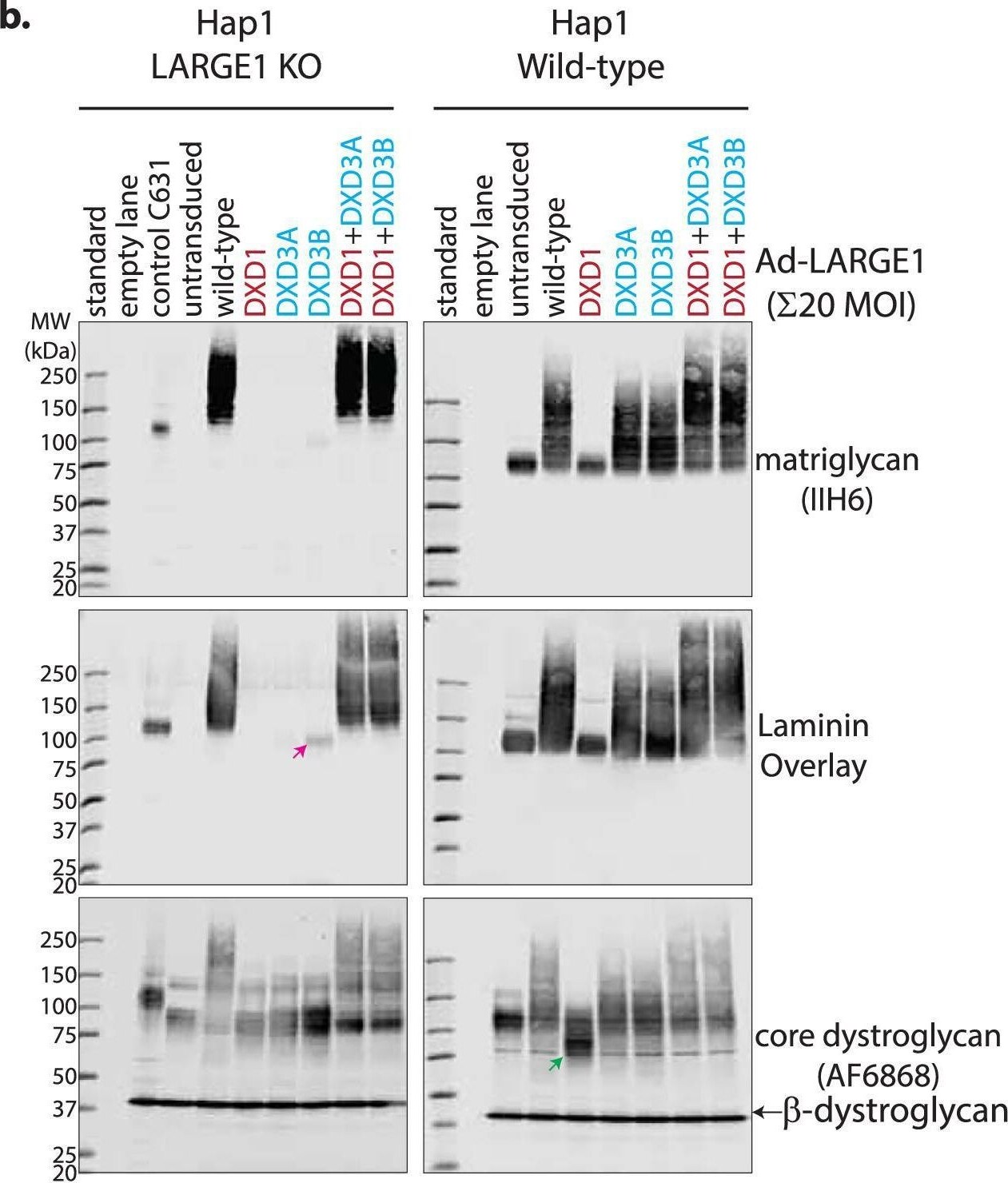
Detection of Dystroglycan by Western Blot
Mixtures of LARGE1(dTM) active site mutants can polymerize matriglycan.a Schematic of wild-type and active site mutants of LARGE1 that either lack xylose transferase activity (DXD1) or glucuronate transferase activity (DXD3). b Western blot of LARGE1 KO and wild-type Hap1 cell lines transduced with adenovirus encoding LARGE1 (20 MOI): wild-type (WT), xylose transfer mutant (DXD1; D242N/D244N), glucuronate transfer mutant (DXD3A/B; D563N or D565N, respectively) or a combination of both (DXD1 + DXD3A/B). Matriglycan was detected using an anti-matriglycan antibody (IIH6) or laminin overlay; core dystroglycan protein was detected using the AF6868 antibody. Pink arrow indicates a faint matriglycan-positive band in LARGE1 KO Hap1 cells transduced with DXD3B only. Green arrow indicates a perturbation in the migration of dystroglycan in wild-type Hap1 cells transduced with DXD1. Similar experimental results were obtained independently at least three times. c Anion exchange chromatogram of matriglycan polymerized on 4-methylumbelliferone-glucuronate-xylose by LARGE1dTM or active site mutants DXD1, DXD3, or DXD1 + DXD3. Similar experimental results were obtained independently at least three times. Source data are provided as a Source Data file. d Western blot of matriglycan polymerized on prodystroglycan constructs (as indicated) by a combination of LARGE1dTM active site mutants in the presence of UDP-sugars over time (left panel). Western blot of matriglycan polymerized on prodystroglycan (DAG128-398) after 24 h by wild-type LARGE1dTM, DXD1:DXD3 heterodimers, and a mixture of DXD12 and DXD32 mutants. Similar experimental results were obtained independently at least seven times. Image collected and cropped by CiteAb from the following open publication (https://www.nature.com/articles/s41467-025-64080-z), licensed under a CC-BY license. Not internally tested by R&D Systems.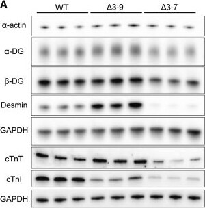
Detection of Dystroglycan by Western Blot
Western blotting analysis(A) Western blot analysis showing protein levels of dystrophin-glycoprotein complex composing proteins, including alpha -DG, beta -DG, alpha -actin, desmin, cTnT, and cardiac cTnI, in day 48 WT, delta 3–7, and delta 3–9 hiPSC-CMs. GAPDH was used as a loading control. (B) Quantification of protein expression in (A). (C) Western blot analysis showing protein levels of dystrophin-associated cardioprotective proteins, including AHNAK, Cypher, and CRYAB, in day 48 WT, delta 3–7, and delta 3–9 hiPSC-CMs. GAPDH was used as a loading control. (D) Quantification of protein expression in (C). (E) Western blot analysis of alpha -actin, desmin, TnT, AHNAK, Cypher, and CRYAB in WT and delta 3–9 hiPSC-CM lysates (input) and dystrophin IP samples. (F) Signal intensity ratio of alpha -actin, desmin, TnT, AHNAK, Cypher, and CRYAB to Dp427m in dystrophin IP samples in (E). Data are presented as mean ± SEM. ∗p < 0.05, ∗∗p < 0.01, ∗∗∗p < 0.005. Image collected and cropped by CiteAb from the following open publication (https://pubmed.ncbi.nlm.nih.gov/38028197), licensed under a CC-BY license. Not internally tested by R&D Systems.Applications for Human Dystroglycan Antibody
Immunohistochemistry
Sample: Immersion fixed paraffin-embedded sections of human skeletal muscle
Knockout Validated
Western Blot
Sample: MCF‑7 human breast cancer cell line, SH‑SY5Y human neuroblastoma cell line, human muscle tissue, and human placenta tissue
Formulation, Preparation, and Storage
Purification
Reconstitution
Sterile PBS to a final concentration of 0.2 mg/mL. For liquid material, refer to CoA for concentration.
Formulation
Shipping
Stability & Storage
- 12 months from date of receipt, -20 to -70 °C as supplied.
- 1 month, 2 to 8 °C under sterile conditions after reconstitution.
- 6 months, -20 to -70 °C under sterile conditions after reconstitution.
Calculators
Background: Dystroglycan
Long Name
Alternate Names
Gene Symbol
UniProt
Additional Dystroglycan Products
Product Documents for Human Dystroglycan Antibody
Certificate of Analysis
To download a Certificate of Analysis, please enter a lot or batch number in the search box below.
Note: Certificate of Analysis not available for kit components.
Product Specific Notices for Human Dystroglycan Antibody
For research use only
Related Research Areas
Citations for Human Dystroglycan Antibody
 Powered by Bioz
Customer Reviews for Human Dystroglycan Antibody
There are currently no reviews for this product. Be the first to review Human Dystroglycan Antibody and earn rewards!
Have you used Human Dystroglycan Antibody?
Submit a review and receive an Amazon gift card!
$25/€18/£15/$25CAN/¥2500 for a review with an image
$10/€7/£6/$10CAN/¥1110 for a review without an image
Submit a review
Protocols
Find general support by application which include: protocols, troubleshooting, illustrated assays, videos and webinars.
- Antigen Retrieval Protocol (PIER)
- Antigen Retrieval for Frozen Sections Protocol
- Appropriate Fixation of IHC/ICC Samples
- Cellular Response to Hypoxia Protocols
- Chromogenic IHC Staining of Formalin-Fixed Paraffin-Embedded (FFPE) Tissue Protocol
- Chromogenic Immunohistochemistry Staining of Frozen Tissue
- ClariTSA™ Fluorophore Kits
- Detection & Visualization of Antibody Binding
- Fluorescent IHC Staining of Frozen Tissue Protocol
- Graphic Protocol for Heat-induced Epitope Retrieval
- Graphic Protocol for the Preparation and Fluorescent IHC Staining of Frozen Tissue Sections
- Graphic Protocol for the Preparation and Fluorescent IHC Staining of Paraffin-embedded Tissue Sections
- Graphic Protocol for the Preparation of Gelatin-coated Slides for Histological Tissue Sections
- IHC Sample Preparation (Frozen sections vs Paraffin)
- Immunofluorescent IHC Staining of Formalin-Fixed Paraffin-Embedded (FFPE) Tissue Protocol
- Immunohistochemistry (IHC) and Immunocytochemistry (ICC) Protocols
- Immunohistochemistry Frozen Troubleshooting
- Immunohistochemistry Paraffin Troubleshooting
- Preparing Samples for IHC/ICC Experiments
- Preventing Non-Specific Staining (Non-Specific Binding)
- Primary Antibody Selection & Optimization
- Protocol for Heat-Induced Epitope Retrieval (HIER)
- Protocol for Making a 4% Formaldehyde Solution in PBS
- Protocol for VisUCyte™ HRP Polymer Detection Reagent
- Protocol for the Preparation & Fixation of Cells on Coverslips
- Protocol for the Preparation and Chromogenic IHC Staining of Frozen Tissue Sections
- Protocol for the Preparation and Chromogenic IHC Staining of Frozen Tissue Sections - Graphic
- Protocol for the Preparation and Chromogenic IHC Staining of Paraffin-embedded Tissue Sections
- Protocol for the Preparation and Chromogenic IHC Staining of Paraffin-embedded Tissue Sections - Graphic
- Protocol for the Preparation and Fluorescent IHC Staining of Frozen Tissue Sections
- Protocol for the Preparation and Fluorescent IHC Staining of Paraffin-embedded Tissue Sections
- Protocol for the Preparation of Gelatin-coated Slides for Histological Tissue Sections
- R&D Systems Quality Control Western Blot Protocol
- TUNEL and Active Caspase-3 Detection by IHC/ICC Protocol
- The Importance of IHC/ICC Controls
- Troubleshooting Guide: Immunohistochemistry
- Troubleshooting Guide: Western Blot Figures
- Western Blot Conditions
- Western Blot Protocol
- Western Blot Protocol for Cell Lysates
- Western Blot Troubleshooting
- Western Blot Troubleshooting Guide
- View all Protocols, Troubleshooting, Illustrated assays and Webinars
Associated Pathways