Receptors for the Fc region of IgG (Fc gamma Rs) are members of the Ig superfamily that function in the activation or inhibition of immune responses such as degranulation, phagocytosis, ADCC (antibody-dependent cellular toxicity), cytokine release, and B cell proliferation (1‑3). The Fc gamma Rs have been divided into three classes based on close relationships in their extracellular domains; these groups are designated Fc gamma RI (also known as CD64), Fc gamma RII (CD32), and Fc gamma RIII (CD16). Each group may be encoded by multiple genes and exist in different isoforms depending on species and cell type. The CD64 proteins are high affinity receptors
(~10‑8 ‑ 10‑9 M) capable of binding monomeric IgG, whereas the CD16 and CD32 proteins bind IgG with lower affinities (~10‑6 ‑ 10‑7 M) only recognizing IgG aggregates surrounding multivalent antigens (1, 4). Fc gamma Rs that deliver an activating signal either have an intrinsic immunoreceptor tyrosine-based activation motif (ITAM) within their cytoplasmic domains or associate with one of the ITAM-bearing adapter subunits, Fc R gamma or zeta (3, 5). The only inhibitory member in human and mouse, Fc gamma RIIb, has an intrinsic cytoplasmic immunoreceptor tyrosine-based inhibitory motif (ITIM). The coordinated functioning of activating and inhibitory receptors is necessary for successful initiation, amplification, and termination of immune responses (5). Three highly homologous genes (A, B, and C) sharing 98% identity at the nucleotide level have been identified for the human CD64 group (1). Fc gamma RI is transmembrane protein with three extracellular Ig-like domains, and it delivers an activating signal via the associatedFc R gamma accessory chain. The genes for Fc gamma RIB and Fc gamma RIC contain stop codons within their membrane proximal Ig-like domains indicating possible secreted receptors (1, 6). An mRNA splice variant of Fc gamma RIB has a deletion of the membrane-proximal Ig-like domain and encodes a putative transmembrane receptor (6). The high affinity recognition of IgG by Fc gamma RI permits the triggering of effector responses at low IgG concentrations typical of early immune responses (2). Fc gamma RI is expressed constitutively on monocytes and macrophages and can be induced on neutrophils and eosinophils (1, 4). Its expression is up‑regulated during bacterial infections and sepsis.
Human Fc gamma RI/CD64 Antibody (10.1)
R&D Systems | Catalog # MAB1257
Clone 10.1 was used by HLDA to establish CD designation

Key Product Details
Species Reactivity
Validated:
Human
Cited:
Human
Applications
Validated:
Flow Cytometry, Immunocytochemistry, CyTOF-ready
Cited:
Immunohistochemistry-Frozen, Neutralization, Bioassay, ELISA Detection
Label
Unconjugated
Antibody Source
Monoclonal Mouse IgG1 Clone # 10.1
Loading...
Product Specifications
Immunogen
Rheumatoid
synovial fluid cells and fibronectin purified monocytes
Specificity
Detects human Fc gamma RI/CD64.
Clonality
Monoclonal
Host
Mouse
Isotype
IgG1
Scientific Data Images for Human Fc gamma RI/CD64 Antibody (10.1)
Detection of Fc gamma RI/CD64 in Human Blood Monocytes by Flow Cytometry.
Human peripheral blood monocytes were stained with Mouse Anti-Human Fc gamma RI/CD64 Monoclonal Antibody (Catalog # MAB1257, filled histogram) or isotype control antibody (Catalog # MAB002, open histogram), followed by Phycoerythrin-conjugated Anti-Mouse IgG Secondary Antibody (Catalog # F0102B). View our protocol for Staining Membrane-associated Proteins.
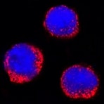
Fc gamma RI/CD64 in U937 Human Cell Line.
Fc gamma RI/CD64 was detected in immersion fixed U937 human histiocytic lymphoma cell line using Mouse Anti-Human Fc gamma RI/CD64 Monoclonal Antibody (Catalog # MAB1257) at 25 µg/mL for 3 hours at room temperature. Cells were stained using the NorthernLights™ 557-conjugated Anti-Mouse IgG Secondary Antibody (red; Catalog # NL007) and counterstained with DAPI (blue). Specific staining was localized to cytoplasm. View our protocol for Fluorescent ICC Staining of Non-adherent Cells.Applications for Human Fc gamma RI/CD64 Antibody (10.1)
Application
Recommended Usage
CyTOF-ready
Ready to be labeled using established conjugation methods. No BSA or other carrier proteins that could interfere with conjugation.
Flow Cytometry
0.25 µg/106 cells
Sample: Human peripheral blood monocytes
Sample: Human peripheral blood monocytes
Immunocytochemistry
8-25 µg/mL
Sample: Immersion fixed U937 human histiocytic lymphoma cell line and whole blood monocytes
Sample: Immersion fixed U937 human histiocytic lymphoma cell line and whole blood monocytes
Reviewed Applications
Read 1 review rated 5 using MAB1257 in the following applications:
Flow Cytometry Panel Builder
Bio-Techne Knows Flow Cytometry
Save time and reduce costly mistakes by quickly finding compatible reagents using the Panel Builder Tool.
Advanced Features
- Spectra Viewer - Custom analysis of spectra from multiple fluorochromes
- Spillover Popups - Visualize the spectra of individual fluorochromes
- Antigen Density Selector - Match fluorochrome brightness with antigen density

Formulation, Preparation, and Storage
Purification
Protein A or G purified from hybridoma culture supernatant
Reconstitution
Reconstitute at 0.5 mg/mL in sterile PBS. For liquid material, refer to CoA for concentration.
Loading...
Formulation
Lyophilized from a 0.2 μm filtered solution in PBS with Trehalose. *Small pack size (SP) is supplied either lyophilized or as a 0.2 µm filtered solution in PBS.
Shipping
Lyophilized product is shipped at ambient temperature. Liquid small pack size (-SP) is shipped with polar packs. Upon receipt, store immediately at the temperature recommended below.
Stability & Storage
Use a manual defrost freezer and avoid repeated freeze-thaw cycles.
- 12 months from date of receipt, -20 to -70 °C as supplied.
- 1 month, 2 to 8 °C under sterile conditions after reconstitution.
- 6 months, -20 to -70 °C under sterile conditions after reconstitution.
Calculators
Background: Fc gamma RI/CD64
References
- Van de Winkel, J. and P. Capes (1993) Immunol. Today 14:215.
- Raghaven, M. and P. Bjorkman (1996) Annu. Rev. Cell Dev. Biol. 12:181.
- Ravetch, J. and S. Bolland (2001) Annu. Rev. Immunol. 19:275.
- Takai, T. (2002) Nature Rev. Immunol. 2:580.
- Ravetch, J. and L. Lanier (2000) Science 290:84.
- Ernst, L. et al. (1998) Mol Immunol. 35:943.
Long Name
Fc gamma Receptor I
Alternate Names
CD64, CD64a, FCGR1, FcgRI, FcgRIA, FCRI, FcRIA
Gene Symbol
FCGR1A
Additional Fc gamma RI/CD64 Products
Product Documents for Human Fc gamma RI/CD64 Antibody (10.1)
Certificate of Analysis
To download a Certificate of Analysis, please enter a lot or batch number in the search box below.
Note: Certificate of Analysis not available for kit components.
Product Specific Notices for Human Fc gamma RI/CD64 Antibody (10.1)
For research use only
Related Research Areas
Citations for Human Fc gamma RI/CD64 Antibody (10.1)
 Powered by Bioz
Customer Reviews for Human Fc gamma RI/CD64 Antibody (10.1) (1)
5 out of 5
1 Customer Rating
Have you used Human Fc gamma RI/CD64 Antibody (10.1)?
Submit a review and receive an Amazon gift card!
$25/€18/£15/$25CAN/¥2500 Yen for a review with an image
$10/€7/£6/$10CAN/¥1110 Yen for a review without an image
Submit a review
Customer Images
Showing
1
-
1 的
1 review
Showing All
Filter By:
-
Application: Immunocytochemistry/ImmunofluorescenceSample Tested: U937 Human Cell LineSpecies: HumanVerified Customer | Posted 02/09/2022

There are no reviews that match your criteria.
Protocols
Find general support by application which include: protocols, troubleshooting, illustrated assays, videos and webinars.
- 7-Amino Actinomycin D (7-AAD) Cell Viability Flow Cytometry Protocol
- Appropriate Fixation of IHC/ICC Samples
- Cellular Response to Hypoxia Protocols
- ClariTSA™ Fluorophore Kits
- Detection & Visualization of Antibody Binding
- Extracellular Membrane Flow Cytometry Protocol
- Flow Cytometry Protocol for Cell Surface Markers
- Flow Cytometry Protocol for Staining Membrane Associated Proteins
- Flow Cytometry Staining Protocols
- Flow Cytometry Troubleshooting Guide
- ICC Cell Smear Protocol for Suspension Cells
- ICC Immunocytochemistry Protocol Videos
- ICC for Adherent Cells
- Immunocytochemistry (ICC) Protocol
- Immunocytochemistry Troubleshooting
- Immunofluorescence of Organoids Embedded in Cultrex Basement Membrane Extract
- Immunohistochemistry (IHC) and Immunocytochemistry (ICC) Protocols
- Intracellular Flow Cytometry Protocol Using Alcohol (Methanol)
- Intracellular Flow Cytometry Protocol Using Detergents
- Intracellular Nuclear Staining Flow Cytometry Protocol Using Detergents
- Intracellular Staining Flow Cytometry Protocol Using Alcohol Permeabilization
- Intracellular Staining Flow Cytometry Protocol Using Detergents to Permeabilize Cells
- Preparing Samples for IHC/ICC Experiments
- Preventing Non-Specific Staining (Non-Specific Binding)
- Primary Antibody Selection & Optimization
- Propidium Iodide Cell Viability Flow Cytometry Protocol
- Protocol for Liperfluo
- Protocol for VisUCyte™ HRP Polymer Detection Reagent
- Protocol for the Characterization of Human Th22 Cells
- Protocol for the Characterization of Human Th9 Cells
- Protocol for the Fluorescent ICC Staining of Cell Smears - Graphic
- Protocol for the Fluorescent ICC Staining of Cultured Cells on Coverslips - Graphic
- Protocol for the Preparation and Fluorescent ICC Staining of Cells on Coverslips
- Protocol for the Preparation and Fluorescent ICC Staining of Non-adherent Cells
- Protocol for the Preparation and Fluorescent ICC Staining of Stem Cells on Coverslips
- Protocol for the Preparation of a Cell Smear for Non-adherent Cell ICC - Graphic
- Protocol: Annexin V and PI Staining by Flow Cytometry
- Protocol: Annexin V and PI Staining for Apoptosis by Flow Cytometry
- TUNEL and Active Caspase-3 Detection by IHC/ICC Protocol
- The Importance of IHC/ICC Controls
- Troubleshooting Guide: Fluorokine Flow Cytometry Kits
- View all Protocols, Troubleshooting, Illustrated assays and Webinars
Loading...