
Key Product Details
Species Reactivity
Validated:
Human
Cited:
Human
Applications
Validated:
Immunohistochemistry, ELISA Capture (Matched Antibody Pair), Neutralization
Cited:
Immunohistochemistry-Frozen, Neutralization
Label
Unconjugated
Antibody Source
Monoclonal Mouse IgG1 Clone # 28105
Loading...
Product Specifications
Immunogen
Sf 21-derived recombinant human gp130 extracellular domain
Leu24-Glu619 (Glu619-Asp, predicted)
Accession # P40189
Leu24-Glu619 (Glu619-Asp, predicted)
Accession # P40189
Specificity
Detects human gp130 in ELISAs. In sandwich immunoassays, no significant cross-reactivity or interference with recombinant human (rh) CNTF, rhIL-6, rhIL-6 R, recombinant mouse (rm) IL-6, rhIL-11, rhLIF, rhLIF R, rmLIF, or rhOSM is observed. This antibody blocks the human gp130-mediated bioactivities induced by IL-6, IL-11, LIF, OSM, and CNTF.
Clonality
Monoclonal
Host
Mouse
Isotype
IgG1
Endotoxin Level
<0.10 EU per 1 μg of the antibody by the LAL method.
Scientific Data Images for Human gp130 Antibody (28105)
gp130 in Human Pancreas Tissue.
gp130 was detected in immersion fixed paraffin-embedded sections of human pancreas tissue using Mouse Anti-Human gp130 Monoclonal Antibody (Catalog # MAB628) at 5 µg/mL overnight at 4 °C. Before incubation with the primary antibody, tissue was subjected to heat-induced epitope retrieval using Antigen Retrieval Reagent-Basic (Catalog # CTS013). Tissue was stained using the Anti-Mouse IgG VisUCyte™ HRP Polymer Antibody (brown; Catalog # VC001) and counterstained with hematoxylin (blue). Specific staining was localized to cytoplasm in exocrine cells. View our protocol for IHC Staining with VisUCyte HRP Polymer Detection Reagents.
Cell Proliferation Induced by Oncostatin M/OSM and Neutralization by Human gp130 Antibody.
Recombinant Human Oncostatin M/OSM stimulates proliferation in the TF-1 human erythroleukemic cell line in a dose-dependent manner (orange line), as measured by Resazurin (Catalog # AR002). Proliferation elicited by Recombinant Human Oncostatin M/OSM (0.8 ng/mL) is neutralized (green line) by increasing concentrations of Mouse Anti-Human gp130 Monoclonal Antibody (Catalog # MAB628). The ND50 is typically 0.02-0.1 µg/mL.
Human gp130 ELISA Standard Curve
Recombinant Human gp130 (Catalog # 228-GP) was serially diluted and captured by Mouse Anti-Human gp130 Monoclonal Antibody (Catalog # MAB628) coated on a Clear Polystyrene Microplate (Catalog # DY990). Goat Anti-Human gp130 Antigen Affinity-purified Polyclonal Antibody (Catalog # AF-228-NA) was biotinylated and incubated with the protein captured on the plate. Detection of the standard curve was achieved by incubating Streptavidin-HRP (Catalog # DY998)Applications for Human gp130 Antibody (28105)
Application
Recommended Usage
Immunohistochemistry
5-25 µg/mL
Sample: Immersion fixed paraffin-embedded sections of human pancreas tissue
Sample: Immersion fixed paraffin-embedded sections of human pancreas tissue
Neutralization
Measured by its ability to neutralize Oncostatin M/OSM-induced proliferation in the TF‑1 human erythroleukemic cell line. Kitamura, T. et al. (1989) J. Cell Physiol. 140:323. The Neutralization Dose (ND50) is typically 0.02-0.1 µg/mL in the presence of 0.8 ng/mL Recombinant Human Oncostatin M/OSM.
Human gp130 Sandwich Immunoassay
Please Note: Optimal dilutions of this antibody should be experimentally determined.
Reviewed Applications
Read 4 reviews rated 4 using MAB628 in the following applications:
Formulation, Preparation, and Storage
Purification
Protein A or G purified from ascites
Reconstitution
Reconstitute at 0.5 mg/mL in sterile PBS. For liquid material, refer to CoA for concentration.
Loading...
Formulation
Lyophilized from a 0.2 μm filtered solution in PBS. *Small pack size (SP) is supplied either lyophilized or as a 0.2 µm filtered solution in PBS.
Shipping
Lyophilized product is shipped at ambient temperature. Liquid small pack size (-SP) is shipped with polar packs. Upon receipt, store immediately at the temperature recommended below.
Stability & Storage
Use a manual defrost freezer and avoid repeated freeze-thaw cycles.
- 12 months from date of receipt, -20 to -70 °C as supplied.
- 1 month, 2 to 8 °C under sterile conditions after reconstitution.
- 6 months, -20 to -70 °C under sterile conditions after reconstitution.
Calculators
Background: gp130
Long Name
Glycoprotein 130
Alternate Names
CD130, IL6ST
Gene Symbol
IL6ST
UniProt
Additional gp130 Products
Product Documents for Human gp130 Antibody (28105)
Certificate of Analysis
To download a Certificate of Analysis, please enter a lot or batch number in the search box below.
Note: Certificate of Analysis not available for kit components.
Product Specific Notices for Human gp130 Antibody (28105)
For research use only
Citations for Human gp130 Antibody (28105)
 Powered by Bioz
Customer Reviews for Human gp130 Antibody (28105) (4)
4 out of 5
4 Customer Ratings
Have you used Human gp130 Antibody (28105)?
Submit a review and receive an Amazon gift card!
$25/€18/£15/$25CAN/¥2500 Yen for a review with an image
$10/€7/£6/$10CAN/¥1110 Yen for a review without an image
Submit a review
Customer Images
Showing
1
-
4 的
4 reviews
Showing All
Filter By:
-
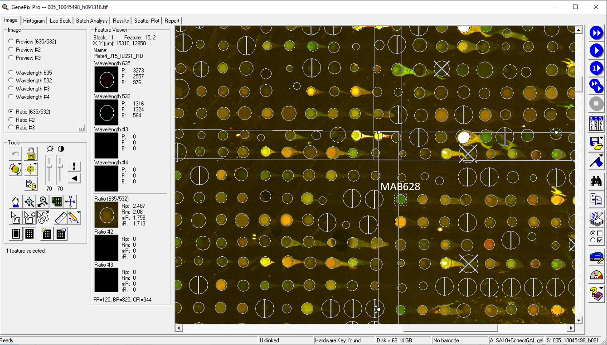
Application: MicroarraysSample Tested: EDTA PlasmaSpecies: HumanVerified Customer | Posted 06/10/2020

-
Application: MicroarraySample Tested: EDTA PlasmaSpecies: HumanVerified Customer | Posted 01/09/2019
-
Application: ELISASample Tested: Plasma and SerumSpecies: Human and MouseVerified Customer | Posted 11/08/2018
-
Application: MicroarraysSample Tested: EDTA PlasmaSpecies: HumanVerified Customer | Posted 11/07/2018
There are no reviews that match your criteria.
Protocols
Find general support by application which include: protocols, troubleshooting, illustrated assays, videos and webinars.
- Antigen Retrieval Protocol (PIER)
- Antigen Retrieval for Frozen Sections Protocol
- Appropriate Fixation of IHC/ICC Samples
- Cellular Response to Hypoxia Protocols
- Chromogenic IHC Staining of Formalin-Fixed Paraffin-Embedded (FFPE) Tissue Protocol
- Chromogenic Immunohistochemistry Staining of Frozen Tissue
- ClariTSA™ Fluorophore Kits
- Detection & Visualization of Antibody Binding
- Fluorescent IHC Staining of Frozen Tissue Protocol
- Graphic Protocol for Heat-induced Epitope Retrieval
- Graphic Protocol for the Preparation and Fluorescent IHC Staining of Frozen Tissue Sections
- Graphic Protocol for the Preparation and Fluorescent IHC Staining of Paraffin-embedded Tissue Sections
- Graphic Protocol for the Preparation of Gelatin-coated Slides for Histological Tissue Sections
- IHC Sample Preparation (Frozen sections vs Paraffin)
- Immunofluorescent IHC Staining of Formalin-Fixed Paraffin-Embedded (FFPE) Tissue Protocol
- Immunohistochemistry (IHC) and Immunocytochemistry (ICC) Protocols
- Immunohistochemistry Frozen Troubleshooting
- Immunohistochemistry Paraffin Troubleshooting
- Preparing Samples for IHC/ICC Experiments
- Preventing Non-Specific Staining (Non-Specific Binding)
- Primary Antibody Selection & Optimization
- Protocol for Heat-Induced Epitope Retrieval (HIER)
- Protocol for Making a 4% Formaldehyde Solution in PBS
- Protocol for VisUCyte™ HRP Polymer Detection Reagent
- Protocol for the Preparation & Fixation of Cells on Coverslips
- Protocol for the Preparation and Chromogenic IHC Staining of Frozen Tissue Sections
- Protocol for the Preparation and Chromogenic IHC Staining of Frozen Tissue Sections - Graphic
- Protocol for the Preparation and Chromogenic IHC Staining of Paraffin-embedded Tissue Sections
- Protocol for the Preparation and Chromogenic IHC Staining of Paraffin-embedded Tissue Sections - Graphic
- Protocol for the Preparation and Fluorescent IHC Staining of Frozen Tissue Sections
- Protocol for the Preparation and Fluorescent IHC Staining of Paraffin-embedded Tissue Sections
- Protocol for the Preparation of Gelatin-coated Slides for Histological Tissue Sections
- TUNEL and Active Caspase-3 Detection by IHC/ICC Protocol
- The Importance of IHC/ICC Controls
- Troubleshooting Guide: Immunohistochemistry
- View all Protocols, Troubleshooting, Illustrated assays and Webinars


