Interleukin 4 receptor (IL-4 R) is the transmembrane ligand binding subunit of the heterodimeric IL-4 receptor complex. In the type I receptor complex, IL-4 R associates with the common gamma chain, whereas in the type II receptor complex, it associates with IL-13 R alpha 1. IL-4 R is expressed by B cells, T cells, monocytes and macrophages.
Human IL-4R alpha Antibody (25463)
R&D Systems | Catalog # MAB230

Key Product Details
Species Reactivity
Validated:
Human
Cited:
Human, Mouse, Rat
Applications
Validated:
Immunohistochemistry, Western Blot, Neutralization, Flow Cytometry, Immunocytochemistry, CyTOF-ready
Cited:
Immunohistochemistry, Immunohistochemistry-Paraffin, Immunohistochemistry-Frozen, Western Blot, Neutralization, Flow Cytometry, Immunocytochemistry, Binding Assay, Bioassay, Blocking, In vivo assay
Label
Unconjugated
Antibody Source
Monoclonal Mouse IgG2A Clone # 25463
Loading...
Product Specifications
Immunogen
S. frugiperda insect ovarian cell line Sf 21-derived recombinant human IL‑4 R
Gly24-His232
Accession # P24394
Gly24-His232
Accession # P24394
Specificity
Detects human IL‑4 R alpha in direct ELISAs and Western blots. In direct ELISAs, no cross-reactivity with recombinant human (rh) IL-9 R, recombinant mouse IL-4 R, rhIL-5 R alpha, rhIL-5 R beta, rhIL-13 R alpha 1, or rhIL-13 R alpha 2 is observed.
Clonality
Monoclonal
Host
Mouse
Isotype
IgG2A
Endotoxin Level
<0.10 EU per 1 μg of the antibody by the LAL method.
Scientific Data Images for Human IL-4R alpha Antibody (25463)
Detection of IL‑4 R alpha in Daudi Human Cell Line by Flow Cytometry.
Daudi human Burkitt's lymphoma cell line was stained with Mouse Anti-Human IL-4 Ra Monoclonal Antibody (Catalog # MAB230, filled histogram) or isotype control antibody (Catalog # MAB003, open histogram), followed by Phycoerythrin-conjugated Anti-Mouse IgG Secondary Antibody (Catalog # F0102B).
IL‑4 R alpha in Human Lymph Node.
IL-4 Ra was detected in immersion fixed paraffin-embedded sections of human lymph node using Mouse Anti-Human IL-4 Ra Monoclonal Antibody (Catalog # MAB230) at 25 µg/mL overnight at 4 °C. Tissue was stained using the Anti-Mouse HRP-DAB Cell & Tissue Staining Kit (brown; Catalog # CTS002) and counterstained with hematoxylin (blue). Specific staining was localized to plasma cells. View our protocol for Chromogenic IHC Staining of Paraffin-embedded Tissue Sections.
IL‑4 R alpha in HDLM‑2 Human Cell Line.
IL-4 Ra was detected in immersion fixed HDLM-2 human Hodgkin's lymphoma cell line using Mouse Anti-Human IL-4 Ra Monoclonal Antibody (Catalog # MAB230) at 8 µg/mL for 3 hours at room temperature. Cells were stained using the NorthernLights™ 557-conjugated Anti-Mouse IgG Secondary Antibody (red; Catalog # NL007) and counterstained with DAPI (blue). Specific staining was localized to cytoplasm. View our protocol for Fluorescent ICC Staining of Non-adherent Cells.
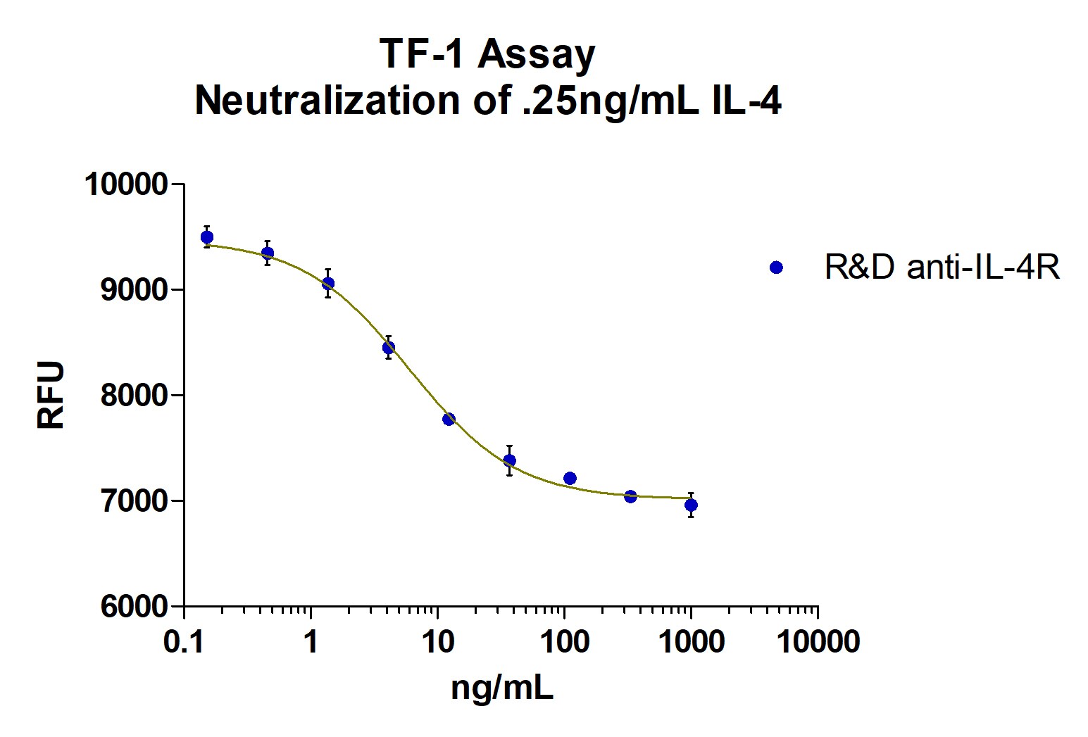
Cell Proliferation Induced by IL‑4 and Neutralization by Human IL‑4 R alpha Antibody.
Recombinant Human IL‑4 (Catalog # 204-IL) stimulates proliferation in the TF‑1 human erythroleukemic cell line in a dose-dependent manner (orange line). Proliferation elicited by Recombinant Human IL‑4 (0.2 ng/mL) is neutralized (green line) by increasing concentrations of Mouse Anti-Human IL‑4 Ra Monoclonal Antibody (Catalog # MAB230). The ND50 is typically 3-6 ng/mL.
Detection of Human IL-4R alpha by Flow Cytometry
In vitro binding assays of [V14]6 and [AP1-V12]6 polymer.(A, B) H226, (C, D) MDA-MB-231 and (E, F) H460 cells were incubated with 10 µM of [V14]6, [AP1-V12]6 and AP1 for 1 h at 4°C. Cell binding was determined using flow cytometry. Histograms are representative of three independent experiments. Graphical bars (on right) represent the percent of Alexa 488 labeled polymer bound to cells as mean ±SD of data obtained from three separate experiments performed in triplicates. ***P<0.0001, **P<0.001, and *P<0.05, one-way ANOVA; n = 3. (G, H) H226 cells (3×10⁵ cells) were pre- incubated with different concentrations (1, 5 and 10 µg/mL) of anti-IL-4 receptor antibody followed by 1 h incubation with 10 µM Alexa-labeled [AP1-V12]6 at 4°C. The cells were further suspended in 300 µL of PBS after washing and analyzed using flow cytometry. Histograms are representative of three independent experiments. Graphical bars represent the percent of Alexa 488 labeled polymer bound to cells as mean ±SD of data obtained from three separate experiments performed in triplicates. ***P<0.0001, One way ANOVA; n = 3. Image collected and cropped by CiteAb from the following publication (https://dx.plos.org/10.1371/journal.pone.0081891), licensed under a CC-BY license. Not internally tested by R&D Systems.
Detection of Human IL-4R alpha by Immunocytochemistry/Immunofluorescence
In vivo, ex vivo imaging and biodistribution of [AP1-V12]6 polymers.(A) Cy5.5-labeled [V14]6 and [AP1-V12]6 (8 mg/kg) were intravenously injected into MDA-MB-231 tumor xenografted nude mice. Biodistribution was determined by collecting in vivo fluorescence images at different time points. Scale bar indicates normalized fluorescence intensity (NC). Representative optical images of three experiments. (B) Quantitation of fluorescence intensities in tumor sites at respective time points (n = 3). (C) Analysis of fluorescence intensities for tumors and organs from ex vivo images (n = 3). (D) Histological analysis of [AP1-V12]6 polymer (red) localization in tumors. Nuclei were stained with DAPI (blue), and IL-4R expression on cells was visualized by anti-IL-4 receptor antibody staining (green). Representative confocal images of three experiments (scale bar 50 µm). Image collected and cropped by CiteAb from the following publication (https://dx.plos.org/10.1371/journal.pone.0081891), licensed under a CC-BY license. Not internally tested by R&D Systems.Applications for Human IL-4R alpha Antibody (25463)
Application
Recommended Usage
CyTOF-ready
Ready to be labeled using established conjugation methods. No BSA or other carrier proteins that could interfere with conjugation.
Flow Cytometry
0.25 µg/106 cells
Sample: Daudi human Burkitt's lymphoma cell line
Sample: Daudi human Burkitt's lymphoma cell line
Immunocytochemistry
5-15 µg/mL
Sample: Immersion fixed HDLM-2 human Hodgkin's lymphoma cell line
Sample: Immersion fixed HDLM-2 human Hodgkin's lymphoma cell line
Immunohistochemistry
8-25 µg/mL
Sample: Immersion fixed paraffin-embedded sections of human lymph node
Sample: Immersion fixed paraffin-embedded sections of human lymph node
Western Blot
1 µg/mL
Sample: Recombinant Human IL‑4 R alpha (Catalog # 230-4R) under non-reducing conditions only
Sample: Recombinant Human IL‑4 R alpha (Catalog # 230-4R) under non-reducing conditions only
Neutralization
Measured by its ability to neutralize IL‑4-induced proliferation in the TF‑1 human erythroleukemic cell line. Kitamura, T. et al. (1989) J. Cell Physiol. 140:323. The Neutralization Dose (ND50) is typically 3-6 ng/mL in the presence of 0.2 ng/mL Recombinant Human IL‑4.
Reviewed Applications
Read 4 reviews rated 4.5 using MAB230 in the following applications:
Flow Cytometry Panel Builder
Bio-Techne Knows Flow Cytometry
Save time and reduce costly mistakes by quickly finding compatible reagents using the Panel Builder Tool.
Advanced Features
- Spectra Viewer - Custom analysis of spectra from multiple fluorochromes
- Spillover Popups - Visualize the spectra of individual fluorochromes
- Antigen Density Selector - Match fluorochrome brightness with antigen density

Formulation, Preparation, and Storage
Purification
Protein A or G purified from ascites
Reconstitution
Reconstitute at 0.5 mg/mL in sterile PBS. For liquid material, refer to CoA for concentration.
Loading...
Formulation
Lyophilized from a 0.2 μm filtered solution in PBS with Trehalose. *Small pack size (SP) is supplied either lyophilized or as a 0.2 µm filtered solution in PBS.
Shipping
Lyophilized product is shipped at ambient temperature. Liquid small pack size (-SP) is shipped with polar packs. Upon receipt, store immediately at the temperature recommended below.
Stability & Storage
Use a manual defrost freezer and avoid repeated freeze-thaw cycles.
- 12 months from date of receipt, -20 to -70 °C as supplied.
- 1 month, 2 to 8 °C under sterile conditions after reconstitution.
- 6 months, -20 to -70 °C under sterile conditions after reconstitution.
Calculators
Background: IL-4R alpha
Long Name
Interleukin 4 Receptor alpha
Alternate Names
CD124, IL-4 R alpha, IL-4Ra, IL4R, IL4R alpha
Gene Symbol
IL4R
UniProt
Additional IL-4R alpha Products
Product Documents for Human IL-4R alpha Antibody (25463)
Certificate of Analysis
To download a Certificate of Analysis, please enter a lot or batch number in the search box below.
Note: Certificate of Analysis not available for kit components.
Product Specific Notices for Human IL-4R alpha Antibody (25463)
For research use only
Related Research Areas
Citations for Human IL-4R alpha Antibody (25463)
 Powered by Bioz
Customer Reviews for Human IL-4R alpha Antibody (25463) (4)
4.5 out of 5
4 Customer Ratings
Have you used Human IL-4R alpha Antibody (25463)?
Submit a review and receive an Amazon gift card!
$25/€18/£15/$25CAN/¥2500 Yen for a review with an image
$10/€7/£6/$10CAN/¥1110 Yen for a review without an image
Submit a review
Customer Images
Showing
1
-
4 的
4 reviews
Showing All
Filter By:
-
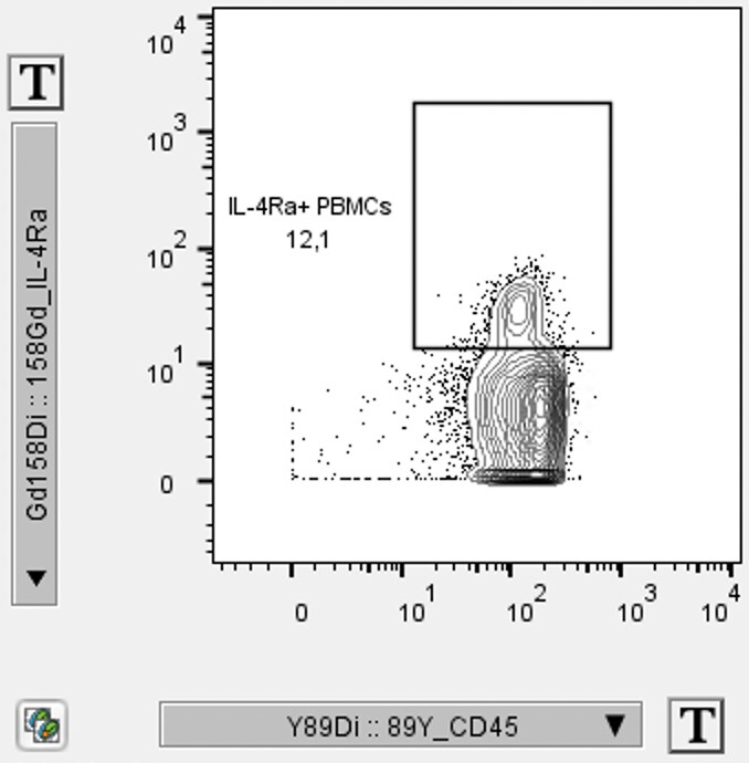
Application: CyTOFSample Tested: Peripheral blood mononuclear cells (PBMCs)Species: HumanVerified Customer | Posted 05/04/2022Gated on CD45+ PBMCs

-
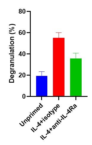
Application: Block/NeutralizeSample Tested: LAD2 human mast cellsSpecies: HumanVerified Customer | Posted 04/07/2022
-
Application: Western BlotSample Tested: Colon cancer cellsSpecies: HumanVerified Customer | Posted 08/19/2021
-
Application: Block/NeutralizeSample Tested: Recombinant proteinSpecies: HumanVerified Customer | Posted 12/06/2017
There are no reviews that match your criteria.
Protocols
Find general support by application which include: protocols, troubleshooting, illustrated assays, videos and webinars.
- 7-Amino Actinomycin D (7-AAD) Cell Viability Flow Cytometry Protocol
- Antigen Retrieval Protocol (PIER)
- Antigen Retrieval for Frozen Sections Protocol
- Appropriate Fixation of IHC/ICC Samples
- Cellular Response to Hypoxia Protocols
- Chromogenic IHC Staining of Formalin-Fixed Paraffin-Embedded (FFPE) Tissue Protocol
- Chromogenic Immunohistochemistry Staining of Frozen Tissue
- ClariTSA™ Fluorophore Kits
- Detection & Visualization of Antibody Binding
- Extracellular Membrane Flow Cytometry Protocol
- Flow Cytometry Protocol for Cell Surface Markers
- Flow Cytometry Protocol for Staining Membrane Associated Proteins
- Flow Cytometry Staining Protocols
- Flow Cytometry Troubleshooting Guide
- Fluorescent IHC Staining of Frozen Tissue Protocol
- Graphic Protocol for Heat-induced Epitope Retrieval
- Graphic Protocol for the Preparation and Fluorescent IHC Staining of Frozen Tissue Sections
- Graphic Protocol for the Preparation and Fluorescent IHC Staining of Paraffin-embedded Tissue Sections
- Graphic Protocol for the Preparation of Gelatin-coated Slides for Histological Tissue Sections
- ICC Cell Smear Protocol for Suspension Cells
- ICC Immunocytochemistry Protocol Videos
- ICC for Adherent Cells
- IHC Sample Preparation (Frozen sections vs Paraffin)
- Immunocytochemistry (ICC) Protocol
- Immunocytochemistry Troubleshooting
- Immunofluorescence of Organoids Embedded in Cultrex Basement Membrane Extract
- Immunofluorescent IHC Staining of Formalin-Fixed Paraffin-Embedded (FFPE) Tissue Protocol
- Immunohistochemistry (IHC) and Immunocytochemistry (ICC) Protocols
- Immunohistochemistry Frozen Troubleshooting
- Immunohistochemistry Paraffin Troubleshooting
- Intracellular Flow Cytometry Protocol Using Alcohol (Methanol)
- Intracellular Flow Cytometry Protocol Using Detergents
- Intracellular Nuclear Staining Flow Cytometry Protocol Using Detergents
- Intracellular Staining Flow Cytometry Protocol Using Alcohol Permeabilization
- Intracellular Staining Flow Cytometry Protocol Using Detergents to Permeabilize Cells
- Preparing Samples for IHC/ICC Experiments
- Preventing Non-Specific Staining (Non-Specific Binding)
- Primary Antibody Selection & Optimization
- Propidium Iodide Cell Viability Flow Cytometry Protocol
- Protocol for Heat-Induced Epitope Retrieval (HIER)
- Protocol for Liperfluo
- Protocol for Making a 4% Formaldehyde Solution in PBS
- Protocol for VisUCyte™ HRP Polymer Detection Reagent
- Protocol for the Characterization of Human Th22 Cells
- Protocol for the Characterization of Human Th9 Cells
- Protocol for the Fluorescent ICC Staining of Cell Smears - Graphic
- Protocol for the Fluorescent ICC Staining of Cultured Cells on Coverslips - Graphic
- Protocol for the Preparation & Fixation of Cells on Coverslips
- Protocol for the Preparation and Chromogenic IHC Staining of Frozen Tissue Sections
- Protocol for the Preparation and Chromogenic IHC Staining of Frozen Tissue Sections - Graphic
- Protocol for the Preparation and Chromogenic IHC Staining of Paraffin-embedded Tissue Sections
- Protocol for the Preparation and Chromogenic IHC Staining of Paraffin-embedded Tissue Sections - Graphic
- Protocol for the Preparation and Fluorescent ICC Staining of Cells on Coverslips
- Protocol for the Preparation and Fluorescent ICC Staining of Non-adherent Cells
- Protocol for the Preparation and Fluorescent ICC Staining of Stem Cells on Coverslips
- Protocol for the Preparation and Fluorescent IHC Staining of Frozen Tissue Sections
- Protocol for the Preparation and Fluorescent IHC Staining of Paraffin-embedded Tissue Sections
- Protocol for the Preparation of Gelatin-coated Slides for Histological Tissue Sections
- Protocol for the Preparation of a Cell Smear for Non-adherent Cell ICC - Graphic
- Protocol: Annexin V and PI Staining by Flow Cytometry
- Protocol: Annexin V and PI Staining for Apoptosis by Flow Cytometry
- R&D Systems Quality Control Western Blot Protocol
- TUNEL and Active Caspase-3 Detection by IHC/ICC Protocol
- The Importance of IHC/ICC Controls
- Troubleshooting Guide: Fluorokine Flow Cytometry Kits
- Troubleshooting Guide: Immunohistochemistry
- Troubleshooting Guide: Western Blot Figures
- Western Blot Conditions
- Western Blot Protocol
- Western Blot Protocol for Cell Lysates
- Western Blot Troubleshooting
- Western Blot Troubleshooting Guide
- View all Protocols, Troubleshooting, Illustrated assays and Webinars
Loading...
Associated Pathways


