
Nurr1/NGFI-B beta/NR4A2 Antibody
R&D Systems | Catalog # AF2156


Scientific images may be resized, cropped, or adjusted for brightness or contrast for presentation purposes. These updates are limited to presentation and do not affect the underlying scientific interpretation of the data. Where available, additional source data may be provided upon request.
Key Product Details
Species Reactivity
Validated:
Cited:
Applications
Validated:
Cited:
Label
Antibody Source
Product Specifications
Immunogen
Val332-Lys558
Accession # Q06219
Specificity
Clonality
Host
Isotype
Scientific Data Images for Nurr1/NGFI-B beta/NR4A2 Antibody
Nurr1/NGFI‑B beta /NR4A2 in HeLa Human Cell Line.
Nurr1/NGFI‑B beta /NR4A2 was detected in immersion fixed HeLa human cervical epithelial carcinoma cell line using Goat Anti-Human/Mouse Nurr1/NGFI‑B beta /NR4A2 Antigen Affinity-purified Polyclonal Antibody (Catalog # AF2156) at 10 µg/mL for 3 hours at room temperature. Cells were stained using the NorthernLights™ 557-conjugated Anti-Goat IgG Secondary Antibody (red; NL001) and counterstained with DAPI (blue). Specific staining was localized to cytoplasm and nuclei. Staining was performed using our protocol for Fluorescent ICC Staining of Non-adherent Cells.
Nurr1/NGFI‑B beta /NR4A2 in Mouse Brain (Hippocampus).
Nurr1/NGFI‑B beta /NR4A2 was detected in immersion fixed paraffin-embedded sections of mouse brain (hippocampus) using Goat Anti-Human/Mouse Nurr1/NGFI‑B beta /NR4A2 Antigen Affinity-purified Polyclonal Antibody (Catalog # AF2156) at 15 µg/mL for 1 hour at room temperature followed by incubation with the Anti-Goat IgG VisUCyte™ HRP Polymer Antibody (VC004). Before incubation with the primary antibody, tissue was subjected to heat-induced epitope retrieval using Antigen Retrieval Reagent-Basic (CTS013). Tissue was stained using DAB (brown) and counterstained with hematoxylin (blue). Specific staining was localized to cytoplasm in neurons. Staining was performed using our protocol for IHC Staining with VisUCyte HRP Polymer Detection Reagents.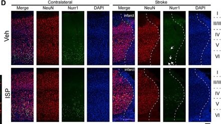
Detection of fincorouzovyalomrgkg.jpg by Immunocytochemistry/ Immunofluorescence
RNA-seq from the peri-infarct cortex of ISP treated versus Veh-treated strokemice shows differentially regulated genes(a) DEGs that can be clustered into GO pathways (B) such as regulatorsof apoptotic signaling pathways, axon development pathways, and pathways thatare involved in responses to stress.(C) Validation of the top selected gene expression by qRT-PCR (n = 7 foreach group).(D–G) Nurr1 (Nr4a2) expression is decreased in peri-infarctcortex in stroke mice (arrows in D) but partially restored in ISP-treated mice(n = 4 for each group). Nurr1 expression is mainly detected in theNeuN+ neurons in the peri-infarct zone.(H–J) Bcl2 expression is upregulated in ISP-treated peri-infarctzone enriched in Iba1+ cells not GFAP+ reactive astrocytes(n = 4 for each group). *p < 0.05, **p < 0.01, ***p <0.001, Student’s t test for (C) and 2-way ANOVA for (J) and (G). Scalebar=50 μm.See Table S2for complete list of DEGs. Image collected and cropped by CiteAb from the following open publication (https://linkinghub.elsevier.com/retrieve/pii/S2211124722009469), licensed under a CC-BY license. Not internally tested by R&D Systems.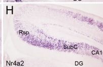
Detection of jjgjbaa1wgvxctil5wru.jpg by Immunohistochemistry
Rsp neurons lose their identity in Satb2 CKO mice. FN1 is restricted to the SubC of controls (a) but expressed by neurons in the Rsp region of Satb2 CKO mice at P4 (b). c–h ER81, Citp2, and Nr4a2 are expressed in the SubC and specific layers of the Rsp in control mice (c, e, g). In Satb2 CKO mice, however, they are expressed throughout the Rsp regions while remain largely unchanged in the SubC (d, f, h). Cbln1 shows restricted expression in the Rsp of control mice (i), but its expression is lost in Satb2 CKO mice (j). Zbtb20 is expressed in superficial Rsp and CA1 of control mice (k), but undetectable in the Rsp region while remains unchanged in the CA1 of Satb2 CKO mice (l). Note that ectopic Zbtb20+ cells (arrows) are present in the subicular region of Satb2 CKO mice. Mef2C is intensely expressed in the Rsp and weakly in the SubC of control mice (m), but its expression was largely reduced in the Rsp region to a comparable cellular density of the SubC in Satb2 CKO mice (n). Dkk3 is strongly expressed in upper Rsp of control mice (o) but absent in the Rsp region of Satb2 CKO mice (p). Intense Lmo4 expression is reduced in the Rsp regions with a similar intensity to that in the SubC in Satb2 CKO mice (r) compared with controls (q). s, t Diagram of Rsp fate change in Satb2 CKO mice. DG dentate gyrus. n = 3 mice for each genotype. Scale bar = 500 μm Image collected and cropped by CiteAb from the following open publication (https://pubmed.ncbi.nlm.nih.gov/31666685), licensed under a CC-BY license. Not internally tested by R&D Systems.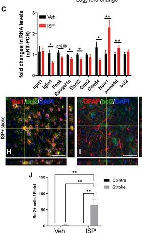
Detection of Nurr1/NGFI-B beta/NR4A2 by Immunohistochemistry
RNA-seq from the peri-infarct cortex of ISP treated versus Veh-treated strokemice shows differentially regulated genes(a) DEGs that can be clustered into GO pathways (B) such as regulatorsof apoptotic signaling pathways, axon development pathways, and pathways thatare involved in responses to stress.(C) Validation of the top selected gene expression by qRT-PCR (n = 7 foreach group).(D–G) Nurr1 (Nr4a2) expression is decreased in peri-infarctcortex in stroke mice (arrows in D) but partially restored in ISP-treated mice(n = 4 for each group). Nurr1 expression is mainly detected in theNeuN+ neurons in the peri-infarct zone.(H–J) Bcl2 expression is upregulated in ISP-treated peri-infarctzone enriched in Iba1+ cells not GFAP+ reactive astrocytes(n = 4 for each group). *p < 0.05, **p < 0.01, ***p <0.001, Student’s t test for (C) and 2-way ANOVA for (J) and (G). Scalebar=50 μm.See Table S2for complete list of DEGs. Image collected and cropped by CiteAb from the following open publication (https://linkinghub.elsevier.com/retrieve/pii/S2211124722009469), licensed under a CC-BY license. Not internally tested by R&D Systems.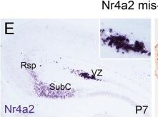
Detection of Nurr1/NGFI-B beta/NR4A2 by Immunohistochemistry
Satb2 determines Rsp fate by repressing Ctip2 and Nr4a2. a Diagram of Satb2-binding MARs in Nr4a2 genomic locus. In vivo ChIP assay using embryonic mouse cortex shows a remarkable enrichment of Satb2-binding MARs in Nr4a2 locus. *p < 0.05, **p < 0.01 (n = 3), error bars represent S.E.M. b Luciferase reporter assay shows that the transcriptional activities of Ctip2-A4 (positive control) and four Nr4a2 MARs are significantly reduced in the presence of Satb2. **p < 0.01 (n = 3), error bars represent S.E.M. c–i′ Ctip2- and/or Nr4a2-expression plasmids together with GFP are delivered into Rsp neurons by in utero electroporation in wild-type mice at E14.5 and pups are analyzed at P7. To make signals more clearly to compare, mRNA signals were photographed first, and then immunostaining of GFP was performed and photographed again. c, c′ Ctip2 is misexpressed in wild-type Rsp, and some of them are clumped. Inserts show Ctip2 expression in GFP+ cells (arrowheads). d, d′ A few Ctip2-misexpressed neurons contain FN1 transcripts and they often reside in ventral Rsp (arrowheads). Most of Rsp neurons with misexpression of Nr4a2 are arrested in VZ (e), and none of them contains FN1 transcripts in wild-type Rsp (f). Rsp neurons with co-misexpression of Ctip2 and Nr4a2 initiate FN1 expression in the VZ of Rsp (g, g′) and neocortical region (h, h′). i, i′ EphA6 is also ectopically induced in wild-type Rsp neurons when Ctip2 and Nr4a2 are co-misexpressed. VZ ventricular zone; Ncx neocortex. n = 3 mice for each group. Scale bars = 200 μm in c′, d′, and i′, 50 μm in inserts of c′, d′, and i′, 400 μm in f, and 100 μm in inserts of f Image collected and cropped by CiteAb from the following open publication (https://pubmed.ncbi.nlm.nih.gov/31666685), licensed under a CC-BY license. Not internally tested by R&D Systems.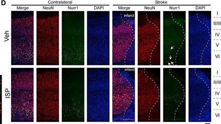
Detection of Nurr1/NGFI-B beta/NR4A2 by Immunocytochemistry/ Immunofluorescence
RNA-seq from the peri-infarct cortex of ISP treated versus Veh-treated strokemice shows differentially regulated genes(a) DEGs that can be clustered into GO pathways (B) such as regulatorsof apoptotic signaling pathways, axon development pathways, and pathways thatare involved in responses to stress.(C) Validation of the top selected gene expression by qRT-PCR (n = 7 foreach group).(D–G) Nurr1 (Nr4a2) expression is decreased in peri-infarctcortex in stroke mice (arrows in D) but partially restored in ISP-treated mice(n = 4 for each group). Nurr1 expression is mainly detected in theNeuN+ neurons in the peri-infarct zone.(H–J) Bcl2 expression is upregulated in ISP-treated peri-infarctzone enriched in Iba1+ cells not GFAP+ reactive astrocytes(n = 4 for each group). *p < 0.05, **p < 0.01, ***p <0.001, Student’s t test for (C) and 2-way ANOVA for (J) and (G). Scalebar=50 μm.See Table S2for complete list of DEGs. Image collected and cropped by CiteAb from the following open publication (https://linkinghub.elsevier.com/retrieve/pii/S2211124722009469), licensed under a CC-BY license. Not internally tested by R&D Systems.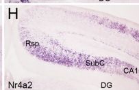
Detection of Nurr1/NGFI-B beta/NR4A2 by Immunohistochemistry
Rsp neurons lose their identity in Satb2 CKO mice. FN1 is restricted to the SubC of controls (a) but expressed by neurons in the Rsp region of Satb2 CKO mice at P4 (b). c–h ER81, Citp2, and Nr4a2 are expressed in the SubC and specific layers of the Rsp in control mice (c, e, g). In Satb2 CKO mice, however, they are expressed throughout the Rsp regions while remain largely unchanged in the SubC (d, f, h). Cbln1 shows restricted expression in the Rsp of control mice (i), but its expression is lost in Satb2 CKO mice (j). Zbtb20 is expressed in superficial Rsp and CA1 of control mice (k), but undetectable in the Rsp region while remains unchanged in the CA1 of Satb2 CKO mice (l). Note that ectopic Zbtb20+ cells (arrows) are present in the subicular region of Satb2 CKO mice. Mef2C is intensely expressed in the Rsp and weakly in the SubC of control mice (m), but its expression was largely reduced in the Rsp region to a comparable cellular density of the SubC in Satb2 CKO mice (n). Dkk3 is strongly expressed in upper Rsp of control mice (o) but absent in the Rsp region of Satb2 CKO mice (p). Intense Lmo4 expression is reduced in the Rsp regions with a similar intensity to that in the SubC in Satb2 CKO mice (r) compared with controls (q). s, t Diagram of Rsp fate change in Satb2 CKO mice. DG dentate gyrus. n = 3 mice for each genotype. Scale bar = 500 μm Image collected and cropped by CiteAb from the following open publication (https://pubmed.ncbi.nlm.nih.gov/31666685), licensed under a CC-BY license. Not internally tested by R&D Systems.Applications for Nurr1/NGFI-B beta/NR4A2 Antibody
Immunocytochemistry
Sample: Immersion fixed HeLa human cervical epithelial carcinoma cell line
Immunohistochemistry
Sample: Immersion fixed paraffin-embedded sections of mouse brain (hippocampus)
Western Blot
Sample: Recombinant Mouse Nurr1/NGFI‑B beta /NR4A2
Formulation, Preparation, and Storage
Purification
Reconstitution
Reconstitute at 0.2 mg/mL in sterile PBS. For liquid material, refer to CoA for concentration.
Formulation
Shipping
Stability & Storage
- 12 months from date of receipt, -20 to -70 °C as supplied.
- 1 month, 2 to 8 °C under sterile conditions after reconstitution.
- 6 months, -20 to -70 °C under sterile conditions after reconstitution.
Calculators
Background: Nurr1/NGFI-B beta/NR4A2
Long Name
Alternate Names
Gene Symbol
UniProt
Additional Nurr1/NGFI-B beta/NR4A2 Products
Product Documents for Nurr1/NGFI-B beta/NR4A2 Antibody
Certificate of Analysis
To download a Certificate of Analysis, please enter a lot or batch number in the search box below.
Note: Certificate of Analysis not available for kit components.
Product Specific Notices for Nurr1/NGFI-B beta/NR4A2 Antibody
For research use only
Related Research Areas
Citations for Nurr1/NGFI-B beta/NR4A2 Antibody
 Powered by Bioz
Customer Reviews for Nurr1/NGFI-B beta/NR4A2 Antibody
There are currently no reviews for this product. Be the first to review Nurr1/NGFI-B beta/NR4A2 Antibody and earn rewards!
Have you used Nurr1/NGFI-B beta/NR4A2 Antibody?
Submit a review and receive an Amazon gift card!
$25/€18/£15/$25CAN/¥2500 for a review with an image
$10/€7/£6/$10CAN/¥1110 for a review without an image
Submit a review
Protocols
Find general support by application which include: protocols, troubleshooting, illustrated assays, videos and webinars.
- Antigen Retrieval Protocol (PIER)
- Antigen Retrieval for Frozen Sections Protocol
- Appropriate Fixation of IHC/ICC Samples
- Cellular Response to Hypoxia Protocols
- Chromogenic IHC Staining of Formalin-Fixed Paraffin-Embedded (FFPE) Tissue Protocol
- Chromogenic Immunohistochemistry Staining of Frozen Tissue
- ClariTSA™ Fluorophore Kits
- Detection & Visualization of Antibody Binding
- Fluorescent IHC Staining of Frozen Tissue Protocol
- Graphic Protocol for Heat-induced Epitope Retrieval
- Graphic Protocol for the Preparation and Fluorescent IHC Staining of Frozen Tissue Sections
- Graphic Protocol for the Preparation and Fluorescent IHC Staining of Paraffin-embedded Tissue Sections
- Graphic Protocol for the Preparation of Gelatin-coated Slides for Histological Tissue Sections
- ICC Cell Smear Protocol for Suspension Cells
- ICC Immunocytochemistry Protocol Videos
- ICC for Adherent Cells
- IHC Sample Preparation (Frozen sections vs Paraffin)
- Immunocytochemistry (ICC) Protocol
- Immunocytochemistry Troubleshooting
- Immunofluorescence of Organoids Embedded in Cultrex Basement Membrane Extract
- Immunofluorescent IHC Staining of Formalin-Fixed Paraffin-Embedded (FFPE) Tissue Protocol
- Immunohistochemistry (IHC) and Immunocytochemistry (ICC) Protocols
- Immunohistochemistry Frozen Troubleshooting
- Immunohistochemistry Paraffin Troubleshooting
- Preparing Samples for IHC/ICC Experiments
- Preventing Non-Specific Staining (Non-Specific Binding)
- Primary Antibody Selection & Optimization
- Protocol for Heat-Induced Epitope Retrieval (HIER)
- Protocol for Making a 4% Formaldehyde Solution in PBS
- Protocol for VisUCyte™ HRP Polymer Detection Reagent
- Protocol for the Fluorescent ICC Staining of Cell Smears - Graphic
- Protocol for the Fluorescent ICC Staining of Cultured Cells on Coverslips - Graphic
- Protocol for the Preparation & Fixation of Cells on Coverslips
- Protocol for the Preparation and Chromogenic IHC Staining of Frozen Tissue Sections
- Protocol for the Preparation and Chromogenic IHC Staining of Frozen Tissue Sections - Graphic
- Protocol for the Preparation and Chromogenic IHC Staining of Paraffin-embedded Tissue Sections
- Protocol for the Preparation and Chromogenic IHC Staining of Paraffin-embedded Tissue Sections - Graphic
- Protocol for the Preparation and Fluorescent ICC Staining of Cells on Coverslips
- Protocol for the Preparation and Fluorescent ICC Staining of Non-adherent Cells
- Protocol for the Preparation and Fluorescent ICC Staining of Stem Cells on Coverslips
- Protocol for the Preparation and Fluorescent IHC Staining of Frozen Tissue Sections
- Protocol for the Preparation and Fluorescent IHC Staining of Paraffin-embedded Tissue Sections
- Protocol for the Preparation of Gelatin-coated Slides for Histological Tissue Sections
- Protocol for the Preparation of a Cell Smear for Non-adherent Cell ICC - Graphic
- R&D Systems Quality Control Western Blot Protocol
- TUNEL and Active Caspase-3 Detection by IHC/ICC Protocol
- The Importance of IHC/ICC Controls
- Troubleshooting Guide: Immunohistochemistry
- Troubleshooting Guide: Western Blot Figures
- Western Blot Conditions
- Western Blot Protocol
- Western Blot Protocol for Cell Lysates
- Western Blot Troubleshooting
- Western Blot Troubleshooting Guide
- View all Protocols, Troubleshooting, Illustrated assays and Webinars
Associated Pathways
