
phospho-Ribosomal Protein S6/RPS6 (S235/S236) Antibody
R&D Systems | Catalog # AF3918

Scientific images may be resized, cropped, or adjusted for brightness or contrast for presentation purposes. These updates are limited to presentation and do not affect the underlying scientific interpretation of the data. Where available, additional source data may be provided upon request.
Key Product Details
Validated by
Species Reactivity
Validated:
Cited:
Applications
Validated:
Cited:
Label
Antibody Source
Product Specifications
Immunogen
Specificity
Clonality
Host
Isotype
Scientific Data Images for phospho-Ribosomal Protein S6/RPS6 (S235/S236) Antibody
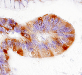
Ribosomal Protein S6 in Human Colon Cancer Tissue.
Ribosomal Protein S6 was detected in immersion fixed paraffin-embedded sections of human colon cancer tissue using 5 µg/mL Rabbit Anti-Human/Mouse/Rat Phospho-Ribosomal Protein S6 (S235/S236) Antigen Affinity-purified Polyclonal Antibody (Catalog # AF3918) overnight at 4 °C. Tissue was stained with the Anti-Rabbit HRP-DAB Cell & Tissue Staining Kit (brown; Catalog # CTS005) and counterstained with hematoxylin (blue). Specific labeling was localized to vascular endothelial cell within glomeruli. View our protocol for Chromogenic IHC Staining of Paraffin-embedded Tissue Sections.
Detection of Human and Mouse Phospho-Ribosomal Protein S6 (S235/S236) by Western Blot.
Western blot shows lysates of MCF-7 human breast cancer cell line untreated (-) or treated (+) with 100 ng/mL Recombinant Human IGF-1 (Catalog # 291-G1) for 20 minutes and NIH-3T3 mouse embryonic fibroblast cell line untreated or treated with 10 ng/mL Human PDGF (Catalog # 120-HD) for 20 minutes. PVDF membrane was probed with 0.5 µg/mL of Rabbit Anti-Human/Mouse/Rat Phospho-Ribosomal Protein S6 (S235/S236) Antigen Affinity-purified Polyclonal Antibody (Catalog # AF3918), followed by HRP-conjugated Anti-Rabbit IgG Secondary Antibody (Catalog # HAF008). A specific band was detected for Phospho-Ribosomal Protein S6 (S235/S236) at approximately 32 kDa (as indicated). This experiment was conducted under reducing conditions and using Immunoblot Buffer Group 1.
Detection of Human Phospho-Ribosomal Protein S6/RPS6 (S235/S236) by Simple WesternTM.
Simple Western lane view shows lysates of MCF-7 human breast cancer cell line untreated (-) or treated (+) with 100 ng/mL Recombinant Human IGF-I (Catalog # 291-G1) for 20 minutes, loaded at 0.5 mg/mL. A specific band was detected for Phospho-Ribosomal Protein S6/RPS6 (S235/S236) at approximately 40 kDa (as indicated) using 5 µg/mL of Rabbit Anti-Human/Mouse/Rat Phospho-Ribosomal Protein S6/RPS6 (S235/S236) Antigen Affinity-purified Polyclonal Antibody (Catalog # AF3918). This experiment was conducted under reducing conditions and using the 12-230 kDa separation system.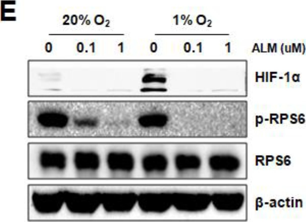
Detection of Human Ribosomal Protein S6/RPS6 by Western Blot
ALM inhibits HIF-1 alpha translation by down-regulating mTOR pathway. (A) Hep3B and PC3 cells were exposed to vehicle or the hypoxia mimics dimethyloxalylglycine (DMOG), cobalt chloride (CoCl2), desferrioxamine (DFX) or MG132 in the presence of ALM at indicated concentrations or vehicle for 24 h and whole cell lysates were subjected to Western blot. (B) Hep3B cells were pretreated for 4 h with vehicle (Vhc), 25 nM rapamycin (Rapa), 20 μg/mL cycloheximide (Chx), or 200 nM ALM, [35S] methionine/cysteine was added for 1 h followed by cell lysis, HIF-1 alpha immunoprecipitation (IP), SDS/PAGE, and autoradiography. (C–E) PC3 cells were cultured at 20% or 1% O2 for 24 h in the presence of ALM (at the indicated concentrations) and whole cell lysates were subjected to for: (C) p-P70, p-Akt, p-mTOR (2448/2481), mTOR, (D) p-4E-BP1, (E) p-RPS6, HIF-1 alpha, or beta -actin. Image collected and cropped by CiteAb from the following publication (https://pubmed.ncbi.nlm.nih.gov/31083403), licensed under a CC-BY license. Not internally tested by R&D Systems.Applications for phospho-Ribosomal Protein S6/RPS6 (S235/S236) Antibody
Immunohistochemistry
Sample: Immersion fixed paraffin-embedded sections of human colon cancer tissue
Simple Western
Sample: MCF‑7 human breast cancer cell line treated with Recombinant Human IGF‑I (Catalog # 291-G1)
Western Blot
Sample: MCF‑7 human breast cancer cell line treated with Recombinant Human IGF‑1 (Catalog # 291-G1) and NIH‑3T3 mouse embryonic fibroblast cell line treated with Human PDGF (Catalog # 120-HD)
Reviewed Applications
Read 2 reviews rated 4.5 using AF3918 in the following applications:
Formulation, Preparation, and Storage
Purification
Reconstitution
Formulation
Shipping
Stability & Storage
- 12 months from date of receipt, -20 to -70 °C as supplied.
- 1 month, 2 to 8 °C under sterile conditions after reconstitution.
- 6 months, -20 to -70 °C under sterile conditions after reconstitution.
Calculators
Background: Ribosomal Protein S6/RPS6
Additional Ribosomal Protein S6/RPS6 Products
Product Documents for phospho-Ribosomal Protein S6/RPS6 (S235/S236) Antibody
Certificate of Analysis
To download a Certificate of Analysis, please enter a lot or batch number in the search box below.
Note: Certificate of Analysis not available for kit components.
Product Specific Notices for phospho-Ribosomal Protein S6/RPS6 (S235/S236) Antibody
For research use only
Related Research Areas
Citations for phospho-Ribosomal Protein S6/RPS6 (S235/S236) Antibody
 Powered by Bioz
Customer Reviews for phospho-Ribosomal Protein S6/RPS6 (S235/S236) Antibody (2)
Have you used phospho-Ribosomal Protein S6/RPS6 (S235/S236) Antibody?
Submit a review and receive an Amazon gift card!
$25/€18/£15/$25CAN/¥2500 for a review with an image
$10/€7/£6/$10CAN/¥1110 for a review without an image
Submit a review
Customer Images


-
Application: Western BlotSample Tested: Bone marrow cellsSpecies: MouseVerified Customer | Posted 11/30/2022Mouse bone marrow stromal cells were lysed with NP-40 buffer for protein extraction. 10 ug of proteins were loaded in a 10% Acrylamide gel.

-
Application: Western BlotSample Tested: Skeletal muscle tissueSpecies: MouseVerified Customer | Posted 11/20/2017Western Blot: Phospho S6/RPS6 [AF3918] - Total protein from mouse skeletal muscle tissue, separated on a 4-12% gel by SDS-PAGE, transferred to nitrocellulose membrane and blocked in 5% non-fat milk for 1h at room temperature. The membrane was probed with anti-phospho S6/RPS6 1:400 in non-fat milk.
There are no reviews that match your criteria.
Protocols
Find general support by application which include: protocols, troubleshooting, illustrated assays, videos and webinars.
- Antigen Retrieval Protocol (PIER)
- Antigen Retrieval for Frozen Sections Protocol
- Appropriate Fixation of IHC/ICC Samples
- Cellular Response to Hypoxia Protocols
- Chromogenic IHC Staining of Formalin-Fixed Paraffin-Embedded (FFPE) Tissue Protocol
- Chromogenic Immunohistochemistry Staining of Frozen Tissue
- ClariTSA™ Fluorophore Kits
- Detection & Visualization of Antibody Binding
- Fluorescent IHC Staining of Frozen Tissue Protocol
- Graphic Protocol for Heat-induced Epitope Retrieval
- Graphic Protocol for the Preparation and Fluorescent IHC Staining of Frozen Tissue Sections
- Graphic Protocol for the Preparation and Fluorescent IHC Staining of Paraffin-embedded Tissue Sections
- Graphic Protocol for the Preparation of Gelatin-coated Slides for Histological Tissue Sections
- IHC Sample Preparation (Frozen sections vs Paraffin)
- Immunofluorescent IHC Staining of Formalin-Fixed Paraffin-Embedded (FFPE) Tissue Protocol
- Immunohistochemistry (IHC) and Immunocytochemistry (ICC) Protocols
- Immunohistochemistry Frozen Troubleshooting
- Immunohistochemistry Paraffin Troubleshooting
- Preparing Samples for IHC/ICC Experiments
- Preventing Non-Specific Staining (Non-Specific Binding)
- Primary Antibody Selection & Optimization
- Protocol for Heat-Induced Epitope Retrieval (HIER)
- Protocol for Making a 4% Formaldehyde Solution in PBS
- Protocol for VisUCyte™ HRP Polymer Detection Reagent
- Protocol for the Preparation & Fixation of Cells on Coverslips
- Protocol for the Preparation and Chromogenic IHC Staining of Frozen Tissue Sections
- Protocol for the Preparation and Chromogenic IHC Staining of Frozen Tissue Sections - Graphic
- Protocol for the Preparation and Chromogenic IHC Staining of Paraffin-embedded Tissue Sections
- Protocol for the Preparation and Chromogenic IHC Staining of Paraffin-embedded Tissue Sections - Graphic
- Protocol for the Preparation and Fluorescent IHC Staining of Frozen Tissue Sections
- Protocol for the Preparation and Fluorescent IHC Staining of Paraffin-embedded Tissue Sections
- Protocol for the Preparation of Gelatin-coated Slides for Histological Tissue Sections
- R&D Systems Quality Control Western Blot Protocol
- TUNEL and Active Caspase-3 Detection by IHC/ICC Protocol
- The Importance of IHC/ICC Controls
- Troubleshooting Guide: Immunohistochemistry
- Troubleshooting Guide: Western Blot Figures
- Western Blot Conditions
- Western Blot Protocol
- Western Blot Protocol for Cell Lysates
- Western Blot Troubleshooting
- Western Blot Troubleshooting Guide
- View all Protocols, Troubleshooting, Illustrated assays and Webinars
Associated Pathways

