NALP1 (NAcht, Leucine-rich repeat and PYD domain containing protein 1; also NAC, CARD7, DEFCAP and CLR17.1) is a 160-170 kDa member of the NLRP family of molecules. It is expressed in dendritic cells, neutrophils, T and B cells, alveolar macrophages, permatogonia, neurons, plus intestinal columnar epithelium, and is found in both cytoplasm and nucleus. NALP1 promotes apoptosis plus IL-1 beta and IL-18 maturation by activating caspase-1 and -5. It does so by forming a 700 kDa inflammasome comprised of NALP1, ASC plus caspase-1 and -5. Bacterial wall peptidoglycan binds to NALP1, promoting ATP binding, NALP1 oligomerization, and caspase activation. Human NALP1 is 1473 amino acids (aa) in length. It contains an N-terminal DAPIN domain (aa 1-92), a NACHT domain (aa 328-637), seven consecutive LRRs (aa 704-1236) and one CARD region (aa 1374-1463). Alternate splice forms exist that range in size from 70 kDa-150 kDa. Either individually, or in combination, there can be a 19 aa substitution for either aa 1353-1473 or aa 1368-1472, a deletion of aa 91-260, 958-987 or 1262-1305, and a 43 aa insert after Leu785. Over aa 1-323, human NALP1 shares less that 20% aa identity with mouse NALP1.
Human NLRP1/NALP1 Antibody (447916)
R&D Systems | Catalog # MAB6788

Key Product Details
Species Reactivity
Human
Applications
Immunohistochemistry
Label
Unconjugated
Antibody Source
Monoclonal Mouse IgG2B Clone # 447916
Loading...
Product Specifications
Immunogen
E. coli-derived recombinant human NLRP1/NALP1
Gly1331-Leu1429
Accession # Q9C000
Gly1331-Leu1429
Accession # Q9C000
Specificity
Detects human NLRP1/NALP1 in direct ELISAs.
Clonality
Monoclonal
Host
Mouse
Isotype
IgG2B
Scientific Data Images for Human NLRP1/NALP1 Antibody (447916)
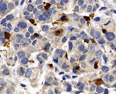
NLRP1/NALP1 in Human Stomach Cancer Tissue.
NLRP1/NALP1 was detected in immersion fixed paraffin-embedded sections of human stomach cancer tissue using Mouse Anti-Human NLRP1/NALP1 Monoclonal Antibody (Catalog # MAB6788) at 0.5 µg/mL for 1 hour at room temperature followed by incubation with the Anti-Mouse IgG VisUCyte™ HRP Polymer Antibody (Catalog # VC001). Before incubation with the primary antibody, tissue was subjected to heat-induced epitope retrieval using Antigen Retrieval Reagent-Basic (Catalog # CTS013). Tissue was stained using DAB (brown) and counterstained with hematoxylin (blue). Specific staining was localized to nuclei. View our protocol for IHC Staining with VisUCyte HRP Polymer Detection Reagents.Applications for Human NLRP1/NALP1 Antibody (447916)
Application
Recommended Usage
Immunohistochemistry
1-25 µg/mL
Sample: Immersion fixed paraffin-embedded sections of human stomach cancer tissue
Sample: Immersion fixed paraffin-embedded sections of human stomach cancer tissue
Reviewed Applications
Read 1 review rated 5 using MAB6788 in the following applications:
Formulation, Preparation, and Storage
Purification
Protein A or G purified from cell culture supernatant
Reconstitution
Reconstitute at 0.5 mg/mL in sterile PBS. For liquid material, refer to CoA for concentration.
Loading...
Formulation
Lyophilized from a 0.2 μm filtered solution in PBS with Trehalose. *Small pack size (SP) is supplied either lyophilized or as a 0.2 µm filtered solution in PBS.
Shipping
Lyophilized product is shipped at ambient temperature. Liquid small pack size (-SP) is shipped with polar packs. Upon receipt, store immediately at the temperature recommended below.
Stability & Storage
Use a manual defrost freezer and avoid repeated freeze-thaw cycles.
- 12 months from date of receipt, -20 to -70 °C as supplied.
- 1 month, 2 to 8 °C under sterile conditions after reconstitution.
- 6 months, -20 to -70 °C under sterile conditions after reconstitution.
Calculators
Background: NLRP1/NALP1
Long Name
NLR Family, Pyrin Domain Containing 1
Alternate Names
CARD7, CLR17.1, DEFCAP, NALP1, PP1044, SLEV1, VAMAS1
Gene Symbol
NLRP1
UniProt
Additional NLRP1/NALP1 Products
Product Documents for Human NLRP1/NALP1 Antibody (447916)
Certificate of Analysis
To download a Certificate of Analysis, please enter a lot or batch number in the search box below.
Note: Certificate of Analysis not available for kit components.
Product Specific Notices for Human NLRP1/NALP1 Antibody (447916)
For research use only
Related Research Areas
Citations for Human NLRP1/NALP1 Antibody (447916)
 Powered by Bioz
Customer Reviews for Human NLRP1/NALP1 Antibody (447916) (1)
5 out of 5
1 Customer Rating
Have you used Human NLRP1/NALP1 Antibody (447916)?
Submit a review and receive an Amazon gift card!
$25/€18/£15/$25CAN/¥2500 Yen for a review with an image
$10/€7/£6/$10CAN/¥1110 Yen for a review without an image
Submit a review
Customer Images
Showing
1
-
1 的
1 review
Showing All
Filter By:
-
Application: ImmunohistochemistrySample Tested: Stomach tissueSpecies: HumanVerified Customer | Posted 09/10/2022

There are no reviews that match your criteria.
Protocols
Find general support by application which include: protocols, troubleshooting, illustrated assays, videos and webinars.
- Antigen Retrieval Protocol (PIER)
- Antigen Retrieval for Frozen Sections Protocol
- Appropriate Fixation of IHC/ICC Samples
- Cellular Response to Hypoxia Protocols
- Chromogenic IHC Staining of Formalin-Fixed Paraffin-Embedded (FFPE) Tissue Protocol
- Chromogenic Immunohistochemistry Staining of Frozen Tissue
- ClariTSA™ Fluorophore Kits
- Detection & Visualization of Antibody Binding
- Fluorescent IHC Staining of Frozen Tissue Protocol
- Graphic Protocol for Heat-induced Epitope Retrieval
- Graphic Protocol for the Preparation and Fluorescent IHC Staining of Frozen Tissue Sections
- Graphic Protocol for the Preparation and Fluorescent IHC Staining of Paraffin-embedded Tissue Sections
- Graphic Protocol for the Preparation of Gelatin-coated Slides for Histological Tissue Sections
- IHC Sample Preparation (Frozen sections vs Paraffin)
- Immunofluorescent IHC Staining of Formalin-Fixed Paraffin-Embedded (FFPE) Tissue Protocol
- Immunohistochemistry (IHC) and Immunocytochemistry (ICC) Protocols
- Immunohistochemistry Frozen Troubleshooting
- Immunohistochemistry Paraffin Troubleshooting
- Preparing Samples for IHC/ICC Experiments
- Preventing Non-Specific Staining (Non-Specific Binding)
- Primary Antibody Selection & Optimization
- Protocol for Heat-Induced Epitope Retrieval (HIER)
- Protocol for Making a 4% Formaldehyde Solution in PBS
- Protocol for VisUCyte™ HRP Polymer Detection Reagent
- Protocol for the Preparation & Fixation of Cells on Coverslips
- Protocol for the Preparation and Chromogenic IHC Staining of Frozen Tissue Sections
- Protocol for the Preparation and Chromogenic IHC Staining of Frozen Tissue Sections - Graphic
- Protocol for the Preparation and Chromogenic IHC Staining of Paraffin-embedded Tissue Sections
- Protocol for the Preparation and Chromogenic IHC Staining of Paraffin-embedded Tissue Sections - Graphic
- Protocol for the Preparation and Fluorescent IHC Staining of Frozen Tissue Sections
- Protocol for the Preparation and Fluorescent IHC Staining of Paraffin-embedded Tissue Sections
- Protocol for the Preparation of Gelatin-coated Slides for Histological Tissue Sections
- TUNEL and Active Caspase-3 Detection by IHC/ICC Protocol
- The Importance of IHC/ICC Controls
- Troubleshooting Guide: Immunohistochemistry
- View all Protocols, Troubleshooting, Illustrated assays and Webinars
Loading...
Associated Pathways
