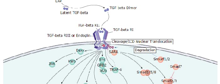
CBFA1, also called runt-related transcription factor 2 (RUNX2), is an essential transcription factor for the regulation of osteoblast differentiation (1). The CBFA1 gene potentially encodes several proteins that differ in their N-terminal sequences and transactivation capacities (2).
Human RUNX2/CBFA1 Antibody (232902)
R&D Systems | Catalog # MAB2006

Scientific images may be resized, cropped, or adjusted for brightness or contrast for presentation purposes. These updates are limited to presentation and do not affect the underlying scientific interpretation of the data. Where available, additional source data may be provided upon request.
Key Product Details
Species Reactivity
Validated:
Human
Cited:
Human, Mouse, Transgenic Mouse
Applications
Validated:
Immunocytochemistry
Cited:
Immunohistochemistry, Immunohistochemistry-Paraffin, Immunohistochemistry-Frozen, Western Blot, Flow Cytometry, Immunocytochemistry
Label
Unconjugated
Antibody Source
Monoclonal Rat IgG2B Clone # 232902
Loading...
Product Specifications
Immunogen
E. coli-derived recombinant human RUNX2/CBFA1 isoform 2
Lys233-Tyr418
Accession # Q13950
Lys233-Tyr418
Accession # Q13950
Specificity
Detects human RUNX2/CBFA1.
Clonality
Monoclonal
Host
Rat
Isotype
IgG2B
Scientific Data Images for Human RUNX2/CBFA1 Antibody (232902)
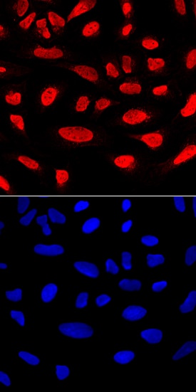
RUNX2/CBFA1 in U2OS Human Cell Line.
RUNX2/CBFA1 was detected in immersion fixed U2OS human osteosarcoma cell line using Rat Anti-Human RUNX2/CBFA1 Monoclonal Antibody (Catalog # MAB2006) at 10 µg/mL for 3 hours at room temperature. Cells were stained using the NorthernLights™ 557-conjugated Anti-Rat IgG Secondary Antibody (red, upper panel; Catalog # NL013) and counterstained with DAPI (blue, lower panel). Specific staining was localized to nuclei. View our protocol for Fluorescent ICC Staining of Cells on Coverslips.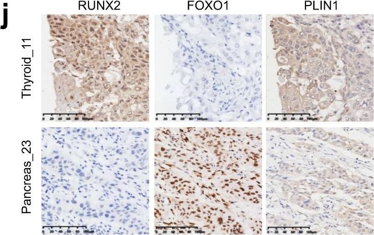
Detection of Human RUNX2/CBFA1 by Immunohistochemistry
Proteomic differences of common SCCs&rare SCCs.j Immunohistochemistry staining for RUNX2, FOXO1,&PLIN1 expression in rare SCCs (one case of thyroid SCC&one case of pancreatic SCC) was concordant with the mass spectrometry findings. Scale bar, 100 μm. Image collected & cropped by CiteAb from the following open publication (https://pubmed.ncbi.nlm.nih.gov/35851595), licensed under a CC-BY license. Not internally tested by R&D Systems.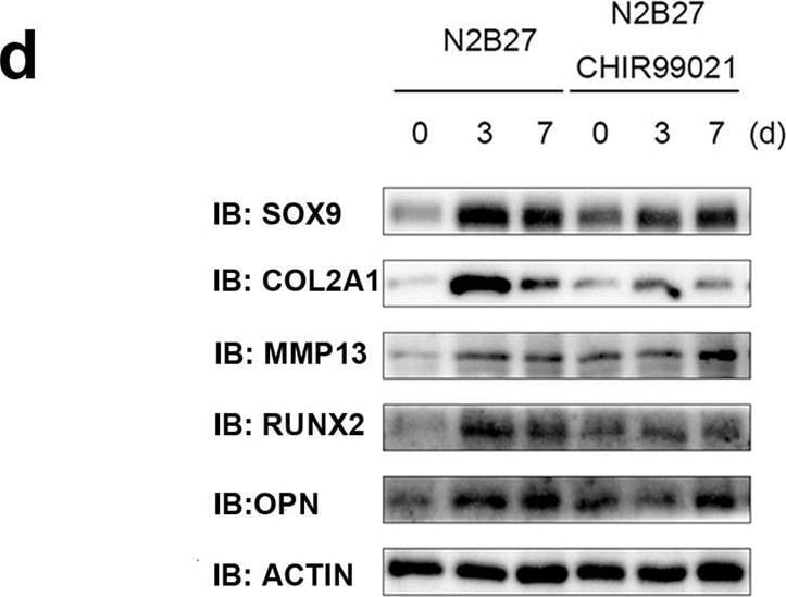
Detection of RUNX2/CBFA1 by Western Blot
The activated Wnt signalling pathway affects chondrogenic differentiation of Cesca KEGG analysis of bulk RNA-seq at different induction points. b The transcription factor SOX9, condylar chondrocyte anabolism markers COL2A1 and COL1A1, and the catabolism marker MMP13 were obviously upregulated 3 days after the removal of CHIR99021. c An obvious downregulation of beta -catenin was detected by western blotting 7 days after the removal of CHIR99021. d The chondrogenic transcription factors SOX9 and RUNX2 were decreased, as well as COL2A1 and OPN, while MMP13 was enhanced in the presence of CHIR99021 in N2B27. e A scheme for the Wnt signalling pathway involved in chondrogenic differentiation of ECCs. Image collected and cropped by CiteAb from the following open publication (https://pubmed.ncbi.nlm.nih.gov/36477591), licensed under a CC-BY license. Not internally tested by R&D Systems.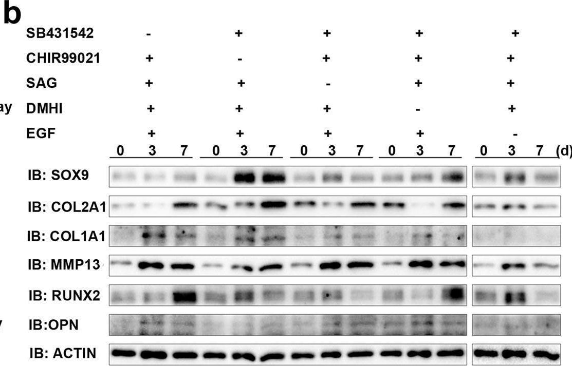
Detection of RUNX2/CBFA1 by Western Blot
The activated Wnt signalling pathway affects chondrogenic differentiation of Cesca KEGG analysis of bulk RNA-seq at different induction points. b The transcription factor SOX9, condylar chondrocyte anabolism markers COL2A1 and COL1A1, and the catabolism marker MMP13 were obviously upregulated 3 days after the removal of CHIR99021. c An obvious downregulation of beta -catenin was detected by western blotting 7 days after the removal of CHIR99021. d The chondrogenic transcription factors SOX9 and RUNX2 were decreased, as well as COL2A1 and OPN, while MMP13 was enhanced in the presence of CHIR99021 in N2B27. e A scheme for the Wnt signalling pathway involved in chondrogenic differentiation of ECCs. Image collected and cropped by CiteAb from the following open publication (https://pubmed.ncbi.nlm.nih.gov/36477591), licensed under a CC-BY license. Not internally tested by R&D Systems.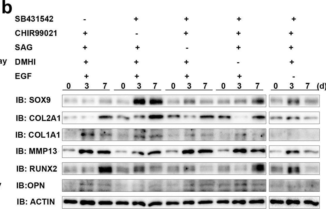
Detection of RUNX2/CBFA1 by Western Blot
The activated Wnt signalling pathway affects chondrogenic differentiation of Cesca KEGG analysis of bulk RNA-seq at different induction points. b The transcription factor SOX9, condylar chondrocyte anabolism markers COL2A1 and COL1A1, and the catabolism marker MMP13 were obviously upregulated 3 days after the removal of CHIR99021. c An obvious downregulation of beta -catenin was detected by western blotting 7 days after the removal of CHIR99021. d The chondrogenic transcription factors SOX9 and RUNX2 were decreased, as well as COL2A1 and OPN, while MMP13 was enhanced in the presence of CHIR99021 in N2B27. e A scheme for the Wnt signalling pathway involved in chondrogenic differentiation of ECCs. Image collected and cropped by CiteAb from the following open publication (https://pubmed.ncbi.nlm.nih.gov/36477591), licensed under a CC-BY license. Not internally tested by R&D Systems.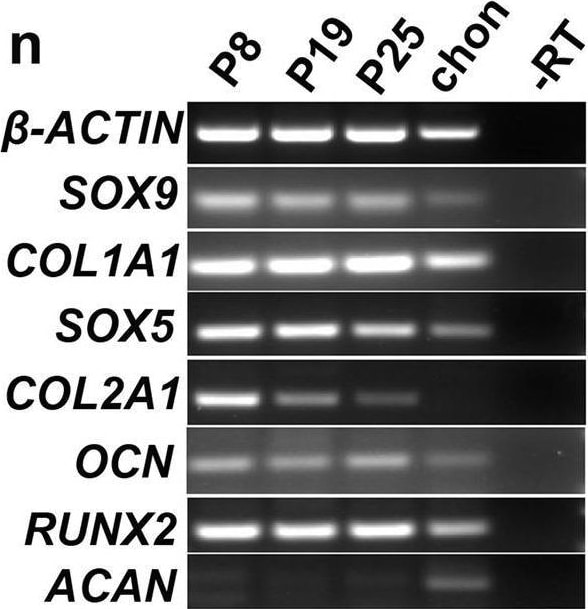
Detection of RUNX2/CBFA1 by Western Blot
CSSEDF-expanded cells stably self-renewed and shared markers of CPCs/C.S.S. A single CSSEDF-expanded cell (passage 5) colony on a Matrigel-coated surface. Scale bars, 50 µm. b–d Immunocytochemistry showing that CSSEDF-expanded cells expressed genes identified as neural ectodermal markers, including SIX1, NESTIN and ETS1. Scale bars, 50 µm. e–i Immunocytochemistry showing that CSSEDF-expanded cells (passage 5) expressed genes identified as CPC/CSC markers, including SOX9, RUNX2, SOX5, TWIST1, and CD29. Scale bars, 50 µm. j–l Immunocytochemistry showing that CSSEDF-expanded cells (passage 5) express genes recently identified as TMJ condylar cartilage markers, including FOXC1, FOXC2 and MSX1. Scale bars, 50 µm. m Flow cytometry analysis showing that CSSEDF-expanded cells stably express cell proliferation and CPC/CSC markers after long-term in vitro expansion. n Gene expression of chondrocytic lineage markers by CSSEDF-expanded cells at different passages and patient TMJ condylar cartilage were analysed by PCR. CPCs/CSCs: cartilaginous progenitor cells/cartilaginous stem cells. Image collected and cropped by CiteAb from the following open publication (https://pubmed.ncbi.nlm.nih.gov/36477591), licensed under a CC-BY license. Not internally tested by R&D Systems.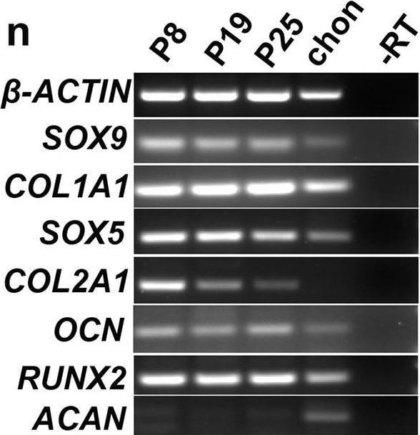
Detection of RUNX2/CBFA1 by Western Blot
CSSEDF-expanded cells stably self-renewed and shared markers of CPCs/C.S.S. A single CSSEDF-expanded cell (passage 5) colony on a Matrigel-coated surface. Scale bars, 50 µm. b–d Immunocytochemistry showing that CSSEDF-expanded cells expressed genes identified as neural ectodermal markers, including SIX1, NESTIN and ETS1. Scale bars, 50 µm. e–i Immunocytochemistry showing that CSSEDF-expanded cells (passage 5) expressed genes identified as CPC/CSC markers, including SOX9, RUNX2, SOX5, TWIST1, and CD29. Scale bars, 50 µm. j–l Immunocytochemistry showing that CSSEDF-expanded cells (passage 5) express genes recently identified as TMJ condylar cartilage markers, including FOXC1, FOXC2 and MSX1. Scale bars, 50 µm. m Flow cytometry analysis showing that CSSEDF-expanded cells stably express cell proliferation and CPC/CSC markers after long-term in vitro expansion. n Gene expression of chondrocytic lineage markers by CSSEDF-expanded cells at different passages and patient TMJ condylar cartilage were analysed by PCR. CPCs/CSCs: cartilaginous progenitor cells/cartilaginous stem cells. Image collected and cropped by CiteAb from the following open publication (https://pubmed.ncbi.nlm.nih.gov/36477591), licensed under a CC-BY license. Not internally tested by R&D Systems.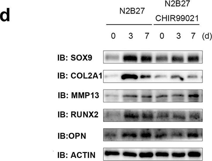
Detection of RUNX2/CBFA1 by Western Blot
The activated Wnt signalling pathway affects chondrogenic differentiation of Cesca KEGG analysis of bulk RNA-seq at different induction points. b The transcription factor SOX9, condylar chondrocyte anabolism markers COL2A1 and COL1A1, and the catabolism marker MMP13 were obviously upregulated 3 days after the removal of CHIR99021. c An obvious downregulation of beta -catenin was detected by western blotting 7 days after the removal of CHIR99021. d The chondrogenic transcription factors SOX9 and RUNX2 were decreased, as well as COL2A1 and OPN, while MMP13 was enhanced in the presence of CHIR99021 in N2B27. e A scheme for the Wnt signalling pathway involved in chondrogenic differentiation of ECCs. Image collected and cropped by CiteAb from the following open publication (https://pubmed.ncbi.nlm.nih.gov/36477591), licensed under a CC-BY license. Not internally tested by R&D Systems.Applications for Human RUNX2/CBFA1 Antibody (232902)
Application
Recommended Usage
Immunocytochemistry
8-25 µg/mL
Sample: Immersion fixed U2OS human osteosarcoma cell line
Sample: Immersion fixed U2OS human osteosarcoma cell line
Reviewed Applications
Read 3 reviews rated 4 using MAB2006 in the following applications:
Formulation, Preparation, and Storage
Purification
Protein A or G purified from hybridoma culture supernatant
Reconstitution
Reconstitute at 0.5 mg/mL in sterile PBS. For liquid material, refer to CoA for concentration.
Loading...
Formulation
Lyophilized from a 0.2 μm filtered solution in PBS with Trehalose. *Small pack size (SP) is supplied either lyophilized or as a 0.2 µm filtered solution in PBS.
Shipping
Lyophilized product is shipped at ambient temperature. Liquid small pack size (-SP) is shipped with polar packs. Upon receipt, store immediately at the temperature recommended below.
Stability & Storage
Use a manual defrost freezer and avoid repeated freeze-thaw cycles.
- 12 months from date of receipt, -20 to -70 °C as supplied.
- 1 month, 2 to 8 °C under sterile conditions after reconstitution.
- 6 months, -20 to -70 °C under sterile conditions after reconstitution.
Calculators
Background: RUNX2/CBFA1
References
- Ducy, P. et al. (1997) Cell 89:747.
- Xiao, Z.S. et al. (1998) Gene 214:187.
- Sato, M. et al. (1998) Oncogene 17:1517.
Long Name
Runt-related Transcription Factor 2
Alternate Names
CBFA1
Entrez Gene IDs
860 (Human)
Gene Symbol
RUNX2
UniProt
Additional RUNX2/CBFA1 Products
Product Documents for Human RUNX2/CBFA1 Antibody (232902)
Certificate of Analysis
To download a Certificate of Analysis, please enter a lot or batch number in the search box below.
Note: Certificate of Analysis not available for kit components.
Product Specific Notices for Human RUNX2/CBFA1 Antibody (232902)
For research use only
Citations for Human RUNX2/CBFA1 Antibody (232902)
 Powered by Bioz
Customer Reviews for Human RUNX2/CBFA1 Antibody (232902) (3)
4 out of 5
3 Customer Ratings
Have you used Human RUNX2/CBFA1 Antibody (232902)?
Submit a review and receive an Amazon gift card!
$25/€18/£15/$25CAN/¥2500 for a review with an image
$10/€7/£6/$10CAN/¥1110 for a review without an image
Submit a review
Customer Images
Showing
1
-
3 的
3 reviews
Showing All
Filter By:
-
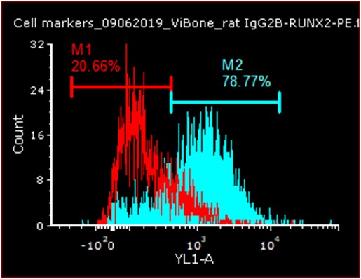
Application: Flow CytometrySample Tested: Bone ExtractsSpecies: HumanVerified Customer | Posted 02/13/2020

-
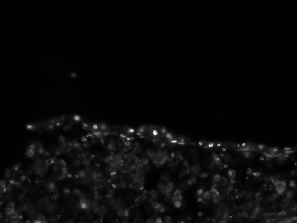
Application: Immunocytochemistry/ImmunofluorescenceSample Tested: bone marrowSpecies: MouseVerified Customer | Posted 08/31/2018
-
Application: Flow CytometrySample Tested: See PMID 22034088Species: HumanVerified Customer | Posted 02/16/2015
There are no reviews that match your criteria.
Protocols
Find general support by application which include: protocols, troubleshooting, illustrated assays, videos and webinars.
- Appropriate Fixation of IHC/ICC Samples
- Cellular Response to Hypoxia Protocols
- ClariTSA™ Fluorophore Kits
- Detection & Visualization of Antibody Binding
- ICC Cell Smear Protocol for Suspension Cells
- ICC Immunocytochemistry Protocol Videos
- ICC for Adherent Cells
- Immunocytochemistry (ICC) Protocol
- Immunocytochemistry Troubleshooting
- Immunofluorescence of Organoids Embedded in Cultrex Basement Membrane Extract
- Immunohistochemistry (IHC) and Immunocytochemistry (ICC) Protocols
- Preparing Samples for IHC/ICC Experiments
- Preventing Non-Specific Staining (Non-Specific Binding)
- Primary Antibody Selection & Optimization
- Protocol for VisUCyte™ HRP Polymer Detection Reagent
- Protocol for the Fluorescent ICC Staining of Cell Smears - Graphic
- Protocol for the Fluorescent ICC Staining of Cultured Cells on Coverslips - Graphic
- Protocol for the Preparation and Fluorescent ICC Staining of Cells on Coverslips
- Protocol for the Preparation and Fluorescent ICC Staining of Non-adherent Cells
- Protocol for the Preparation and Fluorescent ICC Staining of Stem Cells on Coverslips
- Protocol for the Preparation of a Cell Smear for Non-adherent Cell ICC - Graphic
- TUNEL and Active Caspase-3 Detection by IHC/ICC Protocol
- The Importance of IHC/ICC Controls
- View all Protocols, Troubleshooting, Illustrated assays and Webinars