
Key Product Details
Validated by
Knockout/Knockdown, Biological Validation
Species Reactivity
Validated:
Human
Cited:
Human
Applications
Validated:
Western Blot, Immunocytochemistry, Chromatin Immunoprecipitation (ChIP)
Cited:
Immunohistochemistry, Western Blot, Immunocytochemistry, Simple Western, Immunoprecipitation, Chromatin Immunoprecipitation (ChIP)
Label
Unconjugated
Antibody Source
Polyclonal Goat IgG
Loading...
Product Specifications
Immunogen
E. coli-derived recombinant human Smad4
Pro139-Asp332
Accession # Q13485
Pro139-Asp332
Accession # Q13485
Specificity
Detects human Smad4 in direct ELISAs and Western blots. In direct ELISAs and Western blots, approximately 15% cross-reactivity with recombinant human (rh) Smad1 and less than 5% cross-reactivity with rhSmad9, rhSmad5, and rhSmad7 is observed.
Clonality
Polyclonal
Host
Goat
Isotype
IgG
Scientific Data Images for Human Smad4 Antibody
Detection of Human Smad4 by Western Blot.
Western blot shows lysates of HeLa human cervical epithelial carcinoma cell line, Jurkat human acute T cell leukemia cell line, and K562 human chronic myelogenous leukemia cell line. PVDF membrane was probed with 0.25 µg/mL of Goat Anti-Human Smad4 Antigen Affinity-purified Polyclonal Antibody (Catalog # AF2097) followed by HRP-conjugated Anti-Goat IgG Secondary Antibody (Catalog # HAF019). A specific band was detected for Smad4 at approximately 60 kDa (as indicated). This experiment was conducted under reducing conditions and using Immunoblot Buffer Group 1.
Detection of Smad4-regulated Genes by Chromatin Immunoprecipitation.
Jurkat human acute T cell leukemia cell line treated with 10 ng/mL Recombinant Human IL-12 (Catalog # 219-IL) overnight was fixed using formaldehyde, resuspended in lysis buffer, and sonicated to shear chromatin. Smad4/DNA complexes were immunoprecipitated using 5 µg Goat Anti-Human Smad4 Antigen Affinity-purified Polyclonal Antibody (Catalog # AF2097) or control antibody (Catalog # AB-108-C) for 15 minutes in an ultrasonic bath, followed by Biotinylated Anti-Goat IgG Secondary Antibody (Catalog # BAF109). Immunocomplexes were captured using 50 µL of MagCellect Streptavidin Ferrofluid (Catalog # MAG999) and DNA was purified using chelating resin solution. Thep21promoter was detected by standard PCR.
Smad4 in Human PBMCs.
Smad4 was detected in immersion fixed human peripheral blood mononuclear cells (PBMCs) using Goat Anti-Human Smad4 Antigen Affinity-purified Polyclonal Antibody (Catalog # AF2097) at 15 µg/mL for 3 hours at room temperature. Cells were stained using the NorthernLights™ 557-conjugated Anti-Goat IgG Secondary Antibody (red; Catalog # NL001) and counterstained with DAPI (blue). Specific staining was localized to cytoplasm. View our protocol for Fluorescent ICC Staining of Non-adherent Cells.
Detection of Human Smad4 by Western Blot
siRNA knockdown of Smad1, -2 and 4 does not impact RVFV replication.HSAECs transfected with media alone (Mock) or siRNAs (75nM) directed against a nontargeting control (NTC), Smad1, -2, or -4 were infected with MP12 virus (MOI 0.1). Both extracellular media supernatants (A) and protein lysates (B,C) were harvested at 9 and 24hpi. A) Infectious viral titers were determined by plaque assay. Data plotted as a bar graph of the mean with standard deviation of four replicates. Protein lysates from single (B) and double knockdowns (C) were analyzed by western blot for actin, total Smad1, -2, and -4 expression. Image collected and cropped by CiteAb from the following publication (https://pubmed.ncbi.nlm.nih.gov/29408900), licensed under a CC-BY license. Not internally tested by R&D Systems.
Detection of Human Smad4 by Western Blot
siRNA knockdown of Smad1, -2 and 4 does not impact RVFV replication.HSAECs transfected with media alone (Mock) or siRNAs (75nM) directed against a nontargeting control (NTC), Smad1, -2, or -4 were infected with MP12 virus (MOI 0.1). Both extracellular media supernatants (A) and protein lysates (B,C) were harvested at 9 and 24hpi. A) Infectious viral titers were determined by plaque assay. Data plotted as a bar graph of the mean with standard deviation of four replicates. Protein lysates from single (B) and double knockdowns (C) were analyzed by western blot for actin, total Smad1, -2, and -4 expression. Image collected and cropped by CiteAb from the following publication (https://pubmed.ncbi.nlm.nih.gov/29408900), licensed under a CC-BY license. Not internally tested by R&D Systems.Applications for Human Smad4 Antibody
Application
Recommended Usage
Chromatin Immunoprecipitation (ChIP)
5 µg/5 x 106 cells
Sample: Recombinant Human IL-12 (Catalog # 219-IL) treated Jurkat human acute T cell leukemia cell line chromatin, p21 promoter detected by standard PCR
Sample: Recombinant Human IL-12 (Catalog # 219-IL) treated Jurkat human acute T cell leukemia cell line chromatin, p21 promoter detected by standard PCR
Immunocytochemistry
5-15 µg/mL
Sample: Immersion fixed human peripheral blood mononuclear cells (PBMCs)
Sample: Immersion fixed human peripheral blood mononuclear cells (PBMCs)
Western Blot
0.25 µg/mL
Sample: HeLa human cervical epithelial carcinoma cell line, Jurkat human acute T cell leukemia cell line, and K562 human chronic myelogenous leukemia cell line
Sample: HeLa human cervical epithelial carcinoma cell line, Jurkat human acute T cell leukemia cell line, and K562 human chronic myelogenous leukemia cell line
Reviewed Applications
Read 1 review rated 5 using AF2097 in the following applications:
Formulation, Preparation, and Storage
Purification
Antigen Affinity-purified
Reconstitution
Reconstitute at 0.2 mg/mL in sterile PBS. For liquid material, refer to CoA for concentration.
Loading...
Formulation
Lyophilized from a 0.2 μm filtered solution in PBS with Trehalose. *Small pack size (SP) is supplied either lyophilized or as a 0.2 µm filtered solution in PBS.
Shipping
Lyophilized product is shipped at ambient temperature. Liquid small pack size (-SP) is shipped with polar packs. Upon receipt, store immediately at the temperature recommended below.
Stability & Storage
Use a manual defrost freezer and avoid repeated freeze-thaw cycles.
- 12 months from date of receipt, -20 to -70 °C as supplied.
- 1 month, 2 to 8 °C under sterile conditions after reconstitution.
- 6 months, -20 to -70 °C under sterile conditions after reconstitution.
Calculators
Background: Smad4
Long Name
Mothers Against DPP Homolog 4
Alternate Names
DPC4, MADH4
Entrez Gene IDs
4089 (Human)
Gene Symbol
SMAD4
UniProt
Additional Smad4 Products
Product Documents for Human Smad4 Antibody
Certificate of Analysis
To download a Certificate of Analysis, please enter a lot or batch number in the search box below.
Note: Certificate of Analysis not available for kit components.
Product Specific Notices for Human Smad4 Antibody
For research use only
Citations for Human Smad4 Antibody
 Powered by Bioz
Customer Reviews for Human Smad4 Antibody (1)
5 out of 5
1 Customer Rating
Have you used Human Smad4 Antibody?
Submit a review and receive an Amazon gift card!
$25/€18/£15/$25CAN/¥2500 Yen for a review with an image
$10/€7/£6/$10CAN/¥1110 Yen for a review without an image
Submit a review
Customer Images
Showing
1
-
1 的
1 review
Showing All
Filter By:
-
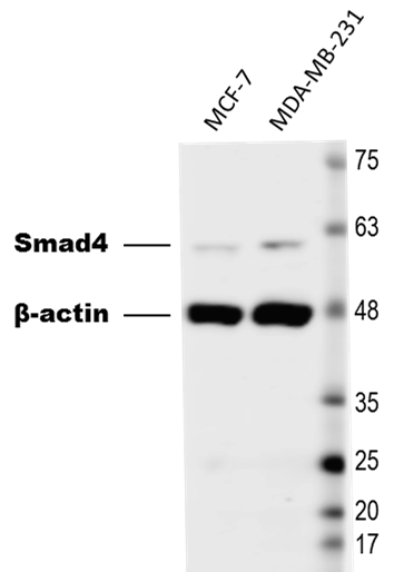
Application: Western BlotSample Tested: Breast cancer cellsSpecies: HumanVerified Customer | Posted 03/16/2017Western Blot of breast cancer cell lysates. The PVDF membrane was probed with 1:1,000 Goat anti-Human Smad4 antibody (#AF2097) followed by 1:5,000 anti-Goat secondary antibody.

There are no reviews that match your criteria.
Protocols
Find general support by application which include: protocols, troubleshooting, illustrated assays, videos and webinars.
- Appropriate Fixation of IHC/ICC Samples
- Cellular Response to Hypoxia Protocols
- ChIP Protocol Video
- Chromatin Immunoprecipitation (ChIP) Protocol
- Chromatin Immunoprecipitation Protocol
- ClariTSA™ Fluorophore Kits
- Detection & Visualization of Antibody Binding
- ICC Cell Smear Protocol for Suspension Cells
- ICC Immunocytochemistry Protocol Videos
- ICC for Adherent Cells
- Immunocytochemistry (ICC) Protocol
- Immunocytochemistry Troubleshooting
- Immunofluorescence of Organoids Embedded in Cultrex Basement Membrane Extract
- Immunohistochemistry (IHC) and Immunocytochemistry (ICC) Protocols
- Preparing Samples for IHC/ICC Experiments
- Preventing Non-Specific Staining (Non-Specific Binding)
- Primary Antibody Selection & Optimization
- Protocol for VisUCyte™ HRP Polymer Detection Reagent
- Protocol for the Fluorescent ICC Staining of Cell Smears - Graphic
- Protocol for the Fluorescent ICC Staining of Cultured Cells on Coverslips - Graphic
- Protocol for the Preparation and Fluorescent ICC Staining of Cells on Coverslips
- Protocol for the Preparation and Fluorescent ICC Staining of Non-adherent Cells
- Protocol for the Preparation and Fluorescent ICC Staining of Stem Cells on Coverslips
- Protocol for the Preparation of a Cell Smear for Non-adherent Cell ICC - Graphic
- R&D Systems Quality Control Western Blot Protocol
- TUNEL and Active Caspase-3 Detection by IHC/ICC Protocol
- The Importance of IHC/ICC Controls
- Troubleshooting Guide: Western Blot Figures
- Western Blot Conditions
- Western Blot Protocol
- Western Blot Protocol for Cell Lysates
- Western Blot Troubleshooting
- Western Blot Troubleshooting Guide
- View all Protocols, Troubleshooting, Illustrated assays and Webinars
Loading...
Associated Pathways

