

Scientific images may be resized, cropped, or adjusted for brightness or contrast for presentation purposes. These updates are limited to presentation and do not affect the underlying scientific interpretation of the data. Where available, additional source data may be provided upon request.
Key Product Details
Species Reactivity
Validated:
Cited:
Applications
Validated:
Cited:
Label
Antibody Source
Product Specifications
Immunogen
Ser249-Ala345
Accession # Q9BT81
Specificity
Clonality
Host
Isotype
Scientific Data Images for Human SOX7 Antibody
Detection of SOX7 by Immunohistochemistry
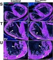
Time‐course morphological analysis of Sox7 endothelial loss‐of‐function embryosA–X(A–C, G–I, M–O, S–U) Morphological characterization of the cardiac defects present in Sox7iECKO hearts compared to control hearts at E11.5 (A–C), E12.5 (G–I), E13.5 (M–O), and E16.5 (S–U) after Cre induction by tamoxifen injection at E9.5 and E10.5, by immunofluorescence with tissue‐specific markers CD31 (endocardium/endothelium; cyan), MF20 (myocardium; magenta), and nuclei (Hoechst; blue). (A, G, M, S) Low‐magnification images showing the general heart morphology. (B, C, H, I, N, O, T, U) High‐magnification images showing the morphology of the compact (arrows) and trabecular (arrowheads) myocardium, and coronary arteries (empty arrow) in the right (RV; B, H, N, T) and left (LV; C, I, O, U) ventricles. White contours in high‐magnification panels highlight the compact myocardium area. Morphological defects are evident from E13.5 (M–O) and maintained at E16.5 (S–U). (D–F, J–L, P–R, V–X) Quantification of total (D, J, P, V), compact and trabecular myocardium (E, F, K, L, Q, R, W, X) area in right (E, K, Q, W) and left (F, L, R, X) ventricular chambers of Sox7iECKO hearts compared to control hearts. Quantification results confirmed no significant difference in tissue area at E11.5 (D–F) and E12.5 (J–L). It also identified a significant increase in total and trabecular myocardium area at E13.5 (P–R) and the maintenance of the E14.5 cardiac defects at E16.5 (V–X). Biological replicates as per the number of data points in the graphs. Mean ± SEM; t‐test. P < 0.05 (*); P < 0.005 (**); P < 0.0005 (***); ns = not significant. Scale bars = 100 μm (A–C), 150 μm (G–I), 200 μm (M–O), and 300 μm (S–U). Image collected and cropped by CiteAb from the following open publication (https://pubmed.ncbi.nlm.nih.gov/37551717), licensed under a CC-BY license. Not internally tested by R&D Systems.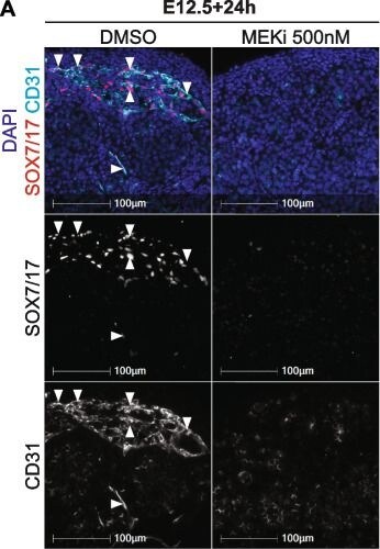
Detection of SOX7 by Immunohistochemistry
SOX7/17 expression depends on MEK1/2 signalling in the developing testis. Section or wholemount immunofluorescent imaging of E12.5 testes cultured for 24 or 72h with DMSO or 500nM of MEKi. A and C Section IF of cultured testes stained with DAPI (blue), SOX7/17 (red) and CD31 (endothelial cells and germ cells; cyan). B Three-dimensional stacked Z-series images of cultured testes showing Oct4-eGFP (germ cells; green), SOX7/17 (blue) and pERK1/2 (red). The DMSO and MEKi images should be viewed in conjunction with Movies S5-8 which reveal the 3D organisation of the cells in the tissue. Scale bar represents 100 μm. Arrows indicate SOX7/17 positive endothelial cells. Biological replicates; n = 3–4 per treatment Image collected and cropped by CiteAb from the following open publication (https://pubmed.ncbi.nlm.nih.gov/39354506), licensed under a CC-BY license. Not internally tested by R&D Systems.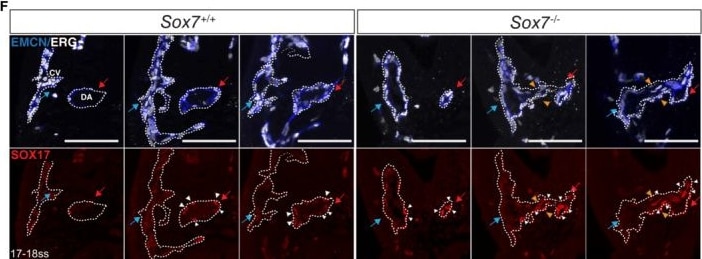
Detection of SOX7 by Immunohistochemistry
Specification of the coronary artery and dorsal aorta is dependent on endothelial Sox7 functionAWhole‐mount immunostaining of control and Sox7iECKO hearts at E14.5 after Cre induction by tamoxifen injection at E9.5, E10.5. Coronary vessels are stained with VEGFR2 (green), coronary arteries (arrows) and trabecular myocardium (TM) are both stained with CX40 (red). Asterisks show where the major distal coronary arteries are lost in the Sox7iECKO heart. The number of embryos showing the illustrated phenotype among the total examined is indicated.BWhole‐mount immunostaining of control and Sox7iECKO hearts at E13.5 after Cre induction at E9.5 and E10.5. Coronary vessels are stained with VEGFR2 (white) and endothelial nuclei with ERG (red). The coronary plexus front is outlined by the dotted lines.C–EGraphs showing quantification of coronary vessel density (C), ERG‐positive endothelial cells (D), and % of H3+ proliferative endothelial cells (E) in the coronary plexus. Scored sibling control, n = 4; Sox7iECKO mutant embryos, n = 4 embryos; Mean ± SEM; Mann–Whitney U‐test. ns = not significant.FSerial sections of a wild‐type and a Sox7−/− embryo at 17–18 ss (E9.0), stained with endomucin (blue) to detect the endothelial lining, ERG (white) for endothelial nuclei, and SOX17 (red) for arterial nuclei. The dorsal aorta (DA) is labeled by the red arrows and white arrowheads, while cardinal vein (CV), blue arrows. Fusion between DA and CV is indicated by orange arrowheads.Data information: Scale bars = 100 μm. Image collected and cropped by CiteAb from the following open publication (https://pubmed.ncbi.nlm.nih.gov/37551717), licensed under a CC-BY license. Not internally tested by R&D Systems.Applications for Human SOX7 Antibody
Immunohistochemistry
Sample: Immersion fixed paraffin-embedded sections of human breast and lung
Western Blot
Sample: Recombinant Human SOX7
Formulation, Preparation, and Storage
Purification
Reconstitution
Reconstitute at 0.2 mg/mL in sterile PBS. For liquid material, refer to CoA for concentration.
Formulation
Shipping
Stability & Storage
- 12 months from date of receipt, -20 to -70 °C as supplied.
- 1 month, 2 to 8 °C under sterile conditions after reconstitution.
- 6 months, -20 to -70 °C under sterile conditions after reconstitution.
Calculators
Background: SOX7
Long Name
Alternate Names
Entrez Gene IDs
Gene Symbol
UniProt
Additional SOX7 Products
Product Documents for Human SOX7 Antibody
Certificate of Analysis
To download a Certificate of Analysis, please enter a lot or batch number in the search box below.
Note: Certificate of Analysis not available for kit components.
Product Specific Notices for Human SOX7 Antibody
For research use only
Related Research Areas
Citations for Human SOX7 Antibody
 Powered by Bioz
Customer Reviews for Human SOX7 Antibody
There are currently no reviews for this product. Be the first to review Human SOX7 Antibody and earn rewards!
Have you used Human SOX7 Antibody?
Submit a review and receive an Amazon gift card!
$25/€18/£15/$25CAN/¥2500 for a review with an image
$10/€7/£6/$10CAN/¥1110 for a review without an image
Submit a review
Protocols
Find general support by application which include: protocols, troubleshooting, illustrated assays, videos and webinars.
- Antigen Retrieval Protocol (PIER)
- Antigen Retrieval for Frozen Sections Protocol
- Appropriate Fixation of IHC/ICC Samples
- Cellular Response to Hypoxia Protocols
- Chromogenic IHC Staining of Formalin-Fixed Paraffin-Embedded (FFPE) Tissue Protocol
- Chromogenic Immunohistochemistry Staining of Frozen Tissue
- ClariTSA™ Fluorophore Kits
- Detection & Visualization of Antibody Binding
- Fluorescent IHC Staining of Frozen Tissue Protocol
- Graphic Protocol for Heat-induced Epitope Retrieval
- Graphic Protocol for the Preparation and Fluorescent IHC Staining of Frozen Tissue Sections
- Graphic Protocol for the Preparation and Fluorescent IHC Staining of Paraffin-embedded Tissue Sections
- Graphic Protocol for the Preparation of Gelatin-coated Slides for Histological Tissue Sections
- IHC Sample Preparation (Frozen sections vs Paraffin)
- Immunofluorescent IHC Staining of Formalin-Fixed Paraffin-Embedded (FFPE) Tissue Protocol
- Immunohistochemistry (IHC) and Immunocytochemistry (ICC) Protocols
- Immunohistochemistry Frozen Troubleshooting
- Immunohistochemistry Paraffin Troubleshooting
- Preparing Samples for IHC/ICC Experiments
- Preventing Non-Specific Staining (Non-Specific Binding)
- Primary Antibody Selection & Optimization
- Protocol for Heat-Induced Epitope Retrieval (HIER)
- Protocol for Making a 4% Formaldehyde Solution in PBS
- Protocol for VisUCyte™ HRP Polymer Detection Reagent
- Protocol for the Preparation & Fixation of Cells on Coverslips
- Protocol for the Preparation and Chromogenic IHC Staining of Frozen Tissue Sections
- Protocol for the Preparation and Chromogenic IHC Staining of Frozen Tissue Sections - Graphic
- Protocol for the Preparation and Chromogenic IHC Staining of Paraffin-embedded Tissue Sections
- Protocol for the Preparation and Chromogenic IHC Staining of Paraffin-embedded Tissue Sections - Graphic
- Protocol for the Preparation and Fluorescent IHC Staining of Frozen Tissue Sections
- Protocol for the Preparation and Fluorescent IHC Staining of Paraffin-embedded Tissue Sections
- Protocol for the Preparation of Gelatin-coated Slides for Histological Tissue Sections
- R&D Systems Quality Control Western Blot Protocol
- TUNEL and Active Caspase-3 Detection by IHC/ICC Protocol
- The Importance of IHC/ICC Controls
- Troubleshooting Guide: Immunohistochemistry
- Troubleshooting Guide: Western Blot Figures
- Western Blot Conditions
- Western Blot Protocol
- Western Blot Protocol for Cell Lysates
- Western Blot Troubleshooting
- Western Blot Troubleshooting Guide
- View all Protocols, Troubleshooting, Illustrated assays and Webinars
Associated Pathways
