Human TACE/ADAM17 Ectodomain Antibody (111633)
R&D Systems | Catalog # MAB9301

Key Product Details
Validated by
Knockout/Knockdown
Species Reactivity
Validated:
Human
Cited:
Human
Applications
Validated:
Knockout Validated, Western Blot, Flow Cytometry, Immunoprecipitation, CyTOF-ready
Cited:
Western Blot, Flow Cytometry, Immunocytochemistry, Proximity Ligation Assay
Label
Unconjugated
Antibody Source
Monoclonal Mouse IgG1 Clone # 111633
Loading...
Product Specifications
Immunogen
Insect ovarian cell line T. ni-derived recombinant human TACE/ADAM17
Pro18-Asn671
Accession # P78536
Pro18-Asn671
Accession # P78536
Specificity
Detects the ectodomain of human TACE/ADAM17 in direct ELISAs and Western blots. In direct ELISAs, less than 5% cross-reactivity with the ectodomain of recombinant human ADAM8, 9, 15 and recombinant mouse ADAM10 is observed.
Clonality
Monoclonal
Host
Mouse
Isotype
IgG1
Scientific Data Images for Human TACE/ADAM17 Ectodomain Antibody (111633)
Detection of TACE/ADAM17 in HeLa Human Cell Line by Flow Cytometry.
HeLa human cervical epithelial carcinoma cell line was stained with Mouse Anti-Human TACE/ADAM17 Ectodomain Monoclonal Antibody (Catalog # MAB9301, filled histogram) or isotype control antibody (MAB002, open histogram), followed by Allophycocyanin-conjugated Anti-Mouse IgG Secondary Antibody (F0101B). View our protocol for Staining Membrane-associated Proteins.
TACE/ADAM17 Specificity is Shown by Flow Cytometry in Knockout Cell Line.
TACE/ADAM17 knockout HeLa epithelial carcinoma cell line was stained with Mouse Anti-Human TACE/ADAM17 Monoclonal Antibody (Catalog # MAB9301, filled histogram) or isotype control antibody (Catalog # MAB002, open histogram) followed by PE-conjugated Goat anti-Mouse IgG Secondary Antibody (Catalog # F0102B). No staining in the TACE/ADAM17 knockout HeLa cell line was observed. View our protocol for Staining Membrane-associated Proteins.
Detection of Human TACE/ADAM17 by Flow Cytometry
Effects of the engineered S197P mutation on CD16a shedding in NK cells.NK92 cells transduced with empty vector (vector only), CD16a, or CD16a/S197P were treated without (Unstim.) or with PMA (100ng/ml) for 30 minutes at 37°C (A), with IL-12 and IL-18 (100ng/ml and 400ng/ml, respectively) for 24 hours at 37°C (B), or with Raji cells and rituximab for 60 min at 37°C (C). Cell surface levels of CD16a were determined by flow cytometry. Isotype-matched negative control antibody staining is indicated by a dotted line. (D) Parent NK92 cells and transduced cells expressing CD16a or CD16a/S197P were treated with Raji cells and rituximab in the presence or absence of the ADAM17 inhibitor BMS566394 (5μM) for 60 min at 37°C. Soluble CD16a levels were determined by ELISA. Each treatment condition was repeated 3 times and the data are representative of 3 independent experiments. Bar graphs show mean ± SD. Statistical significance is indicated as ***P<0.001. (E) NK92 cells expressing CD16a or CD16a/S197P were stained with the anti-ADAM17 mAbs M220, 623, 633, or an isotype-matched negative control antibody, as indicated. (F) CD56+CD45+ NK cells derived from mock-transduced iPSCs (left panel) or iPSCs expressing recombinant CD16a or CD16a/S197P (right panels) were incubated with or without K562 target cells for 4 hours at 37°C. For all histogram plots, the x-axis = Log 10 fluorescence, the y-axis = cell number, and the data are representative of at least 3 independent experiments. Image collected and cropped by CiteAb from the following publication (https://dx.plos.org/10.1371/journal.pone.0121788), licensed under a CC-BY license. Not internally tested by R&D Systems.Applications for Human TACE/ADAM17 Ectodomain Antibody (111633)
Application
Recommended Usage
CyTOF-ready
Ready to be labeled using established conjugation methods. No BSA or other carrier proteins that could interfere with conjugation.
Flow Cytometry
0.25 µg/106 cells
Sample: HeLa human cervical epithelial carcinoma cell line
Sample: HeLa human cervical epithelial carcinoma cell line
Immunoprecipitation
25 µg/mL
Sample: Conditioned cell culture medium spiked with Recombinant Human TACE/ADAM17 (Catalog # 930-ADB), see our available Western blot detection antibodies
Sample: Conditioned cell culture medium spiked with Recombinant Human TACE/ADAM17 (Catalog # 930-ADB), see our available Western blot detection antibodies
Knockout Validated
TACE/ADAM17 is specifically detected in HeLa human carcinoma parental cell line but is not detectable in TACE/ADAM17 knockout HeLa cell line.
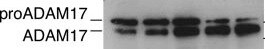
Western Blot
1 µg/mL
Sample: Recombinant Human TACE/ADAM17 Western Blot Standard (Catalog # WBC029) under non-reducing conditions only
Sample: Recombinant Human TACE/ADAM17 Western Blot Standard (Catalog # WBC029) under non-reducing conditions only
Reviewed Applications
Read 2 reviews rated 4.5 using MAB9301 in the following applications:
Flow Cytometry Panel Builder
Bio-Techne Knows Flow Cytometry
Save time and reduce costly mistakes by quickly finding compatible reagents using the Panel Builder Tool.
Advanced Features
- Spectra Viewer - Custom analysis of spectra from multiple fluorochromes
- Spillover Popups - Visualize the spectra of individual fluorochromes
- Antigen Density Selector - Match fluorochrome brightness with antigen density

Formulation, Preparation, and Storage
Purification
Protein A or G purified from hybridoma culture supernatant
Reconstitution
Reconstitute at 0.5 mg/mL in sterile PBS. For liquid material, refer to CoA for concentration.
Loading...
Formulation
Lyophilized from a 0.2 μm filtered solution in PBS with Trehalose. *Small pack size (SP) is supplied either lyophilized or as a 0.2 µm filtered solution in PBS.
Shipping
Lyophilized product is shipped at ambient temperature. Liquid small pack size (-SP) is shipped with polar packs. Upon receipt, store immediately at the temperature recommended below.
Stability & Storage
Use a manual defrost freezer and avoid repeated freeze-thaw cycles.
- 12 months from date of receipt, -20 to -70 °C as supplied.
- 1 month, 2 to 8 °C under sterile conditions after reconstitution.
- 6 months, -20 to -70 °C under sterile conditions after reconstitution.
Calculators
Background: TACE/ADAM17
References
- Black, R.A. and J.D. Becherer (1998) in Tumor Necrosis Factor alpha -Converting Enzyme. Barrett, A.J. et al. (eds): Handbook of Proteolytic Enzymes, San Diego: Academic Press, p. 1315.
- Primakoff, P. and D.G. Myles (2000) Trends in Genetics 16:83.
Long Name
TNF-alpha Converting Enzyme
Alternate Names
ADAM17, CD156b
Gene Symbol
ADAM17
UniProt
Additional TACE/ADAM17 Products
Product Documents for Human TACE/ADAM17 Ectodomain Antibody (111633)
Certificate of Analysis
To download a Certificate of Analysis, please enter a lot or batch number in the search box below.
Note: Certificate of Analysis not available for kit components.
Product Specific Notices for Human TACE/ADAM17 Ectodomain Antibody (111633)
For research use only
Related Research Areas
Citations for Human TACE/ADAM17 Ectodomain Antibody (111633)
 Powered by Bioz
Customer Reviews for Human TACE/ADAM17 Ectodomain Antibody (111633) (2)
4.5 out of 5
2 Customer Ratings
Have you used Human TACE/ADAM17 Ectodomain Antibody (111633)?
Submit a review and receive an Amazon gift card!
$25/€18/£15/$25CAN/¥2500 Yen for a review with an image
$10/€7/£6/$10CAN/¥1110 Yen for a review without an image
Submit a review
Customer Images
Showing
1
-
2 的
2 reviews
Showing All
Filter By:
-
Application: Western BlotSample Tested: A431 cellsSpecies: HumanVerified Customer | Posted 09/30/2021

-
Application: Immunofluorescence in fresh tissueSample Tested: Mesenteric artery-wholeSpecies: MouseVerified Customer | Posted 04/30/2019
There are no reviews that match your criteria.
Protocols
Find general support by application which include: protocols, troubleshooting, illustrated assays, videos and webinars.
- 7-Amino Actinomycin D (7-AAD) Cell Viability Flow Cytometry Protocol
- Cellular Response to Hypoxia Protocols
- Extracellular Membrane Flow Cytometry Protocol
- Flow Cytometry Protocol for Cell Surface Markers
- Flow Cytometry Protocol for Staining Membrane Associated Proteins
- Flow Cytometry Staining Protocols
- Flow Cytometry Troubleshooting Guide
- Immunoprecipitation Protocol
- Intracellular Flow Cytometry Protocol Using Alcohol (Methanol)
- Intracellular Flow Cytometry Protocol Using Detergents
- Intracellular Nuclear Staining Flow Cytometry Protocol Using Detergents
- Intracellular Staining Flow Cytometry Protocol Using Alcohol Permeabilization
- Intracellular Staining Flow Cytometry Protocol Using Detergents to Permeabilize Cells
- Propidium Iodide Cell Viability Flow Cytometry Protocol
- Protocol for Liperfluo
- Protocol for the Characterization of Human Th22 Cells
- Protocol for the Characterization of Human Th9 Cells
- Protocol: Annexin V and PI Staining by Flow Cytometry
- Protocol: Annexin V and PI Staining for Apoptosis by Flow Cytometry
- R&D Systems Quality Control Western Blot Protocol
- Troubleshooting Guide: Fluorokine Flow Cytometry Kits
- Troubleshooting Guide: Western Blot Figures
- Western Blot Conditions
- Western Blot Protocol
- Western Blot Protocol for Cell Lysates
- Western Blot Troubleshooting
- Western Blot Troubleshooting Guide
- View all Protocols, Troubleshooting, Illustrated assays and Webinars
Loading...
Associated Pathways

