TIM-3 (T cell immunoglobulin and mucin domain-3) is a 60 kDa member of the TIM family of immune regulating molecules. TIMs are type I transmembrane glycoproteins with one Ig-like V-type domain and a Ser/Thr-rich mucin stalk (1-3). There are three TIM genes in human and eight in mouse. Mature human TIM-3 consists of a 181 amino acid (aa) extracellular domain (ECD), a 21 aa transmembrane segment, and a 78 aa cytoplasmic tail (4). An alternately spliced isoform is truncated following a short substitution after the Ig-like domain. Within the ECD, human TIM-3 shares 58% aa sequence identity with mouse and rat TIM-3. TIM-3 is expressed on the surface of effector T cells (CD4+ Th1 and CD8+ Tc1) but not on helper T cells (CD4+ Th2 and CD8+ Tc2) (4, 5). In chronic inflammation, autoimmune disorders, and some cancers, TIM-3 is upregulated on several other hematopoietic cell types. It also occurs on hippocampal neurons (7-10). The Ig domain of TIM-3 interacts with a ligand on resting but not activated Th1 and Th2 cells (5, 11). The glycosylated Ig domain of TIM-3 binds cell-associated galectin-9. This induces TIM-3 Tyr phosphorylation and pro-apoptotic signaling (8, 12). TIM-3 functions as a negative regulator of Th1 cell activity. Its blockade results in increased IFN-gamma production, Th1 cell proliferation and cytotoxicity (5, 10, 11, 13), regulatory T cell development (5), and increases in macrophage and neutrophil infiltration into sites of inflammation (14). Soluble mouse TIM-3 constructs which lack the cytoplasmic domain have been shown to inhibit anti-tumor effector T cell responses and to enhance autoimmune reactions (5, 15).

Key Product Details
Validated by
Knockout/Knockdown, Orthogonal Validation
Species Reactivity
Validated:
Human
Cited:
Human
Applications
Validated:
Knockout Validated, Immunohistochemistry, Western Blot, Flow Cytometry, Dual RNAscope ISH-IHC Compatible, CyTOF-ready
Cited:
Immunohistochemistry, Immunohistochemistry-Paraffin, Western Blot, Neutralization, Flow Cytometry, Immunocytochemistry, Immunoprecipitation
Label
Unconjugated
Antibody Source
Polyclonal Goat IgG
Loading...
Product Specifications
Immunogen
Mouse myeloma cell line NS0-derived recombinant human TIM‑3
Ser22-Arg200
Accession # Q8TDQ0
Ser22-Arg200
Accession # Q8TDQ0
Specificity
Detects human TIM-3 in direct ELISAs and Western blots. In direct ELISAs, approximately 5% cross-reactivity with recombinant mouse (rm) TIM‑3 is observed and less than 1% cross-reactivity with recombinant human TIM-1, rmTIM-1, and rmTIM-2 is observed.
Clonality
Polyclonal
Host
Goat
Isotype
IgG
Scientific Data Images for Human TIM-3 Antibody
TIM-3 in Human Tonsil Using Dual RNAscope®ISH and IHC.
TIM-3 mRNA was detected in formalin-fixed paraffin-embedded tissue sections of human tonsil probed with ACD RNAScope®Probe (Catalog # 560681) and stained using ACD RNAscope®2.5 HD Detection Reagents-Red (top image, Catalog # 32260). Adjacent tissue section was processed for immunohistochemistry using R&D Systems Goat Anti-Human TIM-3 Antigen Affinity-purified Polyclonal Antibody (Catalog # AF2365) at 3 ug/mL for 1 hour at room temperature followed by incubation with the Anti-Mouse IgG VisUCyte HRP Polymer Antibody (R&D Systems, Catalog # VC004) and DAB chromogen (lower image, yellow-brown). Tissues were counterstained with hematoxylin (blue).
TIM‑3 in Human Tonsil.
TIM-3 was detected in immersion fixed paraffin-embedded sections of human tonsil using Goat Anti-Human TIM-3 Antigen Affinity-purified Polyclonal Antibody (Catalog # AF2365) at 3 µg/mL for 1 hour at room temperature followed by incubation with the Anti-Goat IgG VisUCyte™ HRP Polymer Antibody (Catalog # VC004). Before incubation with the primary antibody, tissue was subjected to heat-induced epitope retrieval using Antigen Retrieval Reagent-Basic (Catalog # CTS013). Tissue was stained using DAB (brown) and counterstained with hematoxylin (blue). Specific staining was localized to cell membranes and extracellular space. View our protocol for IHC Staining with VisUCyte HRP Polymer Detection Reagents.
Western Blot Shows Human TIM‑3 Specificity by Using Knockout Cell Line.
Western blot shows lysates of HDLM‑2 human Hodgkin’s lymphoma cell line and human TIM-3 knockout HDLM‑2 human Hodgkin’s lymphoma cell line (KO). PVDF membrane was probed with 1 µg/mL of Goat Anti-Human TIM‑3 Antigen Affinity-purified Polyclonal Antibody (Catalog # AF2365) followed by HRP-conjugated Anti-Goat IgG Secondary Antibody (HAF017). A specific band was detected for TIM‑3 at approximately 50 kDa (as indicated) in the parental HDLM‑2 human Hodgkin’s lymphoma cell line, but is not detectable in knockout HDLM‑2 human Hodgkin’s lymphoma cell line. GAPDH (AF5718) is shown as a loading control. This experiment was conducted under reducing conditions and using Western Blot Buffer Group 1.
Detection of Human TIM-3 by Immunohistochemistry
Immunohistochemical staining of peritoneum tissue for CD4, CD8, CD56, OPN, CD68, Gal-9, and TIM-3. HE, hematoxylin and eosin. Magnification, 20×. Arrows indicate loosen tissue at the broken boundary of the granuloma. Image collected and cropped by CiteAb from the following publication (https://www.mdpi.com/1422-0067/18/7/1382), licensed under a CC-BY license. Not internally tested by R&D Systems.
Detection of Human TIM-3 by Simple Western
TIM-3 Association with Intracellular Kinases in CD8+/MART-1+ T cells.TIM-3 co-immunoprecipitation analysis of unactivated and 15 min. stimulation with anti-CD3/CD28 beads (activated). Equivalent amounts of protein (~2mg) were co-immunoprecipitated with pAb anti-TIM-3 antibody and western blot was performed using capillary electrophoresis. Cleared lysate served as a loading control for individual antibody reactivity. Image collected and cropped by CiteAb from the following publication (https://dx.plos.org/10.1371/journal.pone.0140694), licensed under a CC-BY license. Not internally tested by R&D Systems.Applications for Human TIM-3 Antibody
Application
Recommended Usage
CyTOF-ready
Ready to be labeled using established conjugation methods. No BSA or other carrier proteins that could interfere with conjugation.
Dual RNAscope ISH-IHC Compatible
3-25 µg/mL
Sample: Immersion fixed paraffin-embedded sections of human tonsil
Sample: Immersion fixed paraffin-embedded sections of human tonsil
Flow Cytometry
0.25 µg/106 cells
Sample: Human peripheral blood monocytes
Sample: Human peripheral blood monocytes
Immunohistochemistry
3-15 µg/mL
Sample: Immersion fixed paraffin-embedded sections of human tonsil
Sample: Immersion fixed paraffin-embedded sections of human tonsil
Knockout Validated
TIM‑3
is specifically detected in HDLM‑2 human Hodgkin's lymphoma
parental cell line but is not detectable in TIM‑3 knockout
HDLM‑2 human Hodgkin's lymphoma cell line.
Western Blot
0.1 µg/mL
Sample: Recombinant Human TIM‑3 Fc Chimera (Catalog # 2365-TM)
Sample: Recombinant Human TIM‑3 Fc Chimera (Catalog # 2365-TM)
Reviewed Applications
Read 3 reviews rated 4.7 using AF2365 in the following applications:
Flow Cytometry Panel Builder
Bio-Techne Knows Flow Cytometry
Save time and reduce costly mistakes by quickly finding compatible reagents using the Panel Builder Tool.
Advanced Features
- Spectra Viewer - Custom analysis of spectra from multiple fluorochromes
- Spillover Popups - Visualize the spectra of individual fluorochromes
- Antigen Density Selector - Match fluorochrome brightness with antigen density

Formulation, Preparation, and Storage
Purification
Antigen Affinity-purified
Reconstitution
Reconstitute at 0.2 mg/mL in sterile PBS. For liquid material, refer to CoA for concentration.
Loading...
Formulation
Lyophilized from a 0.2 μm filtered solution in PBS with Trehalose. *Small pack size (SP) is supplied either lyophilized or as a 0.2 µm filtered solution in PBS.
Shipping
Lyophilized product is shipped at ambient temperature. Liquid small pack size (-SP) is shipped with polar packs. Upon receipt, store immediately at the temperature recommended below.
Stability & Storage
Use a manual defrost freezer and avoid repeated freeze-thaw cycles.
- 12 months from date of receipt, -20 to -70 °C as supplied.
- 1 month, 2 to 8 °C under sterile conditions after reconstitution.
- 6 months, -20 to -70 °C under sterile conditions after reconstitution.
Calculators
Background: TIM-3
References
- Anderson, A.C. and D.E. Anderson (2006) Curr. Opin. Immunol. 18:665.
- Mariat, C. et al. (2005) Phil. Trans. R. Soc. B. 360:1681.
- Meyers, J.H. et al. (2005) Trends Mol. Med. 11:362.
- Monney, L. et al. (2002) Nature 415:536.
- Sanchez-Fueyo, A. et al. (2003) Nat. Immunol. 4:1093.
- Khademi, M. et al. (2004) J. Immunol. 172:7169.
- Wiener, Z. et al. (2007) J. Invest. Dermatol. 127:906.
- van de Weyer, P.S. et al. (2006) Biochem. Biophys. Res. Commun. 351:571.
- Gielen, A.W. et al. (2005) J. Neuroimmunol. 164:93.
- Oikawa, T. et al. (2006) J. Immunol. 177:4281.
- Sabatos, C.A. et al. (2003) Nat. Immunol. 4:1102.
- Zhu, C. et al. (2005) Nat. Immunol. 6:1245.
- Koguchi, K. et al. (2006) J. Exp. Med. 203:1413.
- Frisancho-Kiss, S. et al. (2006) J. Immunol. 176:6411.
- Geng, H. et al. (2006) J. Immunol. 176:1411.
Long Name
T Cell Immunoglobulin Mucin-3
Alternate Names
CD366, HAVcr-2, HAVCR2, KIM-3, SPTCL, TIM3, TIMD3
Entrez Gene IDs
Gene Symbol
HAVCR2
UniProt
Additional TIM-3 Products
Product Documents for Human TIM-3 Antibody
Certificate of Analysis
To download a Certificate of Analysis, please enter a lot or batch number in the search box below.
Note: Certificate of Analysis not available for kit components.
Product Specific Notices for Human TIM-3 Antibody
For research use only
Citations for Human TIM-3 Antibody
 Powered by Bioz
Customer Reviews for Human TIM-3 Antibody (3)
4.7 out of 5
3 Customer Ratings
Have you used Human TIM-3 Antibody?
Submit a review and receive an Amazon gift card!
$25/€18/£15/$25CAN/¥2500 Yen for a review with an image
$10/€7/£6/$10CAN/¥1110 Yen for a review without an image
Submit a review
Customer Images

Showing
1
-
3 的
3 reviews
Showing All
Filter By:
-
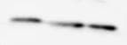
Application: Western BlotSample Tested: JurkatSpecies: HumanVerified Customer | Posted 04/04/2020

-
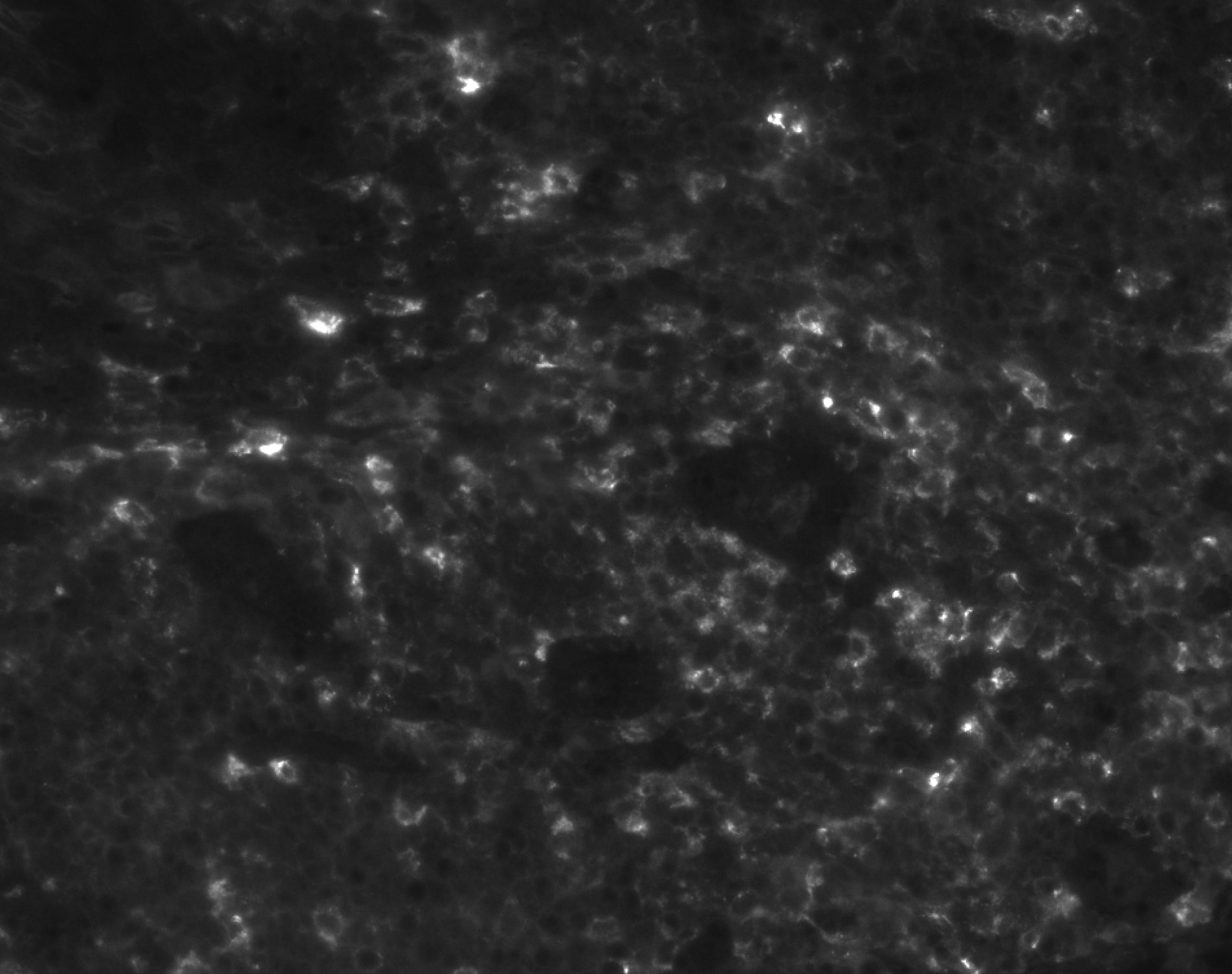
Application: immunofluorescence - paraffinSample Tested: human tonsilSpecies: HumanVerified Customer | Posted 05/02/2019pH 9 heat-induced antigen retrieval
-
Application: Western BlotSample Tested: Jurkat human acute T cell leukemia cell line, RPMI 8226 human multiple myeloma cell line and K562 human chronic myelogenous leukemia cell lineSpecies: HumanVerified Customer | Posted 10/17/2018
There are no reviews that match your criteria.
Protocols
Find general support by application which include: protocols, troubleshooting, illustrated assays, videos and webinars.
- 7-Amino Actinomycin D (7-AAD) Cell Viability Flow Cytometry Protocol
- Antigen Retrieval Protocol (PIER)
- Antigen Retrieval for Frozen Sections Protocol
- Appropriate Fixation of IHC/ICC Samples
- Cellular Response to Hypoxia Protocols
- Chromogenic IHC Staining of Formalin-Fixed Paraffin-Embedded (FFPE) Tissue Protocol
- Chromogenic Immunohistochemistry Staining of Frozen Tissue
- ClariTSA™ Fluorophore Kits
- Detection & Visualization of Antibody Binding
- Extracellular Membrane Flow Cytometry Protocol
- Flow Cytometry Protocol for Cell Surface Markers
- Flow Cytometry Protocol for Staining Membrane Associated Proteins
- Flow Cytometry Staining Protocols
- Flow Cytometry Troubleshooting Guide
- Fluorescent IHC Staining of Frozen Tissue Protocol
- Graphic Protocol for Heat-induced Epitope Retrieval
- Graphic Protocol for the Preparation and Fluorescent IHC Staining of Frozen Tissue Sections
- Graphic Protocol for the Preparation and Fluorescent IHC Staining of Paraffin-embedded Tissue Sections
- Graphic Protocol for the Preparation of Gelatin-coated Slides for Histological Tissue Sections
- IHC Sample Preparation (Frozen sections vs Paraffin)
- ISH-IHC Protocol for Chromogenic Detection on Formalin Fixed Paraffin Embedded (FFPE) Tissue
- Immunofluorescent IHC Staining of Formalin-Fixed Paraffin-Embedded (FFPE) Tissue Protocol
- Immunohistochemistry (IHC) and Immunocytochemistry (ICC) Protocols
- Immunohistochemistry Frozen Troubleshooting
- Immunohistochemistry Paraffin Troubleshooting
- Intracellular Flow Cytometry Protocol Using Alcohol (Methanol)
- Intracellular Flow Cytometry Protocol Using Detergents
- Intracellular Nuclear Staining Flow Cytometry Protocol Using Detergents
- Intracellular Staining Flow Cytometry Protocol Using Alcohol Permeabilization
- Intracellular Staining Flow Cytometry Protocol Using Detergents to Permeabilize Cells
- Preparing Samples for IHC/ICC Experiments
- Preventing Non-Specific Staining (Non-Specific Binding)
- Primary Antibody Selection & Optimization
- Propidium Iodide Cell Viability Flow Cytometry Protocol
- Protocol for Heat-Induced Epitope Retrieval (HIER)
- Protocol for Liperfluo
- Protocol for Making a 4% Formaldehyde Solution in PBS
- Protocol for VisUCyte™ HRP Polymer Detection Reagent
- Protocol for the Characterization of Human Th22 Cells
- Protocol for the Characterization of Human Th9 Cells
- Protocol for the Preparation & Fixation of Cells on Coverslips
- Protocol for the Preparation and Chromogenic IHC Staining of Frozen Tissue Sections
- Protocol for the Preparation and Chromogenic IHC Staining of Frozen Tissue Sections - Graphic
- Protocol for the Preparation and Chromogenic IHC Staining of Paraffin-embedded Tissue Sections
- Protocol for the Preparation and Chromogenic IHC Staining of Paraffin-embedded Tissue Sections - Graphic
- Protocol for the Preparation and Fluorescent IHC Staining of Frozen Tissue Sections
- Protocol for the Preparation and Fluorescent IHC Staining of Paraffin-embedded Tissue Sections
- Protocol for the Preparation of Gelatin-coated Slides for Histological Tissue Sections
- Protocol: Annexin V and PI Staining by Flow Cytometry
- Protocol: Annexin V and PI Staining for Apoptosis by Flow Cytometry
- R&D Systems Quality Control Western Blot Protocol
- TUNEL and Active Caspase-3 Detection by IHC/ICC Protocol
- The Importance of IHC/ICC Controls
- Troubleshooting Guide: Fluorokine Flow Cytometry Kits
- Troubleshooting Guide: Immunohistochemistry
- Troubleshooting Guide: Western Blot Figures
- Western Blot Conditions
- Western Blot Protocol
- Western Blot Protocol for Cell Lysates
- Western Blot Troubleshooting
- Western Blot Troubleshooting Guide
- View all Protocols, Troubleshooting, Illustrated assays and Webinars
Loading...