
Human TROP-2, also called tumor associated calcium signal transducer 2 (TACSTD2), GA733-1, gp50 and T16, is a type I cell surface glycoprotein that is highly expressed on human carcinomas. It was originally identified as an antigen present on human gastrointestinal tumors and is the second of two members of this family. The other family member is GA733-2, also called EpCAM, TROP-1, 17-1A, gp40 and KSA. The TROP-2 gene is unique in that it contains no introns. A study of these two genes suggested that TROP-2 was the result of a retroposition of the EpCAM gene. TROP-2 and EpCAM share approximately 49% amino acid identity and approximately 67% similarity. Human and mouse TROP-2 share 87% similarity. The human TROP-2 protein consists of a putative 26 amino acid (aa) signal sequence, a 248 aa extracellular domain, a 23 aa transmembrane region and a 26 aa cytoplasmic domain. TROP-2 is capable of transducing an intracellular calcium signal and may play a role in tumor growth. It also has adhesive functions.

Scientific images may be resized, cropped, or adjusted for brightness or contrast for presentation purposes. These updates are limited to presentation and do not affect the underlying scientific interpretation of the data. Where available, additional source data may be provided upon request.
Key Product Details
Species Reactivity
Validated:
Cited:
Applications
Validated:
Cited:
Label
Antibody Source
Product Specifications
Immunogen
His27-Thr274
Accession # P09758
Specificity
Clonality
Host
Isotype
Scientific Data Images for Human TROP-2 Antibody (77220)
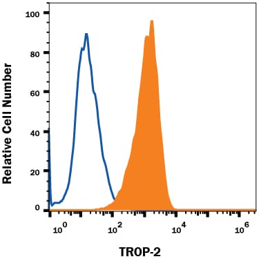
Detection of TROP‑2 in PC-3 Human Cell Line by Flow Cytometry.
PC-3 human prostate cancer cell line was stained with Mouse Anti-Human TROP-2 Monoclonal Antibody (Catalog # MAB650, filled histogram) or isotype control antibody (Catalog # MAB003, open histogram), followed by Phycoerythrin-conjugated Anti-Mouse IgG F(ab')2Secondary Antibody (Catalog # F0102B).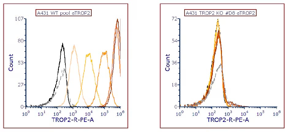
Detection of TROP‑2 in A431 Human Cell Line by Flow Cytometry.
Flow cytometry histograms of A431 wild-type cells (left graph) and A431-TROP2 Knock-out cells (right graph). Both cell lines were incubated with FAB650P before the analysis. Image from a verified customer review. The customer used the PE-conjugated version of MAB650, which is listed under FAB650P.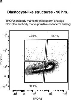
Detection of TROP-2 by Flow Cytometry
Human blastocyst-like structures form analogs of pre-implantation lineages.a. Flow cytometry analysis plot of cells isolated from blastocyst-like structures and stained for lineage-specific surface markers PDGFRa (PrE) and TROP2 (TE). The gates were used to sort analogs of EPI (double negative), TE (TROP2high) and PrE (PDGFR alpha high) to subsequently process for single cell RNA sequencing. Note that the gates did not exclude any cells. This analysis was performed to correlate RNA measures, while ensuring a representation of all cell types. b. UMAPs of the transcriptome of single cells isolated from blastocyst-like structures and displaying the expression levels of genes specific for each of the three blastocyst lineages (TE - Trophectoderm, EPI - Epiblast, and PrE- Primitive endoderm). c-g. UMAPs of single cells isolated from both blastocyst-like structures and from embryos ranging from E3 to E19. c. Coloration of cells originating from In Vitro Fertilization (IVF) embryos isolated on day 3 (E3) to day 7 (E7). This period comprises only pre-implantation stage embryos. d. Coloration of cells originating from IVF embryos isolated on day 6 (E6) to day 12 (E12). These blastocysts (E6) were cultured in vitro. Note that this annotation reflects the number of days in culture rather than the developmental stages. e. Coloration of cells originating from gastrulation-stage embryo isolated on day 17 (E17) to 19 (E19). f. The expression levels of genes specific for each of the three blastocyst lineages (EPI, TE, and PrE). g. Coloration of cells displaying their unsupervised cluster affiliation. Image collected and cropped by CiteAb from the following open publication (https://pubmed.ncbi.nlm.nih.gov/34856602), licensed under a CC-BY license. Not internally tested by R&D Systems.Applications for Human TROP-2 Antibody (77220)
CyTOF-ready
Flow Cytometry
Sample: PC‑3 human prostate cancer cell line
Western Blot
Sample: Recombinant Human TROP‑2 Fc Chimera (Catalog # 650-T2)
Reviewed Applications
Read 3 reviews rated 3.3 using MAB650 in the following applications:
Flow Cytometry Panel Builder
Bio-Techne Knows Flow Cytometry
Save time and reduce costly mistakes by quickly finding compatible reagents using the Panel Builder Tool.
Advanced Features
- Spectra Viewer - Custom analysis of spectra from multiple fluorochromes
- Spillover Popups - Visualize the spectra of individual fluorochromes
- Antigen Density Selector - Match fluorochrome brightness with antigen density

Formulation, Preparation, and Storage
Purification
Reconstitution
Reconstitute at 0.5 mg/mL in sterile PBS. For liquid material, refer to CoA for concentration.
Formulation
Shipping
Stability & Storage
- 12 months from date of receipt, -20 to -70 °C as supplied.
- 1 month, 2 to 8 °C under sterile conditions after reconstitution.
- 6 months, -20 to -70 °C under sterile conditions after reconstitution.
Calculators
Background: TROP-2
References
- Linnenbach, A.J. et al. (1989) Proc. Natl. Acad. Sci. USA 86:27.
- Linnenbach, A.J. et al. (1993) Mol. Cell. Biol. 13:1507.
- Fornaro, M. et al. (1995) Int. J. Cancer 62:610.
- Ripani, E. et al. (1998) Int. J. Cancer 76:671.
- El Sewedy, T. et al. (1998) Int. J. Cancer 75:324.
Long Name
Alternate Names
Gene Symbol
UniProt
Additional TROP-2 Products
Product Documents for Human TROP-2 Antibody (77220)
Certificate of Analysis
To download a Certificate of Analysis, please enter a lot or batch number in the search box below.
Note: Certificate of Analysis not available for kit components.
Product Specific Notices for Human TROP-2 Antibody (77220)
For research use only
Related Research Areas
Citations for Human TROP-2 Antibody (77220)
 Powered by Bioz
Customer Reviews for Human TROP-2 Antibody (77220) (3)
Have you used Human TROP-2 Antibody (77220)?
Submit a review and receive an Amazon gift card!
$25/€18/£15/$25CAN/¥2500 for a review with an image
$10/€7/£6/$10CAN/¥1110 for a review without an image
Submit a review
Customer Images
-
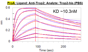
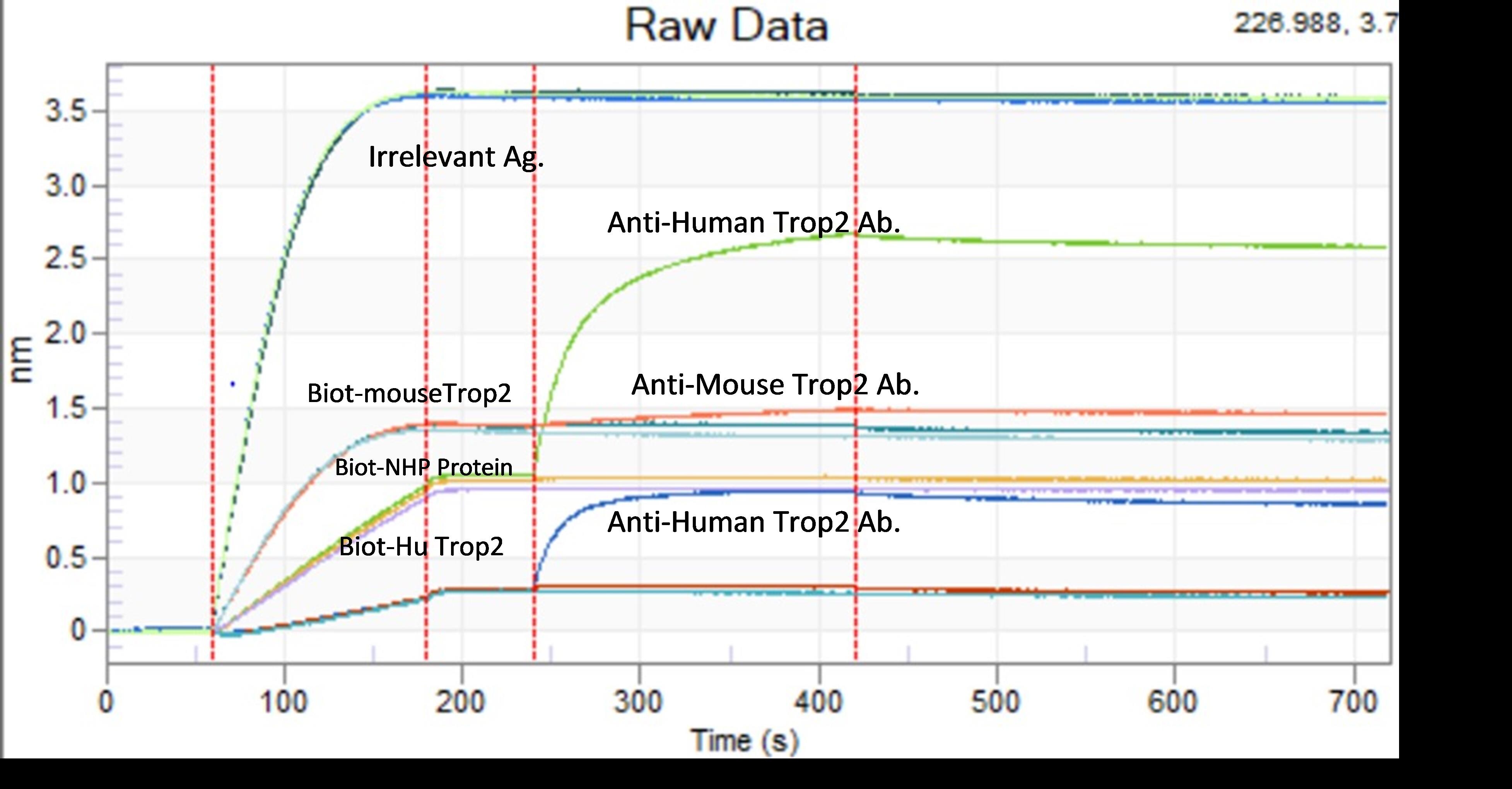
Application: Protein: Protein InteractionSample Tested: Recombinant proteinSpecies: HumanVerified Customer | Posted 08/24/2021Ligand: Anti-Trop2 Analyte: human Trop2-his 1:1 fitting with KD 10nM

-
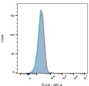
Application: Flow CytometrySample Tested: Breast cancer cell line MCF7Species: HumanVerified Customer | Posted 12/23/2020Detection of TROP2 on human breast cancer cell line MCF7. MCF7 cells were treated with 67 nM of the human TROP2 antibody (catalog # MAB650) or mouse IgG2a isotype control, followed by a secondary antibody goat a-mouse IgG Fc APC. Isotype control (RED), human TROP2 antibody (Blue).
-
Application: Binding by OctetSample Tested: Recombinant proteinSpecies: HumanVerified Customer | Posted 06/08/2020
There are no reviews that match your criteria.
Protocols
Find general support by application which include: protocols, troubleshooting, illustrated assays, videos and webinars.
- 7-Amino Actinomycin D (7-AAD) Cell Viability Flow Cytometry Protocol
- Cellular Response to Hypoxia Protocols
- Extracellular Membrane Flow Cytometry Protocol
- Flow Cytometry Protocol for Cell Surface Markers
- Flow Cytometry Protocol for Staining Membrane Associated Proteins
- Flow Cytometry Staining Protocols
- Flow Cytometry Troubleshooting Guide
- Intracellular Flow Cytometry Protocol Using Alcohol (Methanol)
- Intracellular Flow Cytometry Protocol Using Detergents
- Intracellular Nuclear Staining Flow Cytometry Protocol Using Detergents
- Intracellular Staining Flow Cytometry Protocol Using Alcohol Permeabilization
- Intracellular Staining Flow Cytometry Protocol Using Detergents to Permeabilize Cells
- Propidium Iodide Cell Viability Flow Cytometry Protocol
- Protocol for Liperfluo
- Protocol for the Characterization of Human Th22 Cells
- Protocol for the Characterization of Human Th9 Cells
- Protocol: Annexin V and PI Staining by Flow Cytometry
- Protocol: Annexin V and PI Staining for Apoptosis by Flow Cytometry
- R&D Systems Quality Control Western Blot Protocol
- Troubleshooting Guide: Fluorokine Flow Cytometry Kits
- Troubleshooting Guide: Western Blot Figures
- Western Blot Conditions
- Western Blot Protocol
- Western Blot Protocol for Cell Lysates
- Western Blot Troubleshooting
- Western Blot Troubleshooting Guide
- View all Protocols, Troubleshooting, Illustrated assays and Webinars