


![Flow Cytometry: HypoxiTRAK(TM) [NBP3-18531]](https://resources.rndsystems.com/images/products/HypoxiTRAK-TM-Flow-Cytometry-NBP3-18531-img0001.jpg "Flow Cytometry: HypoxiTRAK(TM) [NBP3-18531]")
Loading...
Key Product Details
Applications
Flow Cytometry, Immunocytochemistry/ Immunofluorescence, Functional Assay
Loading...
Product Specifications
Description
HypoxiTRAK(TM) is a novel, far-red fluorescing dye that reveals hypoxic experience of individual cells yet is non-toxic to normoxic cells. It is suitable for flow cytometry and imaging protocols and is compatible with common vis. range fluors e.g. FITC, R-PE. HypoxiTRAK(TM) enables entirely new assay approaches to functional hypoxia.
HypoxiTRAK(TM) can be used in flow cytometry and fluorescence imaging and is spectrally compatible with common visible range fluors including FITC and R-PE.
HypoxiTRAK(TM) is supplied as a faint blue aqueous solution at a concentration of 10uM. Products are shipped at ambient temperature, but on receipt packs should be stored at 2-8C. DO NOT FREEZE! Otherwise HypoxiTRAK(TM) can come out of solution when frozen and it is difficult (but not impossible) to get it back into solution.
HypoxiTRAK(TM) can be diluted in culture media (e.g. RPMI 1640) and physiological buffers (eg PBS, Hanks's, etc.).
HypoxiTRAK(TM) can be used in flow cytometry and fluorescence imaging and is spectrally compatible with common visible range fluors including FITC and R-PE.
HypoxiTRAK(TM) is supplied as a faint blue aqueous solution at a concentration of 10uM. Products are shipped at ambient temperature, but on receipt packs should be stored at 2-8C. DO NOT FREEZE! Otherwise HypoxiTRAK(TM) can come out of solution when frozen and it is difficult (but not impossible) to get it back into solution.
HypoxiTRAK(TM) can be diluted in culture media (e.g. RPMI 1640) and physiological buffers (eg PBS, Hanks's, etc.).
Application Notes
Flow Cytometry: suitable for dynamic live cell hypoxia study; extract statistical information on the distribution of hypoxic experience over both time and treatment conditions
Immunocytochemistry/Immunofluorescence: dynamic live cell hypoxia study; immuno-fluorescence; gain understanding of the spatial hypoxic experience in micro-tissues and spheroids
Immunocytochemistry/Immunofluorescence: dynamic live cell hypoxia study; immuno-fluorescence; gain understanding of the spatial hypoxic experience in micro-tissues and spheroids
Spectra Viewer
Plan Your Experiments
Use our spectra viewer to interactively plan your experiments, assessing multiplexing options. View the excitation and emission spectra for our fluorescent dye range and other commonly used dyes.
Spectra Viewer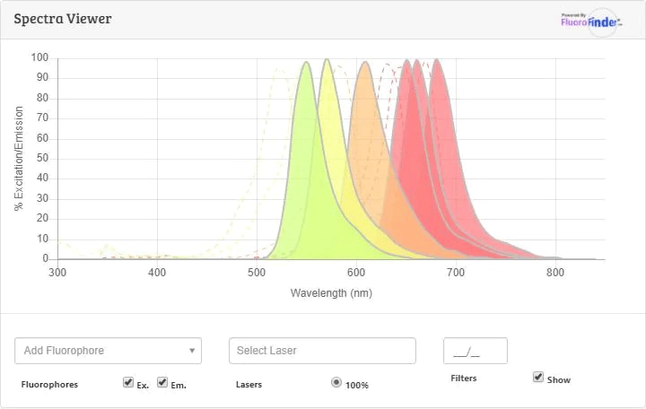
Scientific Data Images for HypoxiTRAK(TM)
Flow Cytometry: HypoxiTRAK(TM) [NBP3-18531]
Flow Cytometry: HypoxiTRAK(TM) [NBP3-18531] - Flow cytometry of -metabolite, blue & red excitation; intact A549 cells under hypoxia (3%, 1%), with & without [4 day co-exposure]![Immunocytochemistry/ Immunofluorescence: HypoxiTRAK(TM) [NBP3-18531]](https://resources.rndsystems.com/images/products/HypoxiTRAK-TM-Immunocytochemistry-Immunofluorescence-NBP3-18531-img0003.jpg "Immunocytochemistry/ Immunofluorescence: HypoxiTRAK(TM) [NBP3-18531]")
Immunocytochemistry/ Immunofluorescence: HypoxiTRAK(TM) [NBP3-18531]
Immunocytochemistry/Immunofluorescence: HypoxiTRAK(TM) [NBP3-18531] - -metabolite accumulates in cytoplasm of A549 cells (ex. 633 nm; em. 680/20 nm) exposed to 100 nM for 4 days 1% O2![Flow Cytometry: HypoxiTRAK(TM) [NBP3-18531]](https://resources.rndsystems.com/images/products/HypoxiTRAK-TM-Flow-Cytometry-NBP3-18531-img0002.jpg "Flow Cytometry: HypoxiTRAK(TM) [NBP3-18531]")
Flow Cytometry: HypoxiTRAK(TM) [NBP3-18531]
Flow Cytometry: HypoxiTRAK(TM) [NBP3-18531] - -metabolite persistence: intact A549 cells retain >70% after washing and 24h incubation in -free medium (ex. 633nm; em. >695nm)![Immunocytochemistry/ Immunofluorescence: HypoxiTRAK(TM) [NBP3-18531]](https://resources.rndsystems.com/images/products/HypoxiTRAK-TM-Immunocytochemistry-Immunofluorescence-NBP3-18531-img0005.jpg "Immunocytochemistry/ Immunofluorescence: HypoxiTRAK(TM) [NBP3-18531]")
Immunocytochemistry/ Immunofluorescence: HypoxiTRAK(TM) [NBP3-18531]
Immunocytochemistry/Immunofluorescence: HypoxiTRAK(TM) [NBP3-18531] - Multi-Cellular Tumor Spheroid (MCTS) generated from 5000 cells (Cal33, HNC) in a ULA well. MCTS grown for 3 days then probed with 50 nM HypoxiTRAK(TM) for a further 3 days. False-color heatmap image shows hypoxia reported in a cortical pattern and with a hypocellular centre (by Hoechst staining consistent with controls, not shown).Courtesy of DA Close & PA Johnston - University of Pittsburgh, School of Pharmacy.Formulation, Preparation, and Storage
NBP3-18531
| Concentration | Please see the protocols for proper use of this product. If no protocol is available, contact technical services for assistance. |
| Shipping | The product is shipped at ambient temperature. Upon receipt, store it immediately at the temperature recommended below. |
| Stability & Storage | Store at 4C in the dark. Do not freeze. |
Background: HypoxiTRAK(TM)
Additional HypoxiTRAK(TM) Products
Product Documents for HypoxiTRAK(TM)
Certificate of Analysis
To download a Certificate of Analysis, please enter a lot or batch number in the search box below.
Product Specific Notices for HypoxiTRAK(TM)
HypoxiTRAK(TM) is a registered trademark of BioStatus Limited.
This product is for research use only and is not approved for use in humans or in clinical diagnosis. Support products are guaranteed for 6 months from date of receipt.
Customer Reviews for HypoxiTRAK(TM)
There are currently no reviews for this product. Be the first to review HypoxiTRAK(TM) and earn rewards!
Have you used HypoxiTRAK(TM)?
Submit a review and receive an Amazon gift card!
$25/€18/£15/$25CAN/¥2500 for a review with an image
$10/€7/£6/$10CAN/¥1110 for a review without an image
Submit a review
Protocols
Find general support by application which include: protocols, troubleshooting, illustrated assays, videos and webinars.
- 7-Amino Actinomycin D (7-AAD) Cell Viability Flow Cytometry Protocol
- Appropriate Fixation of IHC/ICC Samples
- Cellular Response to Hypoxia Protocols
- ClariTSA™ Fluorophore Kits
- Detection & Visualization of Antibody Binding
- Extracellular Membrane Flow Cytometry Protocol
- Flow Cytometry Protocol for Cell Surface Markers
- Flow Cytometry Protocol for Staining Membrane Associated Proteins
- Flow Cytometry Staining Protocols
- Flow Cytometry Troubleshooting Guide
- ICC Cell Smear Protocol for Suspension Cells
- ICC Immunocytochemistry Protocol Videos
- ICC for Adherent Cells
- Immunocytochemistry (ICC) Protocol
- Immunocytochemistry Troubleshooting
- Immunofluorescence of Organoids Embedded in Cultrex Basement Membrane Extract
- Immunohistochemistry (IHC) and Immunocytochemistry (ICC) Protocols
- Intracellular Flow Cytometry Protocol Using Alcohol (Methanol)
- Intracellular Flow Cytometry Protocol Using Detergents
- Intracellular Nuclear Staining Flow Cytometry Protocol Using Detergents
- Intracellular Staining Flow Cytometry Protocol Using Alcohol Permeabilization
- Intracellular Staining Flow Cytometry Protocol Using Detergents to Permeabilize Cells
- Preparing Samples for IHC/ICC Experiments
- Preventing Non-Specific Staining (Non-Specific Binding)
- Primary Antibody Selection & Optimization
- Propidium Iodide Cell Viability Flow Cytometry Protocol
- Protocol for Liperfluo
- Protocol for VisUCyte™ HRP Polymer Detection Reagent
- Protocol for the Characterization of Human Th22 Cells
- Protocol for the Characterization of Human Th9 Cells
- Protocol for the Fluorescent ICC Staining of Cell Smears - Graphic
- Protocol for the Fluorescent ICC Staining of Cultured Cells on Coverslips - Graphic
- Protocol for the Preparation and Fluorescent ICC Staining of Cells on Coverslips
- Protocol for the Preparation and Fluorescent ICC Staining of Non-adherent Cells
- Protocol for the Preparation and Fluorescent ICC Staining of Stem Cells on Coverslips
- Protocol for the Preparation of a Cell Smear for Non-adherent Cell ICC - Graphic
- Protocol: Annexin V and PI Staining by Flow Cytometry
- Protocol: Annexin V and PI Staining for Apoptosis by Flow Cytometry
- TUNEL and Active Caspase-3 Detection by IHC/ICC Protocol
- The Importance of IHC/ICC Controls
- Troubleshooting Guide: Fluorokine Flow Cytometry Kits
- View all Protocols, Troubleshooting, Illustrated assays and Webinars
Loading...