
IgA2 Antibody (RM125)
Novus Biologicals | Catalog # NBP2-62025

Scientific images may be resized, cropped, or adjusted for brightness or contrast for presentation purposes. These updates are limited to presentation and do not affect the underlying scientific interpretation of the data. Where available, additional source data may be provided upon request.
Key Product Details
Species Reactivity
Applications
Label
Antibody Source
Product Specifications
Immunogen
Specificity
Clonality
Host
Isotype
Scientific Data Images for IgA2 Antibody (RM125)
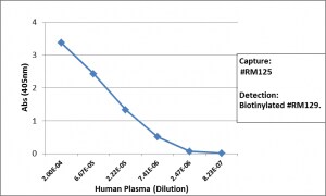
Sandwich ELISA: IgA2 Antibody (RM125) - Azide and BSA Free [NBP2-62025] -
Sandwich ELISA: IgA2 Antibody (RM125) - Azide and BSA Free [NBP2-62025] - Sandwich ELISA using RM125 as the capture antibody (100ng/well), and NBP3-18532 as the detection antibody, followed by an alkaline phosphatase conjugated streptavidin.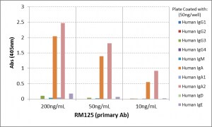
ELISA: IgA2 Antibody (RM125) - Azide and BSA Free [NBP2-62025] -
ELISA: IgA2 Antibody (RM125) - Azide and BSA Free [NBP2-62025] - ELISA of human immunoglobulins shows RM125 only reacts to human IgA2. No cross reactivity with human IgA1, IgG, IgM, IgD, or IgE. The plate was coated with 50 ng/well of different immunoglobulins. 200 ng/mL, 50ng/mL, or 10 ng/mL of RM125 was used as the primary antibody. An alkaline phosphatase conjugated anti-rabbit IgG as the secondary antibody.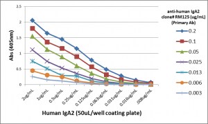
ELISA: IgA2 Antibody (RM125) - Azide and BSA Free [NBP2-62025] -
ELISA: IgA2 Antibody (RM125) - Azide and BSA Free [NBP2-62025] - A titer ELISA using RM125. The plate was coated with different amounts of human IgA2. A serial dilution of RM125 was used as the primary antibody. An alkaline phosphatase conjugated anti-rabbit IgG as the secondary antibody.Applications for IgA2 Antibody (RM125)
Immunocytochemistry/ Immunofluorescence
Immunohistochemistry
Sandwich ELISA Capture
Sandwich ELISA Detection
Reviewed Applications
Read 1 review rated 5 using NBP2-62025 in the following applications:
Formulation, Preparation, and Storage
Purification
Formulation
Preservative
Concentration
Shipping
Stability & Storage
Background: IgA2
Alternate Names
Gene Symbol
Additional IgA2 Products
Product Documents for IgA2 Antibody (RM125)
Certificate of Analysis
To download a Certificate of Analysis, please enter a lot or batch number in the search box below.
Product Specific Notices for IgA2 Antibody (RM125)
This product is for research use only and is not approved for use in humans or in clinical diagnosis. Primary Antibodies are guaranteed for 1 year from date of receipt.
Customer Reviews for IgA2 Antibody (RM125) (1)
Have you used IgA2 Antibody (RM125)?
Submit a review and receive an Amazon gift card!
$25/€18/£15/$25CAN/¥2500 for a review with an image
$10/€7/£6/$10CAN/¥1110 for a review without an image
Submit a review
Customer Images
-
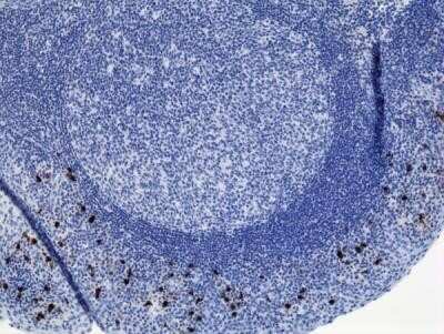
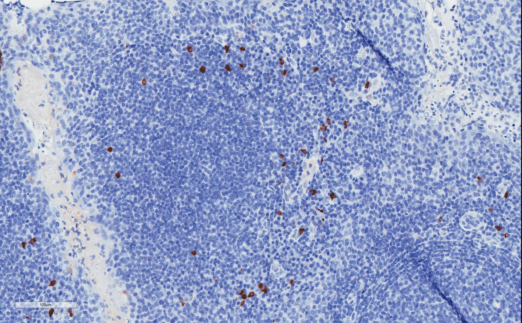
Application: Immunohistochemistry-ParaffinSample Tested: Human Tonsil tissueSpecies: HumanVerified Customer | Posted 11/27/2018IgA2 stained human tonsil FFPE, 20X magnification. Antigen retrieval was performed with citrate pH6 buffer. Primary concentration 1:8000.Antigen retrieval was done with citrate pH6 for 15 mins at 100C and allowed to cool 30 minutes at room temperature. Primary was diluted 1:8000 and incubated at room temperature for 30 mins in Dako antibody diluent (S3022). Secondary ImmPRESS goat anti-rabbit HRP polymer was incubate 30 minutes at room temperature and signal was developed with SIgnalStain DAB chromagen.

There are no reviews that match your criteria.
Protocols
Find general support by application which include: protocols, troubleshooting, illustrated assays, videos and webinars.
- Antigen Retrieval Protocol (PIER)
- Antigen Retrieval for Frozen Sections Protocol
- Appropriate Fixation of IHC/ICC Samples
- Cellular Response to Hypoxia Protocols
- Chromogenic IHC Staining of Formalin-Fixed Paraffin-Embedded (FFPE) Tissue Protocol
- Chromogenic Immunohistochemistry Staining of Frozen Tissue
- ClariTSA™ Fluorophore Kits
- Detection & Visualization of Antibody Binding
- ELISA Sample Preparation & Collection Guide
- ELISA Troubleshooting Guide
- Fluorescent IHC Staining of Frozen Tissue Protocol
- Graphic Protocol for Heat-induced Epitope Retrieval
- Graphic Protocol for the Preparation and Fluorescent IHC Staining of Frozen Tissue Sections
- Graphic Protocol for the Preparation and Fluorescent IHC Staining of Paraffin-embedded Tissue Sections
- Graphic Protocol for the Preparation of Gelatin-coated Slides for Histological Tissue Sections
- How to Run an R&D Systems DuoSet ELISA
- How to Run an R&D Systems Quantikine ELISA
- How to Run an R&D Systems Quantikine™ QuicKit™ ELISA
- ICC Cell Smear Protocol for Suspension Cells
- ICC Immunocytochemistry Protocol Videos
- ICC for Adherent Cells
- IHC Sample Preparation (Frozen sections vs Paraffin)
- Immunocytochemistry (ICC) Protocol
- Immunocytochemistry Troubleshooting
- Immunofluorescence of Organoids Embedded in Cultrex Basement Membrane Extract
- Immunofluorescent IHC Staining of Formalin-Fixed Paraffin-Embedded (FFPE) Tissue Protocol
- Immunohistochemistry (IHC) and Immunocytochemistry (ICC) Protocols
- Immunohistochemistry Frozen Troubleshooting
- Immunohistochemistry Paraffin Troubleshooting
- Preparing Samples for IHC/ICC Experiments
- Preventing Non-Specific Staining (Non-Specific Binding)
- Primary Antibody Selection & Optimization
- Protocol for Heat-Induced Epitope Retrieval (HIER)
- Protocol for Making a 4% Formaldehyde Solution in PBS
- Protocol for VisUCyte™ HRP Polymer Detection Reagent
- Protocol for the Fluorescent ICC Staining of Cell Smears - Graphic
- Protocol for the Fluorescent ICC Staining of Cultured Cells on Coverslips - Graphic
- Protocol for the Preparation & Fixation of Cells on Coverslips
- Protocol for the Preparation and Chromogenic IHC Staining of Frozen Tissue Sections
- Protocol for the Preparation and Chromogenic IHC Staining of Frozen Tissue Sections - Graphic
- Protocol for the Preparation and Chromogenic IHC Staining of Paraffin-embedded Tissue Sections
- Protocol for the Preparation and Chromogenic IHC Staining of Paraffin-embedded Tissue Sections - Graphic
- Protocol for the Preparation and Fluorescent ICC Staining of Cells on Coverslips
- Protocol for the Preparation and Fluorescent ICC Staining of Non-adherent Cells
- Protocol for the Preparation and Fluorescent ICC Staining of Stem Cells on Coverslips
- Protocol for the Preparation and Fluorescent IHC Staining of Frozen Tissue Sections
- Protocol for the Preparation and Fluorescent IHC Staining of Paraffin-embedded Tissue Sections
- Protocol for the Preparation of Gelatin-coated Slides for Histological Tissue Sections
- Protocol for the Preparation of a Cell Smear for Non-adherent Cell ICC - Graphic
- Quantikine HS ELISA Kit Assay Principle, Alkaline Phosphatase
- Quantikine HS ELISA Kit Principle, Streptavidin-HRP Polymer
- Sandwich ELISA (Colorimetric) – Biotin/Streptavidin Detection Protocol
- Sandwich ELISA (Colorimetric) – Direct Detection Protocol
- TUNEL and Active Caspase-3 Detection by IHC/ICC Protocol
- The Importance of IHC/ICC Controls
- Troubleshooting Guide: ELISA
- Troubleshooting Guide: Immunohistochemistry
- View all Protocols, Troubleshooting, Illustrated assays and Webinars